0500
Application of diffusion microstructure imaging in thigh muscle1The Third Affiliated Hospital of Southern Medical University, Guangzhou, China, 2School of Biomedical Engineering, Southern Medical University, GuangZhou, China, 3Guangdong Provincial Key Laboratory of Medical Image Processing and Guangdong Province Engineering Laboratory for Medical Imaging and Diagnostic Technology, Southern Medical University, Guangzhou, China, 4Philips Healthcare, Guangzhou, China, 5Philips Healthcare, Hong Kong, China
Synopsis
Keywords: Muscle, Aging, thigh muscle, Fat/Water Separation
Motivation: Diffusion microstructure imaging (DMI) obtains the distribution of three microstructure compartments of each voxel[1], and more specifically understands the microstructure integrity of tissues.
Goal(s): Quantitative detection of the correlation between the microstructure parameters of thigh muscle and its fat infiltration degree and its relationship with the body composition of healthy individuals.
Approach: We used DMI and mDIXON quant sequences to measure microstructural parameters and fat fraction (FF) of thigh muscle in healthy individuals, and to assess the value of DMI in thigh muscle imaging.
Results: DMI parameters differed significantly during aging and were significantly correlated with intramuscular FF.
Impact: The change of DMI parameters is a sensitive indicator of the physiological changes of muscle fiber structure during aging, which may suggest the microstructure characteristics of local metabolic changes in the body during aging.
Introduction
For the quantitative assessment of thigh muscle, advanced quantitative magnetic resonance imaging is an important tool. Diffusion microstructure imaging (DMI) is an advanced multicompartment technique based on diffusion magnetic resonance imaging, which has been shown to be more comprehensive than diffusion tensor imaging in displaying information about the tissue microstructure [2-5]. Our aim is to investigate the changes in thigh muscle microstructure with age and its correlation with fat fraction. This helps us to further our understanding of the microscopic changes in muscle fibers during the aging process and DMI parameters can be used as imaging biomarkers to detect changes in the integrity or part of the muscle fiber microstructure.Methods
3T-MRI of thigh muscles was performed in 30 healthy individuals using DMI, turbo spin echo (TSE) T1WI sequence, and water fat imaging based on chemical shift imaging techniques. The region of interest (roi) was manually depicted by two radiologists with more than five years of diagnostic experience on the FF maps (InterlliSpaceTM Portal, Philips) provided by Philips, based on T1WI anatomy (Figure 1)[6]. We divided the cohort in an age gradient (20-40 years old, number of subjects (n) = 10; 40-60 years old, n = 10; ≥60 years old, n = 10) to study the correlation of thigh muscle microstructural parameters with FF. DMI was acquired using a single-shot echo planar imaging technique with the following parameters: TR/TE = 2032/64.6 msec; acquisition matrix = 128×128; 20 axial slices, slice thickness = 4 mm with no inter-slice gap; reconstructed voxel size = 2.44mm×2.44 mm×2.03 mm; 25 noncollinear directions for b = 600, 1000 sec/mm2; and three b = 0 reference images. All diffusion-weighted images were preprocessed by MRtrix3 (https://www.mrtrix.org). Firstly, denoising was performed using Marchenko–Pastur principal component analysis. Subsequently, correction for head motion and eddy current-induced distortions was carried out. The diffusion vector table was then transformed based on the motion and distortion correction transformation. The preprocessed data were fitted into the NODDI model using the AMICO package[7]. The DMI parameters for thigh muscle are a microstructural diffusion index estimated in the NODDI model (Figure 2) - free water fraction (V-ISO), and the volume fraction inside of muscular fibers (V-intra)[1]. FF and T2* values were measured in mDixon Quant sequence images. The Shapiro-Wilk test was used to assess the normal distribution of the data. The Mann-Whitney U test was used to assess differences in DMI parameters, FF and T2* values among participants of different genders and different BMI groups. The Kruskal-Wallis test was used to compare between-group differences in DMI parameters, FF and T2* values by dividing the participants into three groups according to age, and Pearson and Spearman (controlling for gender) correlations were used to correlate differences in DMI parameters, FF and T2* values.Results
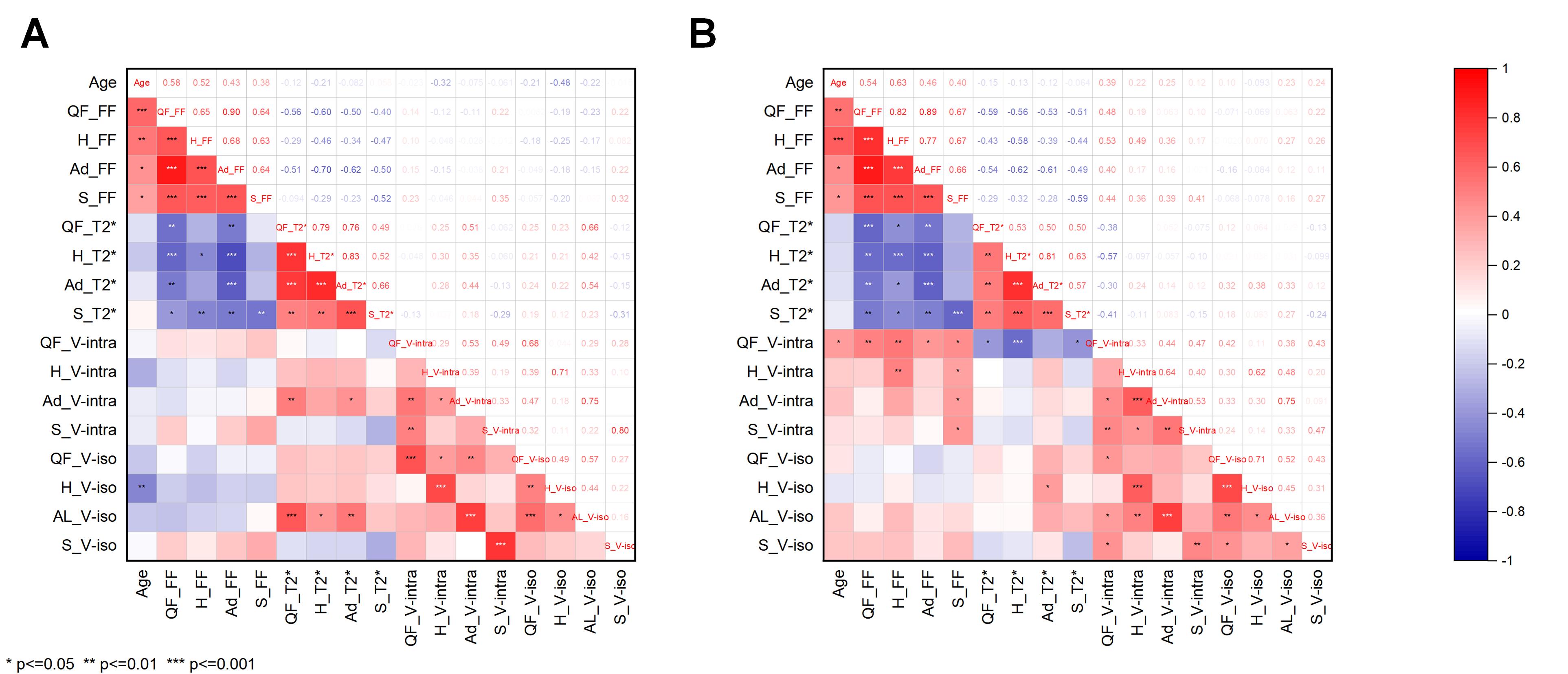
The demographics of the participants can be shown in Table 1. Among 30 participants of different age groups, the intramuscular FF and V-ISO of quadriceps femoris and hamstring muscles of both sides showed differences (p < 0.05) (Table 2). The intramuscular FF of quadriceps femoris, hamstring, adductors, and sartorius in both thighs were positively correlated with age (r = 0.376 to 0.631, p < 0.001), and negatively correlated with T2* value (r = -0.613 to -0.46, p < 0.001). There is a strong correlation between intramuscular FF of the right thigh muscle and V-intra (r = 0.364 to 0.527, p < 0.001) (Figure 3).Discussion & Conclusion
Since directional diffusivity similar to that of neuronal axons was found in muscle, and the sizes of both myogenic fibers (1 to 2μm) and neuronal axons (0.01 to 10μm) were in a similar range, we determined two volume fractions based on a standard model of cerebral white matter- V-ISO and V-intra[1]. Intramuscular FF and T2* values reflect physiological changes in aging. The FF of thigh muscle can not reflect the degree of fat deposition in the whole body well, and the relationship between body mass index and fat varies in different populations. DMI parameters can provide additional information on muscle function and microstructure during the aging process and may serve as a biomarker suggestive of physiological microscopic changes in aging. Diffusion microstructural imaging was successfully applied to non-invasively assess the microstructure of thigh muscles in healthy volunteers. It is observed that the microstructure composition of thigh muscles is different during aging. Muscle microstructure parameters are related to intramuscular FF, which can be used as imaging parameters to evaluate the integrity and function of thigh muscles.Acknowledgements
Xiaodong Zhang and Xinyuan Zhang are both co-corresponding authors.
The study was supported by Present's Fund of the Third Affiliated Hospital of Southern Medical University(grant No. YM2021012).
References
[1] Rau A, Jungmann PM, Diallo TD, et al. Application of diffusion microstructure imaging in musculoskeletal radiology - translation from head to shoulders. Eur Radiol. 2023;33(3):1565-1574.
[2] Rau A, Reisert M, Kellner E, et al. Increased interstitial fluid in periventricular and deep white matter hyperintensities in patients with suspected idiopathic normal pressure hydrocephalus. Sci Rep. 2021;11(1):19552.
[3] Rau A, Jost WH, Demerath T, et al. Diffusion microstructure imaging in progressive supranuclear palsy: reduced axonal volumes in the superior cerebellar peduncles, dentato-rubro-thalamic tracts, ventromedial thalami, and frontomesial white matter.Cereb Cortex. 2022;32(24):5628-5636.
[4] Rau A, Schroeter N, Blazhenets G, et al. Widespread white matter oedema in subacute COVID-19 patients with neurological symptoms. Brain. 2022;145(9):3203-3213.
[5] Würtemberger U, Diebold M, Erny D, et al. Diffusion microstructure imaging to analyze perilesional T2 signal changes in brain metastases and glioblastomas.Cancers (Basel). 2022;14(5):1155.
[6] Mohajer B, Dolatshahi M, Moradi K, et al. Role of Thigh Muscle Changes in Knee Osteoarthritis Outcomes: Osteoarthritis Initiative Data. Radiology. 2022;305(1):169-178.
[7]Tariq M, Schneider T, Alexander DC, et al. Bingham-NODDI: Mapping anisotropic orientation dispersion of neurites using diffusion MRI.Neuroimage. 2016;133:207-223.
Figures
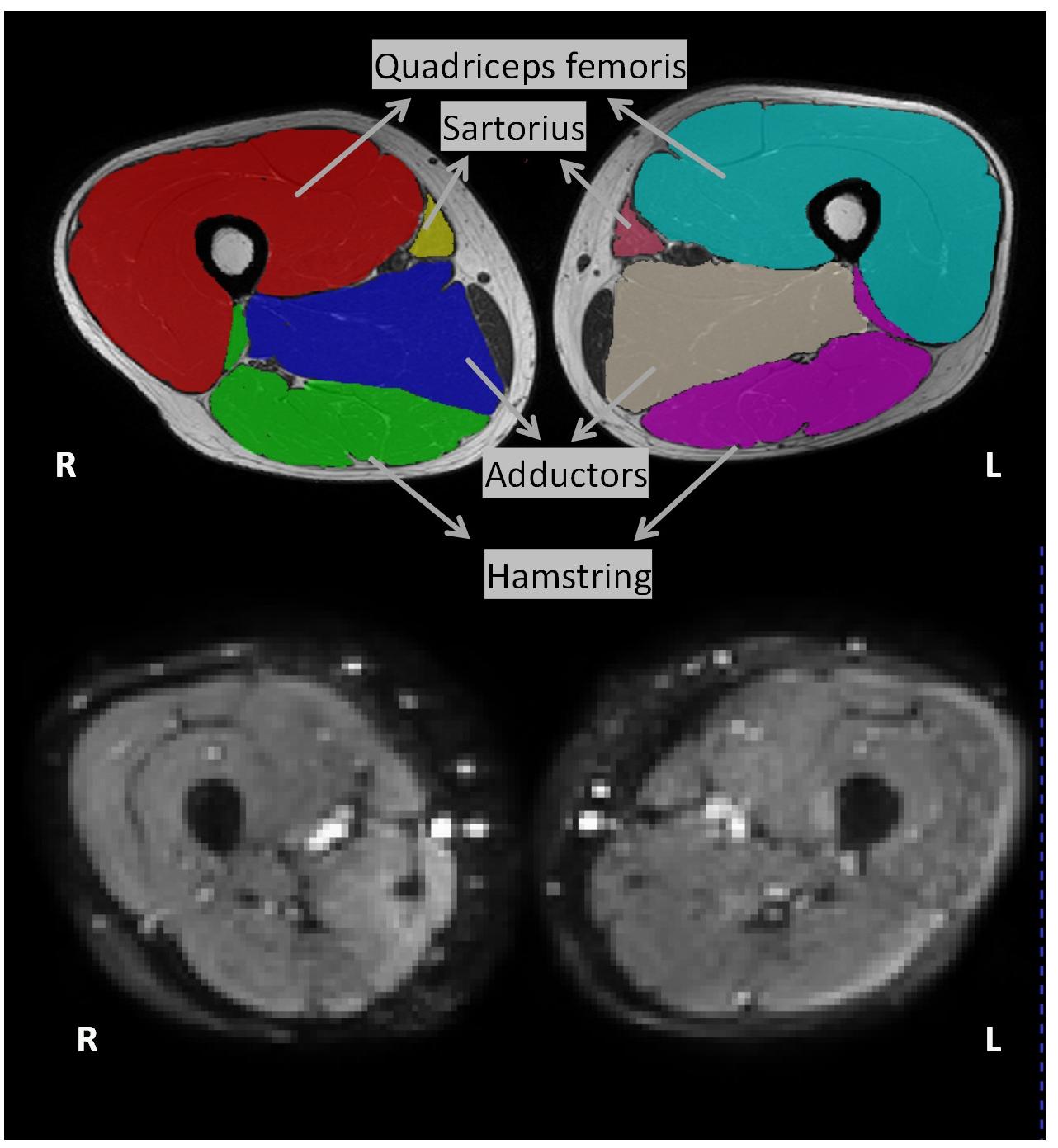
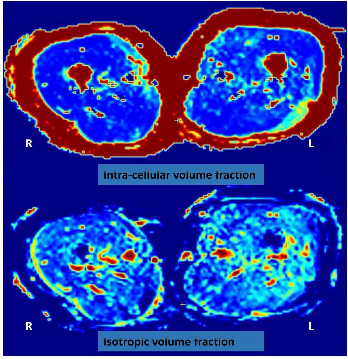
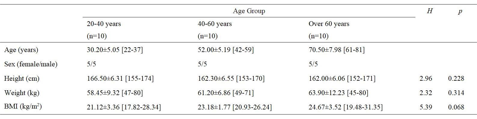
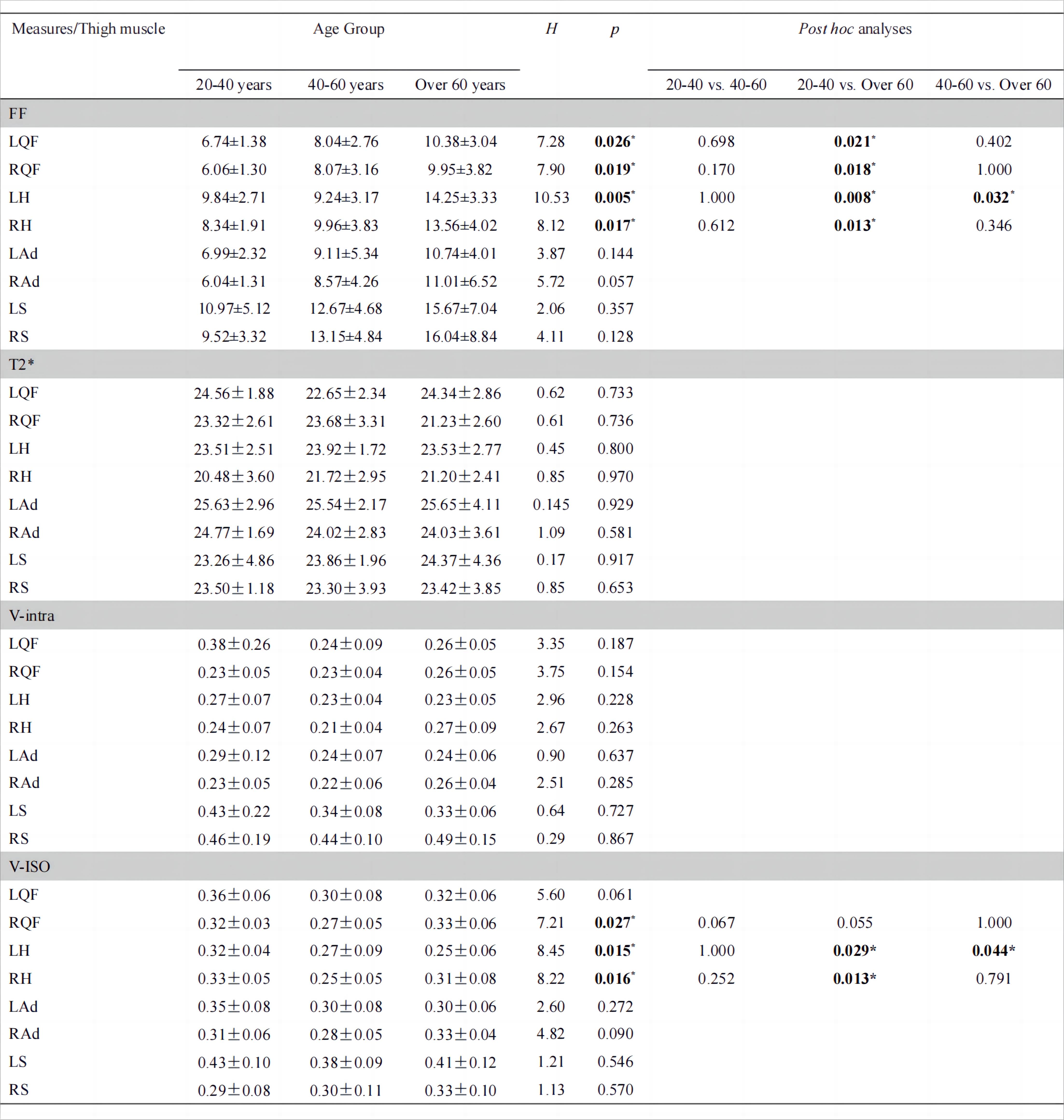
Table 2: The Kruskal-Wallis H test of muscle parameters in different age groups.
Bonferroni correction was done. LQF: Left Quadriceps Femoris, RQF: Right Quadriceps Femoris, LH: Left Hamstring, RH: Right Hamstring, LAd: Left Adductors, RAd: Right Adductors, LS: Left Sartorius, RS: Right Sartorius.