0499
Single point Dixon for water-fat separation in musculoskeletal TSE imaging1Department of Diagnostic and Interventional Radiology, Technical University of Munich, Munich, Germany, 2Philips GmbH Market DACH, Hamburg, Germany
Synopsis
Keywords: Other Musculoskeletal, Fat, Bone, Fat/Water separation
Motivation: TSE Dixon imaging is highly desired in clinical musculoskeletal imaging for its versatile contrasts. However, conventional Dixon imaging needs at least two different echo times, resulting in prolonged scan time.
Goal(s): To develop a single point Dixon imaging technique for TSE acquisition (sTSE-Dixon) that provide fat-separated, water-separated and in phase images from a single echo time acquisition.
Approach: Shifted single echo TSE acquisition was combined with smoothness-constrained non-linear inverse water fat problem.
Results: High quality water-separated, fat-separated and in phase images comparable to those obtained with conventional two-point Dixon were achieved from a single echo time shifted TSE acquisition.
Impact: In this work, a single echo time TSE sequence can deliver three image sets that are of great clinical need, including fat-separated images (comparable to standard T1-weighted), water-separated images (comparable to STIR), in-phase images (comparable to standard T2-weighted)
Introduction
Turbo spin echo (TSE) is widely used in musculoskeletal imaging because of its ability to provide versatile contrasts. Fat suppressed TSE images are often desired for reliable detection of pathology without being obscured by the high intensity of the fat signal. Fat suppression in TSE acquisition can be performed using the short tau inversion recovery (STIR) technique at the cost of losing partially the water signal. Dixon imaging techniques can be applied to TSE acquisition generating not only fat-suppressed (or water-separated) images but also fat-separated images (comparable to standard T1-weighted sequence) and in phase images (comparable to standard T2-weighted sequence) [1]. Traditional Dixon technique however requires the acquisition of at least two different echo times, resulting in either prolonged scan time or increased echo spacing. In this work we develop a single point Dixon imaging technique for TSE acquisition (sTSE-Dixon) that provide fat-separated, water-separated and in phase images from a single echo time acquisition.Methods
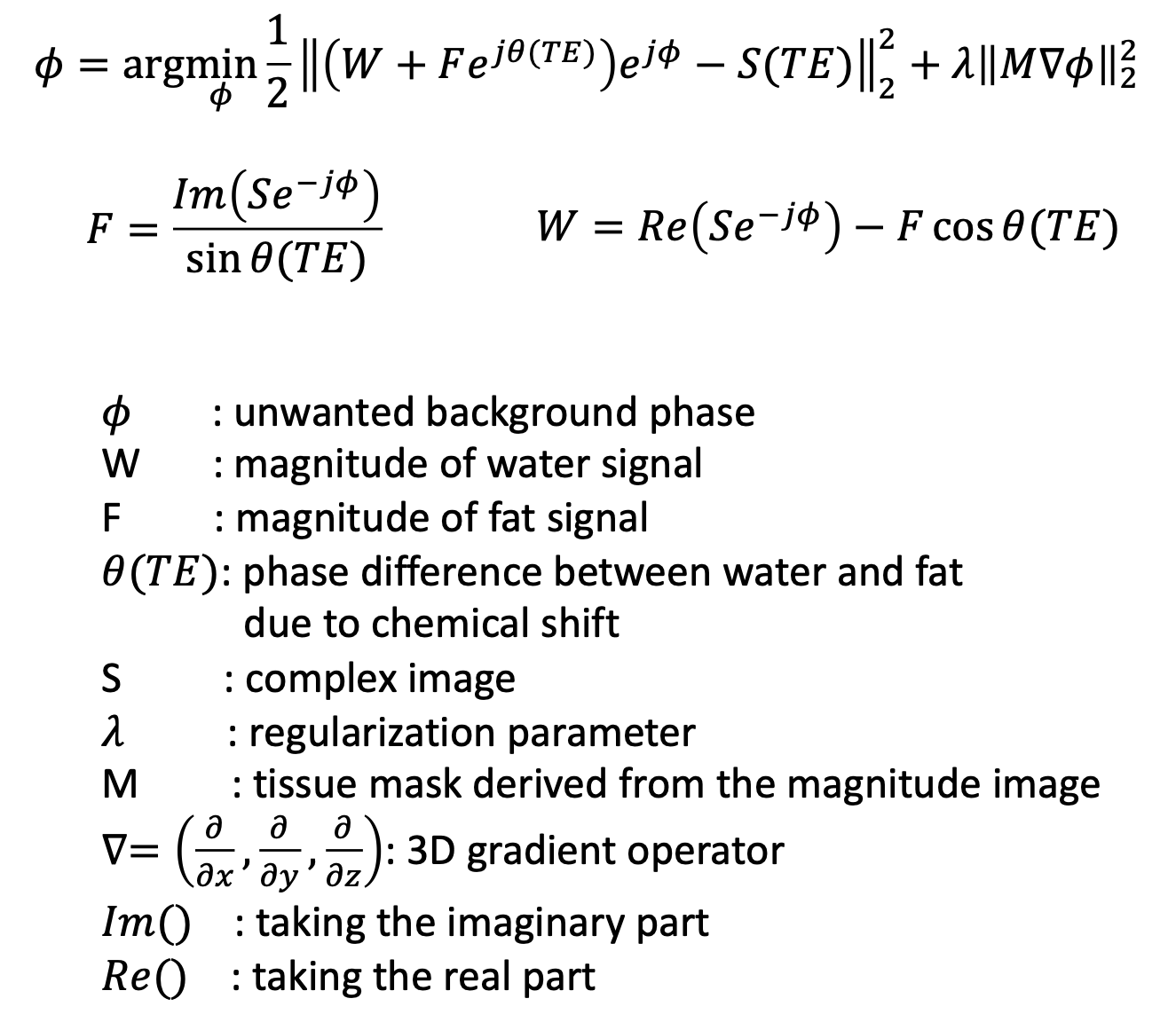
sTSE-Dixon: sTSE-Dixon was inspired by the works on single-point Dixon for ultra-short echo time (sUTE-Dixon) imaging [2-9]. Due to the chemical shift difference, water and fat can be differentiated by their accumulated phases when they are not refocused (at t > 0 for gradient echo or at $$$t \neq TE$$$for spin echo). However, other phases induced by sources such as B0 inhomogeneity, B1, gradient delay can overshadow the water fat phase difference, making the phase-based fat water separation impossible.At ultra-shot echo time, the phase introduced by B0 inhomogeneity for most anatomies can be safely assumed to vary smoothly across the field-of-view (FOV). With the additional assumption that other unwanted phases also spatially smooth, a recent sUTE-Dixon technique estimated the water, the fat, and the unwanted phase simultaneously by solving the smoothness-constrained non-linear inverse water-fat problem shown in Figure 1 [5].
Similar smoothness assumption on unwanted phases can also be made for the case of TSE acquisition where the acquisition is shifted away from the spin echo by a small $$$\Delta TE$$$. Therefore, the sUTE-Dixon water fat separation approach can be applied for the shifted TSE acquisition.
Data acquisition: Ankles of three volunteers were scanned in a 3 T scanner (Ingenia Elition X, Philips Healthcare) using a 16-channel foot/ankle coil. A shifted 2D TSE sequence was employed with $$$\Delta TE = 0.4$$$ ms. Other imaging parameters are TE/TR = 30/3000 ms, FOV (FH x AP) = 200 x 250 mm2, acquisition voxel size 0.51 x 0.67 mm2, 24 slices of 2.5 mm thickness. The total scan time is 1 min 42 seconds. To compare the proposed sTSE-Dixon with the conventional two-point Dixon, a TSE Dixon sequence with $$$\Delta TE = 0$$$ ms and $$$\Delta TE = 1$$$ ms was also acquired. The total scan time here is 3 mins 24 seconds.
Results
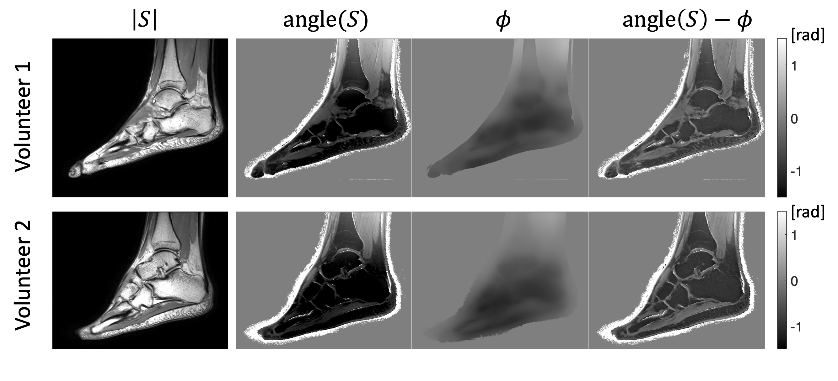
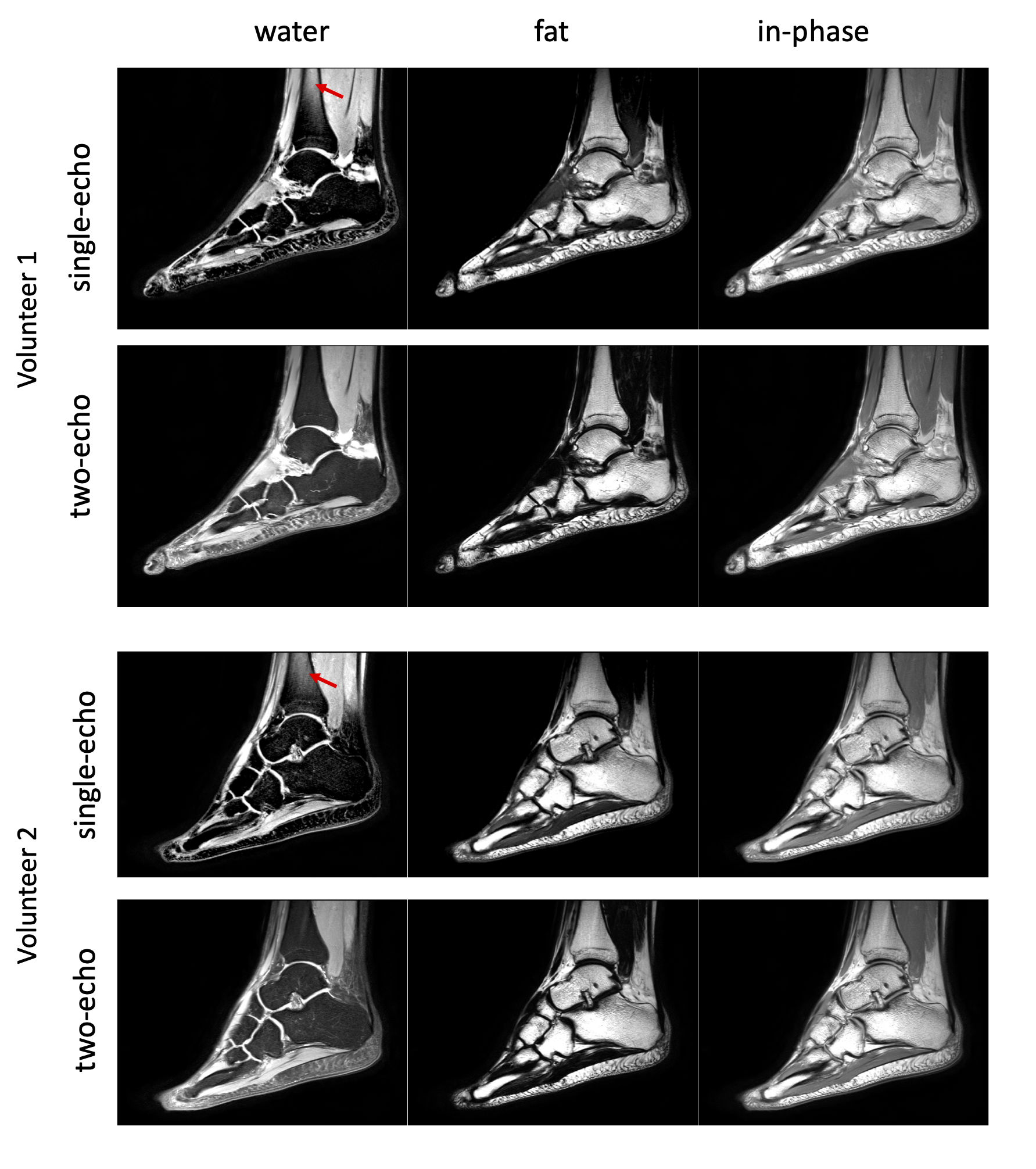
Figure 2 shows the magnitude, $$$|S|$$$, and phase of the image, $$$angle(S)$$$, acquired with the shifted TSE sequence, the estimated unwanted phase, $$$\Phi$$$, and the water-fat phase, $$$angle(S) - \Phi$$$. The unwanted phase shows smoothly varying feature while the water-fat phase gives clear contrast between water and fat regions. Figure 3 presents the water-separated, fat-separated, and in-phase images obtained with single-echo shifted acquisition (sTSE-Dixon) and with two-echo Dixon acquisition in two subjects. In both subjects, sTSE-Dixon yield high quality water only, fat only and in-phase images that are comparable to those from two-echo Dixon. A slight fat-swap can be observed at the top of the water images for both volunteers most likely due to residual B0 effects.Conclusion
By combining a shifted TSE acquisition with single-point Dixon processing, reliable fat-separated, water-separated and in-phase images can be achieved with the acquisition of only one echo.Acknowledgements
The authors acknowledge research support from Philips Healthcare.References
[1] Reeder SB, Pineda AR, Wen Z, Shimakawa A, Yu H, Brittain JH, Gold GE, Beaulieu CH, Pelc NJ. Iterative decomposition of water and fat with echo asymmetry and least-squares estimation (IDEAL): application with fast spin-echo imaging. Magn Reson Med 2005 54(3):636-44.
[2] Carl M, Bydder GM, Du J. UTE imaging with simultaneous water and fat signal suppression using a time-efficient multispoke inversion recovery pulse sequence. Magn Reson Med 2016;76(2):577-582.
[3] Jang H, Carl M, Ma Y, Jerban S, Guo T, Zhao W, Chang EY, Du J. Fat suppression for ultrashort echo time imaging using a single-point Dixon method. NMR Biomed 2019;32(5):e4069.
[4] Ma J. A single-point Dixon technique for fat-suppressed fast 3D gradient-echo imaging with a flexible echo time. J Magn Reson Imaging 2008;27(4):881-890.
[5] Kronthaler S, Boehm C, Feuerriegel G, Bornert P, Katscher U, Weiss K, Makowski MR, Schwaiger BJ, Gersing AS, Karampinos DC. Assessment of vertebral fractures and edema of the thoracolumbar spine based on water-fat and susceptibility-weighted images derived from a single ultra-short echo time scan. Magn Reson Med 2021;87(4):1771-1783.
[6] Feuerriegel GC, Kronthaler S, Boehm C, Renz M, Leonhardt Y, Gassert F, Foreman SC, Weiss K, Wurm M, Liebig T, Makowski MR, Schwaiger BJ, Karampinos DC, Gersing AS. Diagnostic value of water-fat-separated images and CT-like susceptibility-weighted images extracted from a single ultrashort echo time sequence for the evaluation of vertebral fractures and degenerative changes of the spine. Eur Radiol 2022.
[7] Kronthaler S, Feuerriegel GC, Boehm C, Gersing AS, Schwaiger BJ, Makowski MR, Weiss K, Karampinos DC. On the robustness of single UTE-Dixon for simultaneous short T2*, water and fat imaging across skeletal anatomies. Proc ISMRM 2022. p 536.
[8] Van AT, Weiss K, Feuerriegel GC, Braun P, Krishnamoorthy G, Gersing AS, Pipe JG, Karampinos DC. Single UTE-Dixon for simultaneous short T2* water and fat imaging using a FLORET trajectory. Proc ISMRM 2023. p. 1222.
[9] Newbury N, Sedaghat S, Athertya J, Carl M, Du J, Jang H. Ultrashort echo time single point Dixon using 3D phase modeling. Proc ISMRM 2023.p. 121.
Figures