0498
UTE-Based DW-SSFP for Musculoskeletal MRI1Beckman Institute, Biomedical Imaging Center, University of Illinois at Urbana-Champaign, Urbana, IL, United States
Synopsis
Keywords: Tendon/Ligament, Tendon/Ligament, DW-SSFP
Motivation: Fiber tracking of ligaments suffer from a low signal due to their fast T2 relaxation during a long echo time in spin-echo EPI diffusion sequences.
Goal(s): Shorten the echo time of the diffusion sequence and acquire in 3D.
Approach: Use a 3D spiral-in readout in a DW-SSFP sequence.
Results: The 3D spiral-in readout features an enhanced signal, shorter TR or TE, a wider interval for diffusion gradients, reduced geometric distortion, and a minimized echo time shift. It is demonstrated for tracts of knee ligaments using ex vivo hind limbs of piglets.
Impact: The proposed diffusion imaging sequence provides a high sensitivity to musculoskeletal tissue with short T2 relaxation times. This will be useful in studying the muscles, tendons, ligaments, and cartilage.
Introduction
T2 relaxation time of musculoskeletal tissues is known to be small. Hence, the spin-echo diffusion imaging sequence suffers from a low signal due to a long echo time in the spin-echo diffusion sequence with one or two refocusing 180° RF pulses. On the hand, DW-SSFP diffusion imaging sequence forms an echo without the refocusing RF pulse, hence it can be more sensitive to tissues with small T2 relaxation time 1, 2. The DW-SSFP sequence can be further improved by employing the UTE sequence using a 3D spiral-in readout 3.Methods
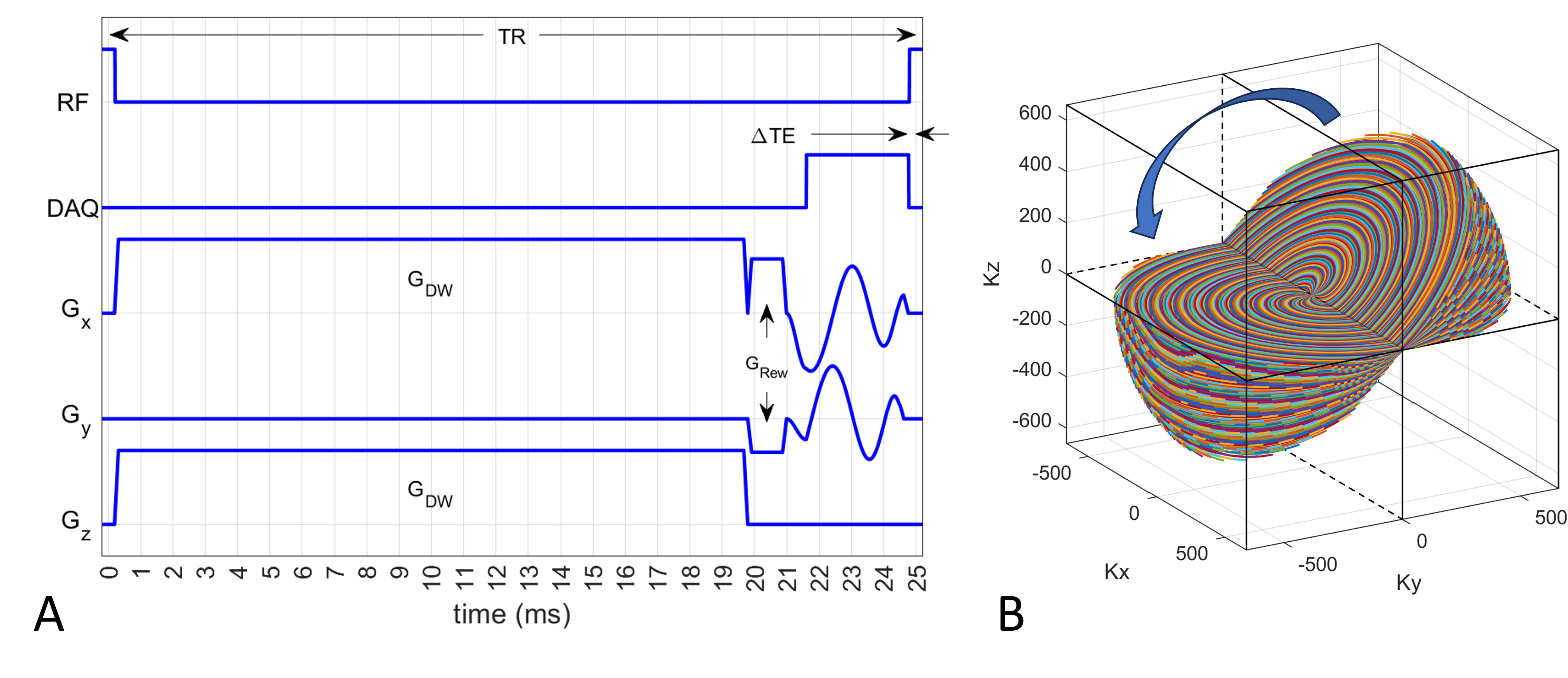
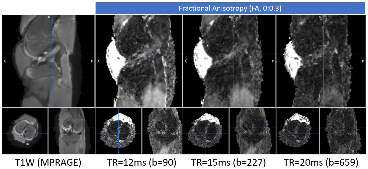
The DW-SSFP sequence with the 3D spiral trajectory is shown in Figure 1. The echo shift (ΔTE) is minimized compared to the EPI readout 4, which can increase the duration of the diffusion encoding gradient (GDW). One b0 volume was obtained with a small b value to spoil the FID component in SSFP 5, followed by 6 diffusion-weighted acquisitions with a large b value. The diffusion sensitivity or b value is dependent on TR and GDW, hence the DW-SSFP sequence was run with 3 different TR’s of 12, 15, and 20ms. The scan parameters for TR=15ms were: echo shift (ΔTE)=0.12ms, voxel=1mm isotropic, duration of GDW=10.1ms, amplitude of GDW=37mT/m, flip angle=30°, and scan time=14:58. The b values at the 3 TR’s are listed in Figure 2. At TR=15ms, the b value for the b0 volume was 0.28 s/mm2. The objects were ex vivo knee samples of 3 to 4 months old porcine. Four knee specimens were scanned using a 15-ch knee RF coil at 3T MRI. A T1W anatomy image was also obtained. The fractional anisotropy (FA) was estimated using DTIFIT of FSL 6. Fiber tracts of anterior and posterior cruciate ligaments (ACL and PCL) were traced and characterized using DSI Studio 7.Results
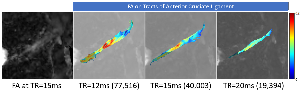
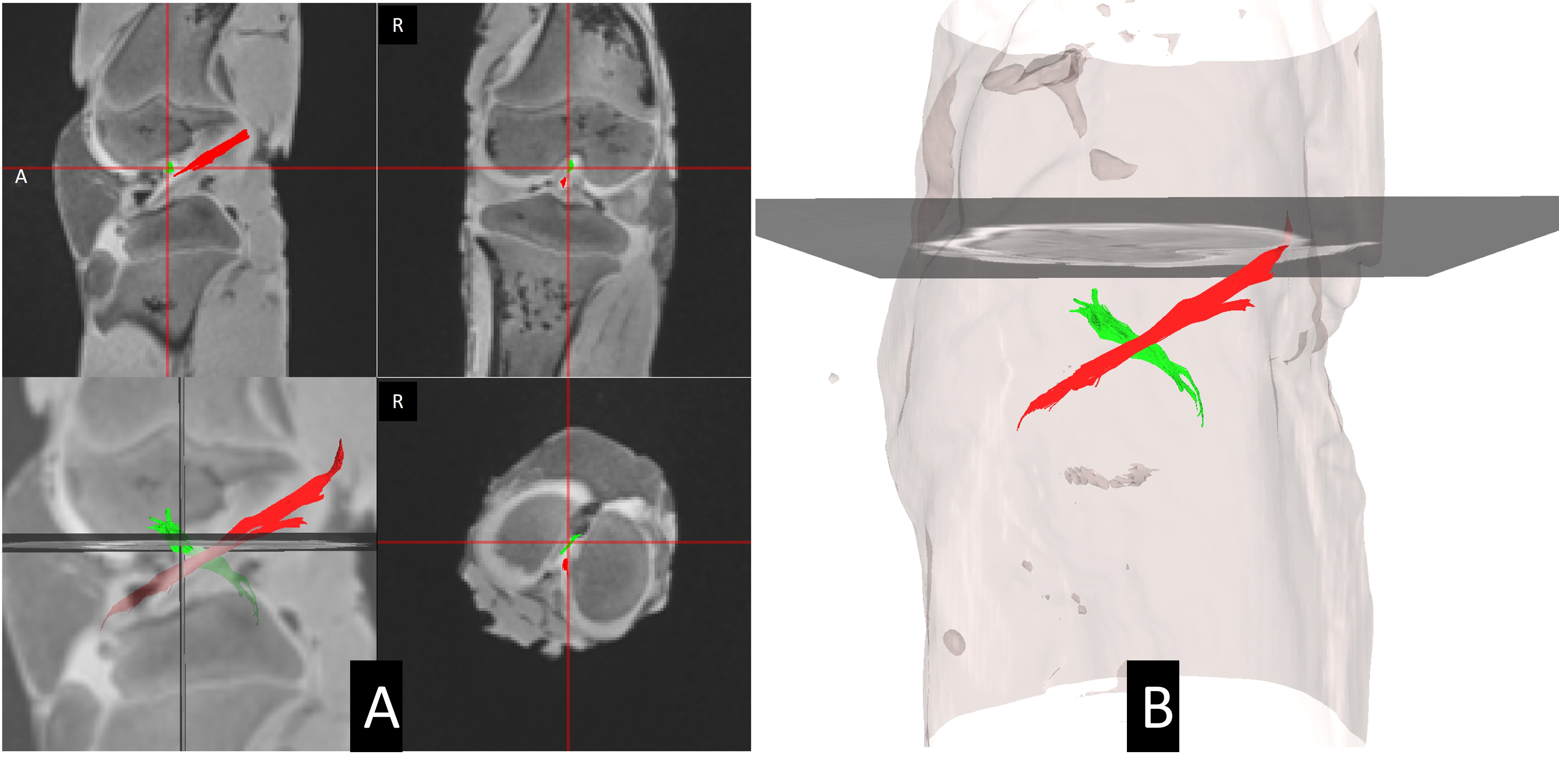
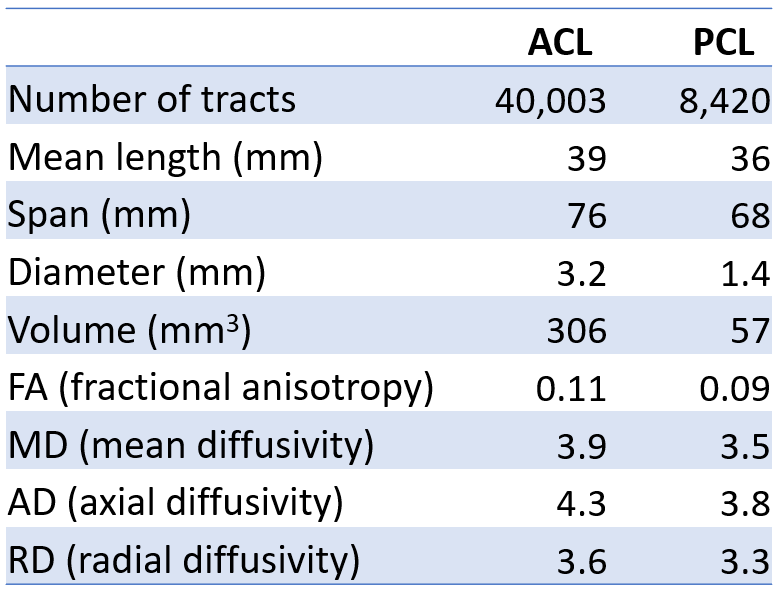
TR and b value had affected FA of ligaments (Figure 2) and accordingly the tracts for the ACL tracts (Figure 3). In these two figures, the FA map and tracts appeared more reasonable at TR=15ms or b=226s/mm2. Tracts of ACL and PCL of one representative specimen at TR=15ms are shown in Figure 4 and their statistics are summarized in Table 1.Discussion
The echo time for diffusion in the DW-SSFP sequence with TR=15ms was 30ms for the echo pathway initiated from the immediately prior TR. This is smaller than the echo time achievable using the spin echo EPI. The spiral readout contributed to widening the diffusion encoding gradient. However, this made the duty cycle of the diffusion gradient greater than 75% at TR=15ms, which limited the allowed maximum gradient amplitude from the scanner. The maximum gradient amplitude in the vendor specification is valid only for a small duty cycle. The spiral readout did not suffer from noticeable geometric distortion, which was a clear advantage over the EPI-based diffusion sequence in tracking the tracts. The 3D acquisition helped not only a higher signal, but also tracking a thin and tilted bundle of ACL and PCL tracts. Although the DW-SSFP is known to be extremely sensitive to motion 8, 9, its in vivo application to knee is expected to be relatively manageable than the brain due to a low b value, no physiologic effect except at arteries, and the intrinsic robustness of spiral trajectory to flow and fluctuations.Conclusion
The echo time of the proposed DW-SSFP sequence could be set within 30ms with a diffusion encoding gradient sensitive enough for tracking knee ligaments in a whole-body MRI. Furthermore, this sequence did not suffer from geometric distortion and provided high signal for tracking knee ligaments.Acknowledgements
The author would like to thank Dr. Anna Dilger in Meat Science Laboratory at University of Illinois at Urbana-Champaign for porcine samples. The author also appreciates Dr. Elahe Ganji and Dr. Mariana Kersh for allowing me to scan these samples.References
1. Miller KL, Hargreaves BA, Gold GE, Pauly JM. Steady-state diffusion-weighted imaging of in vivo knee cartilage. Magn Reson Med. 2004;51(2):394-8.2. Bieri O, Ganter C, Welsch GH, Trattnig S, Mamisch TC, Scheffler K. Fast diffusion-weighted steady state free precession imaging of in vivo knee cartilage. Magnet Reson Med. 2012;67(3):691-700.
3. Jung KJ, Sutton B, editors. Three-Dimensional Sodium MRI Using A Rotation of Spiral Disc (RSD) Trajectory. Int Soc Magn Reson Med; 2021.
4. Miller KL, McNab JA, Jbabdi S, Douaud G. Diffusion tractography of post-mortem human brains: optimization and comparison of spin echo and steady-state free precession techniques. Neuroimage. 2012;59(3):2284-97.
5. Jung KJ. Synthesis methods of multiple phase-cycled SSFP images to reduce the band artifact and noise more reliably. Magn Reson Imaging. 2010;28(1):103-18.
6. Behrens TEJ, Woolrich MW, Jenkinson M, Johansen-Berg H, Nunes RG, Clare S et al. Characterization and propagation of uncertainty in diffusion-weighted MR imaging. Magnet Reson Med. 2003;50(5):1077-88.
7. Yeh FC. Shape analysis of the human association pathways. Neuroimage. 2020;223:117329.
8. Jung KJ, Cho ZH. Reduction of Flow Artifacts in Nmr Diffusion Imaging Using View-Angle Tilted Line-Integral Projection Reconstruction. Magnet Reson Med. 1991;19(2):349-60.
9. McNab JA, Miller KL. Steady-state diffusion-weighted imaging: theory, acquisition and analysis. NMR Biomed. 2010;23(7):781-93.
Figures