0497
Skeletal Muscle phosphocreatine assessment in diabetes and peripheral arterial disease with 1H MRI: a feasibility study1Radiology, Washington University in, Saint Louis, MO, United States, 2Washington University in Saint Louis, Saint Louis, MO, United States, 3Surgery, Washington University in Saint Louis, Saint Louis, MO, United States, 4Program for Physical Therapy, Washington University in Saint Louis, Saint Louis, MO, United States, 5Radi, John Hopkins University, Baltimore, MD, United States, 6Biomedical Engineering, Zhejiang University, Hangzhou, China, 7Medicine, Washington University in Saint Louis, Saint Louis, MO, United States, 8Radiology, Washington University in Saint Louis, Saint Louis, MO, United States
Synopsis
Keywords: Functional/Dynamic, Metabolism, diabetes, peripheral arterial disease, perfusion
Motivation: Both Type 2 Diabetes Mellitus (T2D) and peripheral artery disease (PAD) are linked to impaired mitochondrial function in peripheral tissue that may precede micro-vascular disorders.
Goal(s): The goal is to demonstrate the feasibility of 1H-based MRI for dynamic quantification of skeletal muscle PCr (SMPCr) concentration in vivo on a 3T clinical MRI system, in healthy controls, T2D, and PAD.
Approach: Dynamic 1H-based PCr imaging was developed and evaluated in human subjects in a rest-exercise-recovery protocol, based on Chemical Exchange Saturation Transfer (CEST) technique.
Results: Reproducibility of PCr measurement and declines in measures of mitochondria function in aging and diseases are demonstrated.
Impact: The 1H MRI technique was able to measure differences in assessing mitochondrial function in people with T2D and PAD, without additional hardware. This technical development may allow early diagnosis of complications associated with various peripheral disorders.
Introduction
Both Type 2 Diabetes Mellitus (T2D) and peripheral artery disease (PAD) are linked to impaired mitochondrial function in peripheral tissue that may precede micro-vascular disorders, evidenced by decreased mRNA expression in genes playing a role in oxidative phosphorylation [1,2,3]. 31P magnetic resonance spectroscopy (MRS) offers a direct method of measuring mitochondrial function via phosphocreatine (PCr) concentrations. However, 31P MRS is limited by low spatial resolution and requirement for extra hardware. The objective of this study is to demonstrate the feasibility of 1H-based magnetic resonance imaging (MRI) for dynamic quantification of skeletal muscle PCr (SMPCr) concentration in vivo on a 3T clinical MRI system, in healthy controls, T2D, and PAD.Methods
1H-based PCr imaging is based on the MRI Chemical Exchange Saturation Transfer (CEST) technique. Three 1H pools, i.e., water, SMPCr, and magnetic transfer contrast, were considered in a chemical exchange model and the SMPCr can be calculated using Bloch-McConnell equations.Four groups of individuals were recruited for the evaluation of this approach, including 10 young healthy controls (young HC, 25-27y), 7 older heathy controls (old HC, 54 – 64y), 7 T2D without PAD (50-79y), and 3 PAD (53 – 70y). All were examined by the dynamic 1H MRI method to assess kinetic changes of SMPCr concentrations with a rest-exercise-recovery protocol. The exercise is a standardized isometric plantar flexion contraction with 40% maximal voluntary contraction [4]. The young HC group had repeated scans to assess reproducibility via coefficient of variance (CV) values.
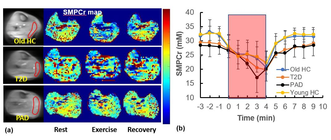
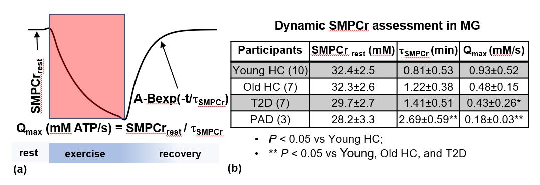
The in vivo calf imaging sessions were performed on a 3T Prisma Siemens whole-body MR system (Siemens Healthineer, Erlangen, Germany). The MRI CEST sequence was a single-slice, saturation pulse (0.6 μT, 800 ms) prepared single-shot rapid acquisition sequence. A total of 31 frequency offsets were acquired to generate a Z-spectrum from 1.3 to 3.5 ppm. Other imaging parameters were field-of-view = 220 x 220 mm2, matrix = 76 x 76, slice thickness = 8 mm, and temporal resolution 60s. A regions-of-interest (ROI) was drawn on the medial gastrocnemius (MG) (Figure 1a). The dynamic SMPCr concentrations were then fitted to first-order kinetics to obtain a time-constant t and Qmax [5].
Results
The overall CV in MG muscle for SMPCr measurement was 8.6%,[6.8%,10.1%]. The reproducibility for rest, exercise, recovery phases were 3.3% 13%, and 6.6%, respectively. Figure 1a and 1b show examples of SMPCr maps and time course of averaged SMPCr. Quantitative endpoints are illustrated in Figure 2. Declines in measures of mitochondria function (increased t and decreased Qmax) are seen with ageing and diseases (T2D or PAD).Conclusion
The dynamic 1H MRI technique was able to measure differences in assessing mitochondrial function in people with T2D and PAD, comparing to those without T2D and PAD, without additional hardware.. Ongoing effort currently focus on acceleration of acquisition speed for more accurate PCr index measurements with the assistance of artificial intelligence. This technical development may allow early diagnosis of complications associated with various peripheral disorders.Acknowledgements
The study was supported in part by a medical student research grant of Radiology Society of North American (RSNA), National institute of health (NIH) DK105322 and AR065672, as well as funding by the Washington University Institute of Clinical and Translational Sciences and Biomedical Magnetic Resonance CenterReferences
1. Sivitz WI, Yorek MA. Mitochondrial dysfunction in diabetes: from molecular mechanisms to functional significance and therapeutic opportunities. Antioxid Redox Signal. 2010;12:537-577.
2. Tay S, Abdulnabi S, Saffaf O, Harroun N, Yang C, Semenkovich CF, Zayed MA. Comprehensive Assessment of Current Management Strategies for Patients With Diabetes and Chronic Limb-Threatening Ischemia. Clin Diabetes. 2021;39:358-388.
3. Anderson JD, Epstein FH, Meyer CH, Hagspiel KD, Wang H, Berr SS, Harthun NL, Weltman A, Dimaria JM, West AM, Kramer CM. Multifactorial determinants of functional capacity in peripheral arterial disease: uncoupling of calf muscle perfusion and metabolism. J Am Coll Cardiol. 2009;54:628-635. 4. Zheng J, Li R, Zayed MA, Yan Y, An H, Hastings MK. Pilot study of contrast-free MRI reveals significantly impaired calf skeletal muscle perfusion in diabetes with incompressible peripheral arteries. Vascular Medicine. 2021;26:367-373.
5. Liu Y, Gu Y, Yu X. Assessing tissue metabolism by phosphorous-31 magnetic resonance spectroscopy and imaging: a methodology review. Quant Imaging Med Surg. 2017;7:707-726.
Figures