0496
A Pilot Study of Insert Nonlinear Diffusion Gradient Coil on Calf Imaging1Department of Biomedical Engineering, Yale University, New Haven, CT, United States, 2Department of Radiology and Biomedical Imaging, Yale University, New Haven, CT, United States, 3Department of Cardiology, Yale University, New Haven, CT, United States
Synopsis
Keywords: Diffusion Acquisition, Diffusion/other diffusion imaging techniques, Nonlinear diffusion gradient
Motivation: A flexible light-weight nonlinear gradient coil was recently proposed and the feasibility for diffusion imaging is to be studied.
Goal(s): To get EPI imaging on calf with the presence of the nonlinear diffusion gradient coil
Approach: We design the experiment, processing pipeline, and an ADC map is shown as preliminary quantitative validation.
Results: This study demonstrates the feasibility of diffusion imaging using an insert diffusion gradient coil.
Impact: This study demonstrates the feasibility of diffusion imaging using an insert diffusion gradient coil, paving the way for further application of muscle and nerve imaging on the lower extremities with high gradient strength.
Introduction
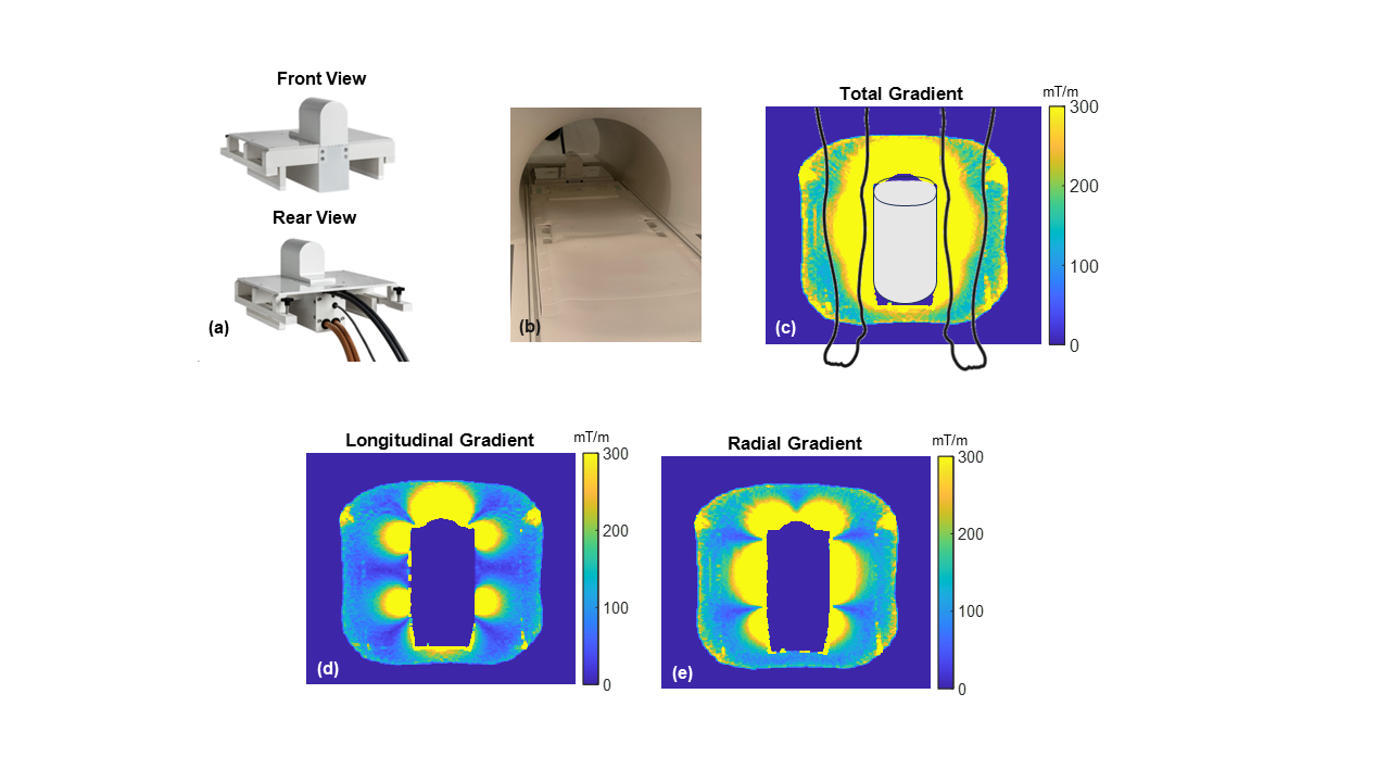
Numerous efforts have been made to increase the gradient strength in diffusion imaging as it is crucial to resolution, signal-to-noise ratio (SNR), contrast-to-noise ratio (CNR), etc1–3. More recently, the concept of a local nonlinear gradient has been proposed, where very high gradient strengths can be achieved over a targeted region4,5.We proposed an inside-out nonlinear gradient coil for diffusion imaging4 that features 1) 10cm compact layout, 2) flexible installation on a scan-by-scan basis, and 3) strong gradients in the range of 200-800mT/m surrounding the cylinder.
It has been reported to improve prostate ADC mapping, but the geometry is also ideal for characterization of skeletal muscle in PAD. The approach achieves a strong gradient particularly in the left-right direction, where restricted diffusion requires high b-values. The overview is shown in Fig. 1. Because the nonlinear gradient can be played in tandem with linear gradients, it is feasible to acquire multidirectional diffusion with short encoding times, revealing muscle fiber diameters as well as higher SNR data for DTI6–8. Evaluation of muscle architecture and injury is important in characterizing patients with PAD and evaluating therapeutic interventions9–11.
The geometry of the nonlinear gradient requires some modifications to EPI acquisition. The gradient proximity to anatomy, and its location at the isocenter, distorts the B0 field. In addition, coupling with the B0 gradient generates an uncommon but benign eddy current. Here we conducted a pilot study of EPI imaging on the calf for feasibility demonstration and quantitative validation. Typical artifacts were examined, and corresponding corrections were applied.
Methods
AcquisitionFor ordinary EPI imaging, single-shot spin-echo EPI data were acquired with the following acquisition setting: FOV = 256x256 mm2; in-plane resolution = 2x2mm2; TE = 45ms; slice thickness = 5mm; acceleration factor = 3; RO = HF, PE = AP. Diffusion-weighted EPI (DW-EPI) imaging shares the same parameters, and the nonlinear gradient waveform has an amplitude of 60% of the max value, with a duration of 5.4ms before and after the 180°pulse. A 4-channel flex coil was wrapped around the calf. The strong mode of spectral attenuated inversion recovery (SPAIR) was applied for fat suppression. For reference ADC map, RESOLVE with linear gradients was acquired with a range of b-value: from 200s/mm2 to 1000s/mm2 with a step size of 100s/mm2.
Larmor Frequency Drift
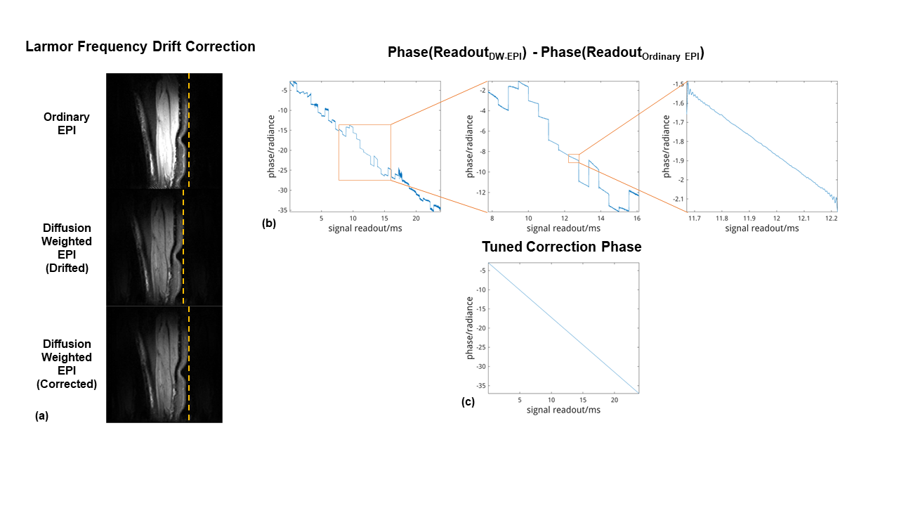
The nonlinear gradient coil was previously found to induce eddy currents in the main field coil, with negligible coupling to the linear gradient coils. Strong coupling to the main field is relatively unique to our setting, as the second-ordered nonlinear field is prone to inducing eddy currents in zeroth-ordered main field coils. However, this uniquely benign eddy current does not cause dephasing and is easily corrected in data prior to processing.
The accrued phase by the drifting frequency was calibrated by a pair of scans with and without nonlinear gradient waveform. The phase jumps due to the phase-encoding blips are ignored and the slope of the consecutive readouts were averaged and tuned for correction phase curve.
Other corrections
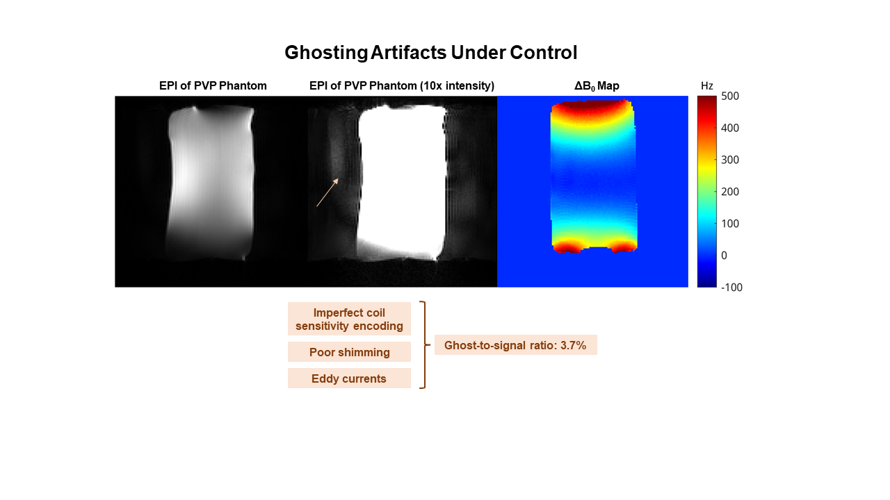
Despite higher B0 inhomogeneity and potential vibration, standard ghost and distortion corrections applied by vendor reconstruction were found sufficient for adequate image quality. The ghosting artifact is pronounced in EPI due to the inconsistency of the bipolar readouts. Besides the linear gradient which can be corrected by the 1D phase correction lines, there are nonlinear field sources, including the field inhomogeneity from the insert coil and the induced eddy currents.
Results and Discussion
Fig. 2 shows shifted DW-EPI caused by the frequency drift. In the absence of correction, this eddy current manifested only as translation in the diffusion weighted images. Averaging the slope and creating a correction phase curve realigns the DW-EPI images.Fig. 3 demonstrates the manageable ghosting artifacts. With careful layout of coils, both ghosting and aliasing can be decreased to a ghost-to-noise ratio of 3.7%.
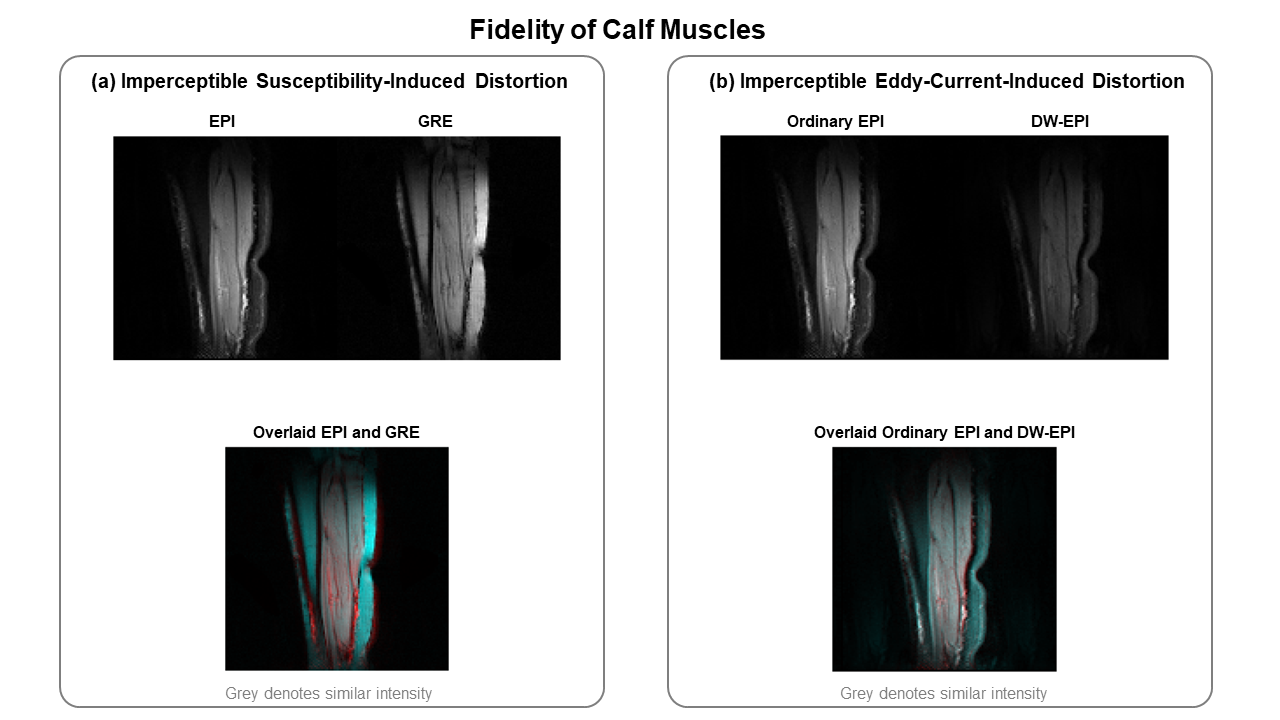
Fig. 4 shows the lack of susceptibility-induced and eddy-current-induced distortion The overlaid images shows no significant shearing or misregistration of alf muscles between EPI and GRE, and between ordinary EPI and DW-EPI.
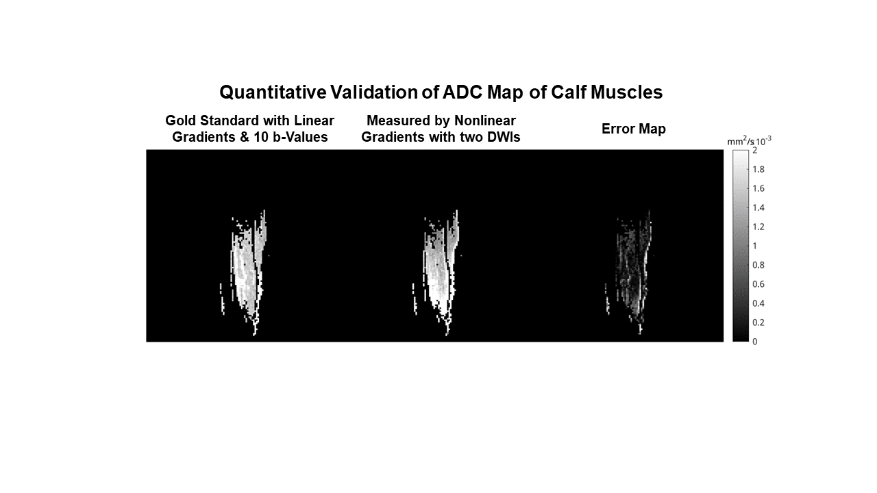
Fig. 5 is the quantitative validation of ADC map against a gold standard RESOLVE. The measured NLG ADC map, based on only two DWIs, shows similar intensity, providing potential for further diffusion analysis.
Conclusion
Our proposed inside-out nonlinear diffusion gradient coil shows feasibility of EPI imaging with corrected frequency drift, controllable ghosting artifacts, and imperceptible distortion on calf muscles from a pilot study. The quantitative ADC map was also validated by the reference. Future work will evaluate timing-matched sequences to quantify SNR gains in ADC as well as multidirectional diffusion encoding.Acknowledgements
No acknowledgement found.References
1. Setsompop K, Kimmlingen R, Eberlein E, et al. Pushing the limits of in vivo diffusion MRI for the Human Connectome Project. NeuroImage. 2013;80:220-233. doi:10.1016/j.neuroimage.2013.05.078
2. Huang SY, Witzel T, Keil B, et al. Connectome 2.0: Developing the next-generation ultra-high gradient strength human MRI scanner for bridging studies of the micro-, meso- and macro-connectome. NeuroImage. 2021;243:118530. doi:10.1016/j.neuroimage.2021.118530
3. Foo TKF, Tan ET, Vermilyea ME, et al. Highly efficient head-only magnetic field insert gradient coil for achieving simultaneous high gradient amplitude and slew rate at 3.0T (MAGNUS) for brain microstructure imaging. Magnetic Resonance in Medicine. 2020;83(6):2356-2369. doi:10.1002/mrm.28087
4. Hoque Bhuiyan E, Dewdney A, Weinreb J, Galiana G. Feasibility of diffusion weighting with a local inside-out nonlinear gradient coil for prostate MRI. Medical Physics. 2021;48(10):5804-5818. doi:10.1002/mp.15100
5. Littin S, Kuder TA, Jia F, et al. Approaching order of magnitude increase of gradient strength: Non-linear breast gradient coil for diffusion encoding. In: Proc. Intl. Soc. Mag. Reson. Med. 30.
6. Galbán CJ, Maderwald S, Uffmann K, Ladd ME. A diffusion tensor imaging analysis of gender differences in water diffusivity within human skeletal muscle. NMR Biomed. 2005;18(8):489-498. doi:10.1002/nbm.975
7. Novikov DS, Fieremans E, Jensen JH, Helpern JA. Random walk with barriers. Nat Phys. 2011;7(6):508-514. doi:10.1038/nphys1936
8. Sigmund EE, Novikov DS, Sui D, et al. Time-dependent diffusion in skeletal muscle with the random permeable barrier model (RPBM): application to normal controls and chronic exertional compartment syndrome patients. NMR Biomed. 2014;27(5):519-528. doi:10.1002/nbm.3087
9. Mazur W, Urbańczyk-Zawadzka M, Banyś R, Obuchowicz R, Trystuła M, Krzyżak AT. Diffusion as a Natural Contrast in MR Imaging of Peripheral Artery Disease (PAD) Tissue Changes. A Case Study of the Clinical Application of DTI for a Patient with Chronic Calf Muscles Ischemia. Diagnostics. 2021;11(1):92. doi:10.3390/diagnostics11010092
10. Stavres J, Wang J, Sica CT, et al. Diffusion tensor imaging indices of acute muscle damage are augmented after exercise in peripheral arterial disease. Eur J Appl Physiol. 2021;121(9):2595-2606. doi:10.1007/s00421-021-04711-7
11. Tan ET, Zochowski KC, Sneag DB. Diffusion MRI fiber diameter for muscle denervation assessment. Quant Imaging Med Surg. 2022;12(1):80-94. doi:10.21037/qims-21-313
Figures