0494
Macromolecule Exchange and Non-Exchange Protons Multiparametric Imaging of Knee: using ZAP/CEST and T2* Mapping with δTE Ultra-Short TE.1Radiology, UC San Diego, La Jolla, CA, United States, 2VA San Diego Healthcare System, San Diego, CA, United States, 3Canon Medical, Ōtawara-shi, Japan
Synopsis
Keywords: Whole Joint, Joints, UTE, ZAP, CEST
Motivation: The study addresses the need for knee injury diagnosis by advancing MRI techniques to capture both structural and biochemical tissue characteristics.
Goal(s): The goal is to develop advanced MRI biomarkers by integrating Z-spectrum/CEST with UTE techniques, improving visualization of knee structures for detection of injuries and pathologies
Approach: We used novel Z-spectrum/CEST and UTE sequences to characterize proton exchange and obtain detailed T2* mapping, aiming to reveal precise biochemical and ultrastructural knee tissue details.
Results: Findings demonstrate improved T2* mapping and proton characterization in knee tissues, suggesting potential biomarkers for early pathology identification, although further validation is necessary for clinical application.
Impact: Multiparametric imaging of macromolecular exchange and non-exchange protons using ZAP/CEST MRI and UTE imaging with tight intervals δTE offers detailed knee tissue visualization, with potential for developing precise diagnostic biomarkers.
Introduction
MRI imaging of the knee stands out as an excellent modality due to its unparalleled capability to visualize soft tissue contrast without ionizing radiation, making it ideal for diagnosing a wide spectrum of knee injuries and conditions. The multiparametric integration of ZAP/CEST and UTE further enhances this capacity by providing both macromolecular exchange protons and non-exchange ultrastructural insights respectively. For tissues with short relaxation times and low proton densities such as ligaments, and meniscus, ultra-short TE (UTE) imaging with a TE of less than 0.1ms is necessary. Z-spectrum analysis protons (ZAP) and chemical exchange saturation transfer (CEST) offer multi-parametric quantitative methods to characterize macromolecule exchange protons. ZAP evaluates the broad spectrum (-100 to 100kHz) of exchange protons estimating the fractions and apparent T2 relaxation times of restricted and freely exchanging protons [1], while CEST focuses on specific frequencies identifying exchange groups like hydroxy (-OH, ~1.0ppm), amine (-NH2, ~2ppm), and amide (-NH, ~3.5ppm) from MTRAsym signal [2]. This study seeks to establish multi-parametric quantitative CEST and ZAP measurements for cartilage, meniscus, ligament, and bone in knee. These quantitative measures have the potential to serve as distinct biomarkers for various injuries and diseases.Methods
Four healthy individuals (35±10 years, 2 males and 2 females) underwent MRI scans on a 3T clinical scanner (Vantage Galan 3T, Canon Medical, Japan). Images were acquired with a 16-ch knee SPEEDER coil. The imaging protocol included: (i) high-resolution Fast-Spin-Echo with TE/TR=36/2300, NEX=1, FOV=16×16cm matrix size 448×448 (ii) a multi-echo 3DUTE with six TEs=0.096/2.3/4.5/6.7/8.9/11.1ms, TR=14.7ms, FA=4°, 1mm thick slices, FOV=20×20cm, sagittal orientation, matrix size 256×256; additional echoes were acquired using (iii) UTE with tight intervals TE=0.4ms (TEs=0.50/0.90/1.30/1.80ms) [3]. (iv) 2D single-shot FSE (TE=10ms, FA=90°) with magnetization transfer (MT) preparation pulses at 55 off-resonance frequencies ranging from –100 to 100kHz, acquired in a centric pattern, FOV=20×20cm, matrix size 320×320, single 5-mm thick slice, and B1rms of 2μT. The total scan time was 18 minutes. Seven regions within the knee—the femur, tibia, tibial and femoral cartilage, anterior and posterior meniscus, and the popliteal ligament—were manually delineated for analysis. per voxel mapping was performed by fitting signal into the bi-exponential model [Eq. 1].$$S(t)=S_s(0)\cdot\exp(-t/T_{2,s}^{*})+S_l(0)\cdot\exp(-t//T_{2,l}^{*})+S' \quad\quad [Eq. 1]$$
where $$$T_{2,s}^{*}$$$ and $$$T_{2,l}^{*}$$$ are short and long relaxation times, S’ is the term to account for noise, fractions of long and short components were calculated as follows:
$$F_{s}=\frac{S_s}{S_s+S_l} \cdot 100\% \quad\quad [Eq. 2.1]$$
$$F_{l}=\frac{S_l}{S_s+S_l} \cdot 100\% \quad\quad [Eq. 2.2]$$
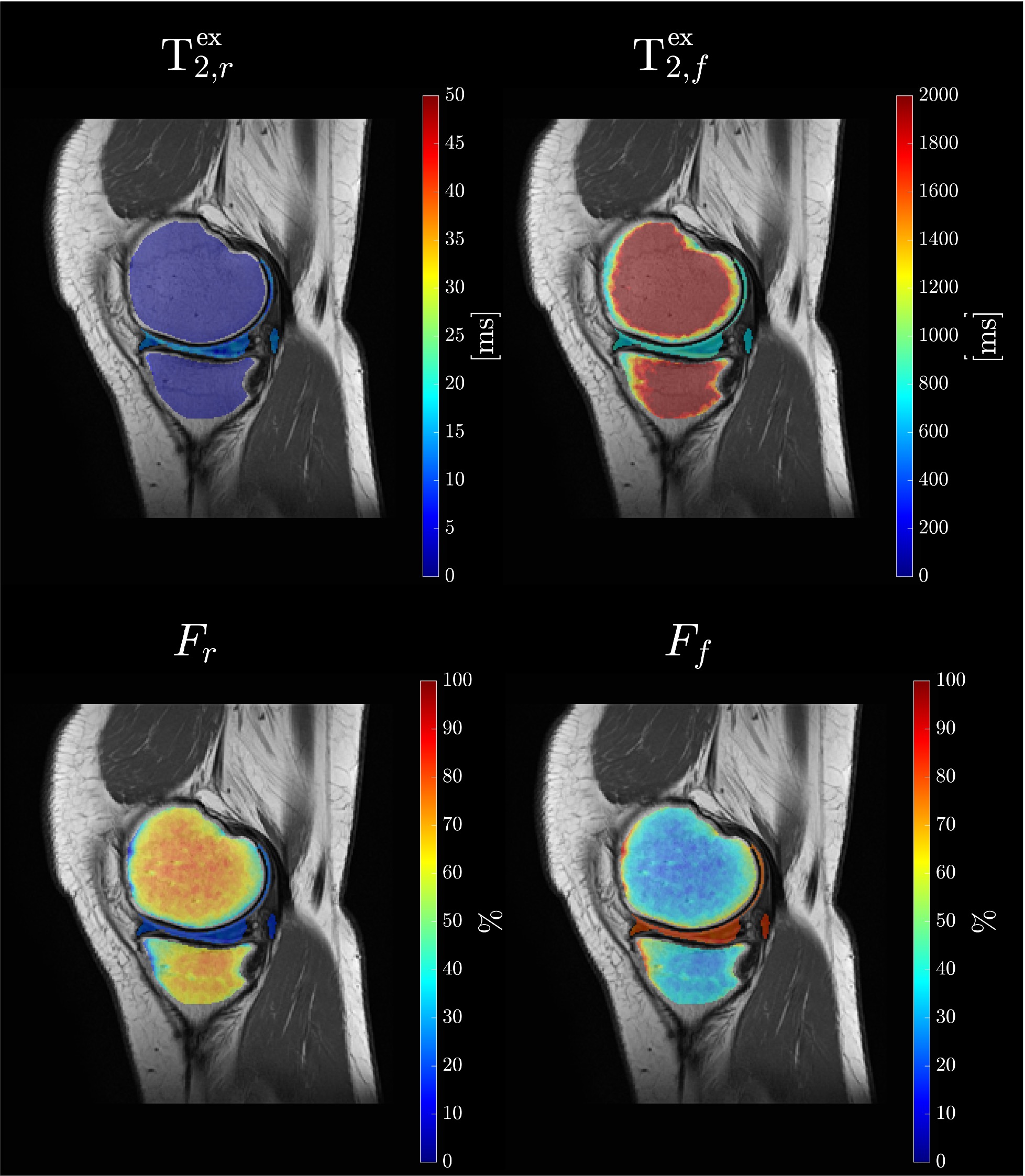
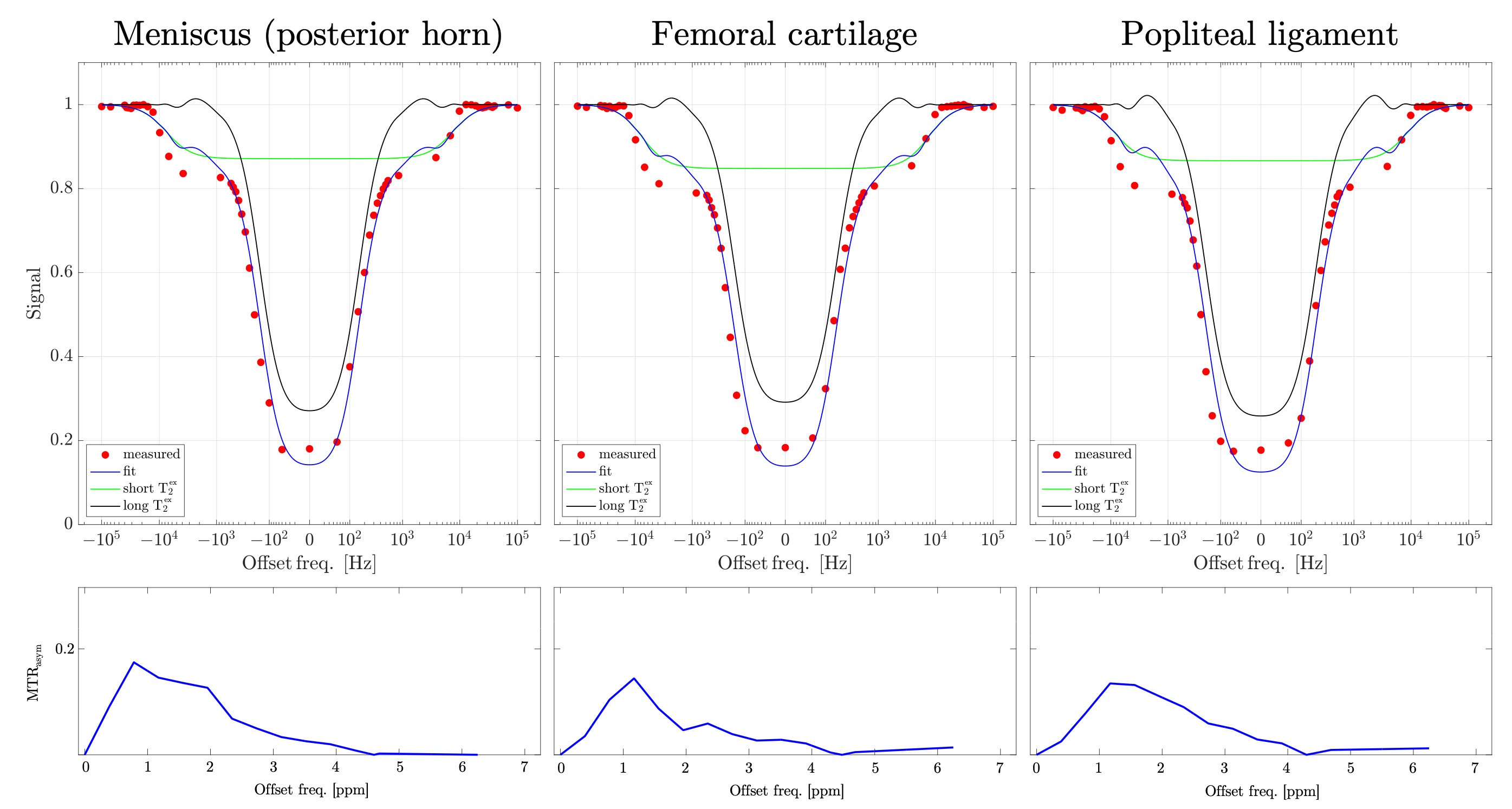
Z-spectrum was fitted using two-Lorentzian compartment model yielding metrics: apparent relaxation times ($$$\mathrm{T}_{2,f}^{\mathrm{ex}}$$$ and $$$\mathrm{T}_{2,r}^{\mathrm{ex}}$$$) in free and restricted proton pools, and their fractions (Ff and Fr). The narrower offset range [-900Hz, 900Hz] was used to measure MTRAsym.
Results
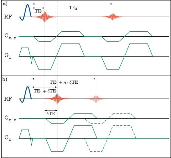
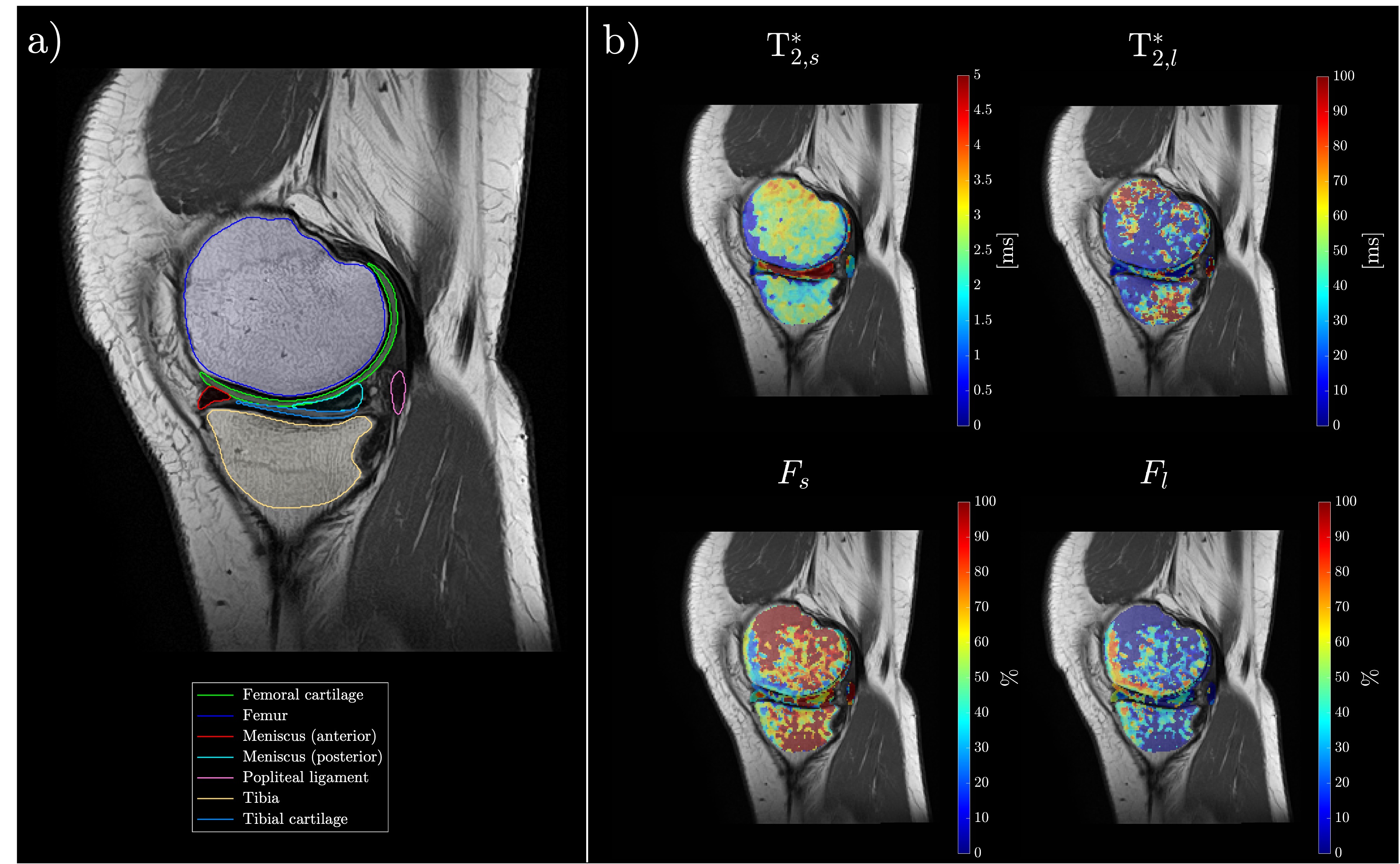
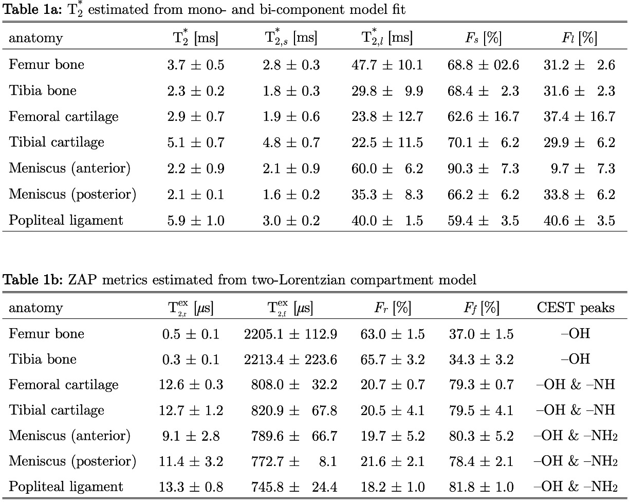
Schematic pulse sequence diagrams of multi-echo UTE and UTE with tight intervals TE are shown in Figures 1a and 1b, respectively. By varying the position of the first echo for different segments in one acquisition, we can significantly improve T2* mapping in tissues with extremely short T2. Figure 2 shows the high-resolution FSE image with identified anatomies of interest (a) and T2* colormaps as calculated from the bi-exponential model. Figure 3 demonstrates colormaps of ZAP metrics. Figure 4 shows logarithmic scale plots of the Z-spectrum for meniscus, cartilage, and ligament with the fits for two Lorentzian compartments: short ($$$\mathrm{T}_{2,r}^{\mathrm{ex}}$$$ (restricted) and long $$$\mathrm{T}_{2,f}^{\mathrm{ex}}$$$ (free) is shown on the top row of Figure 3. The corresponding MTRAsym plots are displayed in the bottom row. Finally, Figure 5 consolidates the quantitative findings from both T2* mapping (Table 1a) and ZAP & CEST analysis (Table 1b).Conclusions
The multiparametric approach of ZAP/CEST and UTE enhances the detection and characterization of knee joint tissues, broadening our understanding of knee pathophysiology in terms of macromolecule exchange and non-exchange protons. This approach shows promise for the emergence of new imaging biomarkers that could significantly advance the diagnosis of knee conditions.Acknowledgements
Authors thank the research support of Canon Medical Systems. Corp., Japan.References
[1] Miyazaki M., et al., PLOS ONE. 10(3), (2015).
[2] van Zijl PC., et al., Magn Reson Med. 65(4), 927-48. (2011).
[3] Malis V., et al., Magn Reson Med. 90(5), 2001-2010. (2023).
[4] Liu J., et al., Biomed Res Int. 8597423. (2019).
[5] Bae WC., et al., Skeletal Radiol. 50(12), (2021).
Figures