0493
Accelerated high-resolution deep learning reconstructed turbo spin echo MRI of the knee at ultra-high field strength1Swiss Center for Musculoskeletal Imaging, Balgrist Campus AG, Zurich, Switzerland, 2Balgrist University Hospital, Zurich, Switzerland, 3Department of Radiology, Balgrist University Hospital, Zurich, Switzerland, 4Advanced Clinical Imaging Technology, Siemens Healthineers International AG, Zurich, Switzerland
Synopsis
Keywords: Whole Joint, Joints
Motivation: High-resolution 7T-MRI using Turbo-Spin-Echoes requires high acceleration factors for reasonable scan times. Deep-Learning (DL) algorithms enable increased data under-sampling compared to state-of-the-art reconstructions.
Goal(s): To explore the feasibility of undersampled data acquisition in combination with DL-reconstruction for high-resolution T1- and PD-weighted knee MRI.
Approach: Volunteers underwent twofold, threefold and fourfold-accelerated 7T knee MRI with and without DL image reconstruction. Three readers rated various aspects of image quality.
Results: Image quality was rated significantly superior for fourfold-accelerated DL reconstructed images compared to images without DL reconstruction, while compared to twofold and threefold accelerated images, no image quality difference was observed.
Impact: This study successfully employed DL reconstructions at ultra-high field strength with promising results regarding image quality compared to conventional image reconstruction. Therefore, DL reconstructions at fourfold acceleration allows an efficient reduction in acquisition time, while still delivering high-quality images.
Introduction
Ultra-high field MRI inherently offers higher contrast-to-noise and signal-to-noise ratio (SNR), which can be utilized to increase spatial resolution. This enables enhanced visibility of anatomical details or subtle lesions which can increase diagnostic confidence and has therefore been advocated for application in musculoskeletal imaging 1-4. Specific absorption rate (SAR) restrictions render the acquisition process challenging, particularly in case of Turbo-Spin-Echo (TSE) imaging. Total acquisition time and SAR limitations make strong under-sampling necessary, however at cost of SNR, which restricts the achievable speed for images of satisfactory quality 5. A promising strategy to overcome this drawback is the implementation of deep learning (DL) algorithms, which offer the possibility of higher acceleration by incorporating denoising techniques in the reconstruction process 6-8. At ultra-high field systems, DL reconstructions have only recently become available. Therefore, the aim of this study was to explore the feasibility of highly undersampled data acquisition in combination with DL reconstruction for high-resolution T1- and PD-weighted knee MRI and comparison to state-of-the-art parallel imaging reconstruction.Methods
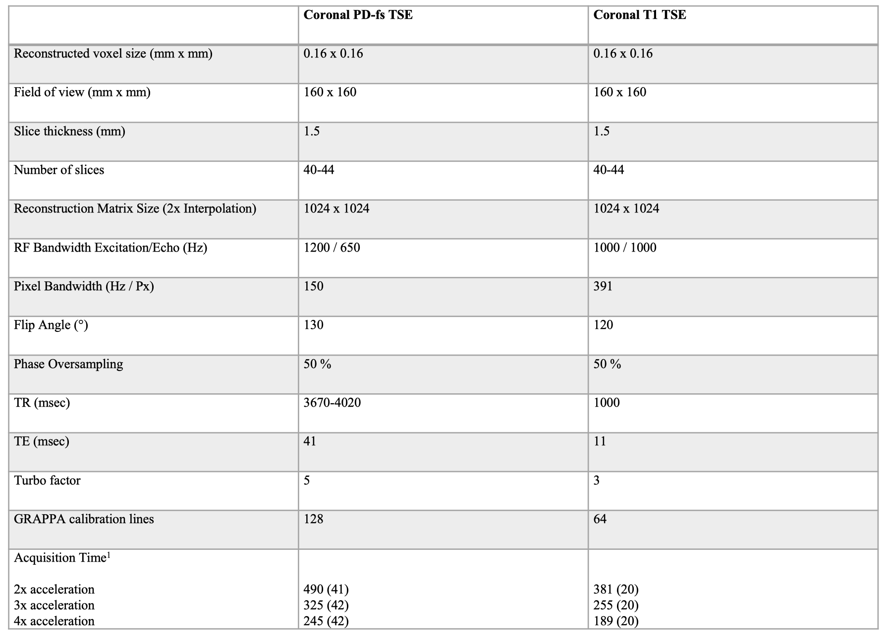
For this prospective study, 20 healthy volunteers (mean age of 32.0±8.1 years, left knee in 10 cases) underwent MRI at 7T (Magnetom Terra.X, Siemens Healthcare, Erlangen, Germany), utilizing a dedicated 1Tx-28Rx knee coil (Quality Electrodynamics QED, Mayfield Village, USA). The protocol consisted of high-resolution (reconstructed voxel volume: 0.16 x 0.16 x 1.5 mm3) fat-suppressed PD-weighted (PD-fs) and T1-weighted TSE sequences in coronal orientation. Cartesian undersampling was used to accelerate both sequences two-, three- and fourfold. The imaging parameters are listed in Table 1. Images were reconstructed employing two manufacturer-implemented algorithms 9. In case of the DL reconstruction (“Deep Resolve Boost”), data consistency was assured in k-space, with a trained regularization in the image domain (iterative SENSE) 10,11. A State-of-the-art GRAPPA reconstruction was used as reference 12. Reviews for aspects of image quality (image contrast, sharpness, noise, artifacts, overall quality) were performed by three fellowship-trained radiologists using a 4-point Likert scale. For comparison of image quality criteria and acquisition time of images acquired with different acceleration factors (AF), Friedman’s two-way analysis of variance by ranks with pairwise post hoc tests was performed. p-values were corrected for multiple comparisons utilizing the Bonferroni procedure. The Wilcoxon signed rank test was used for comparisons of fourfold accelerated acquisitions with and without DL reconstruction.Results
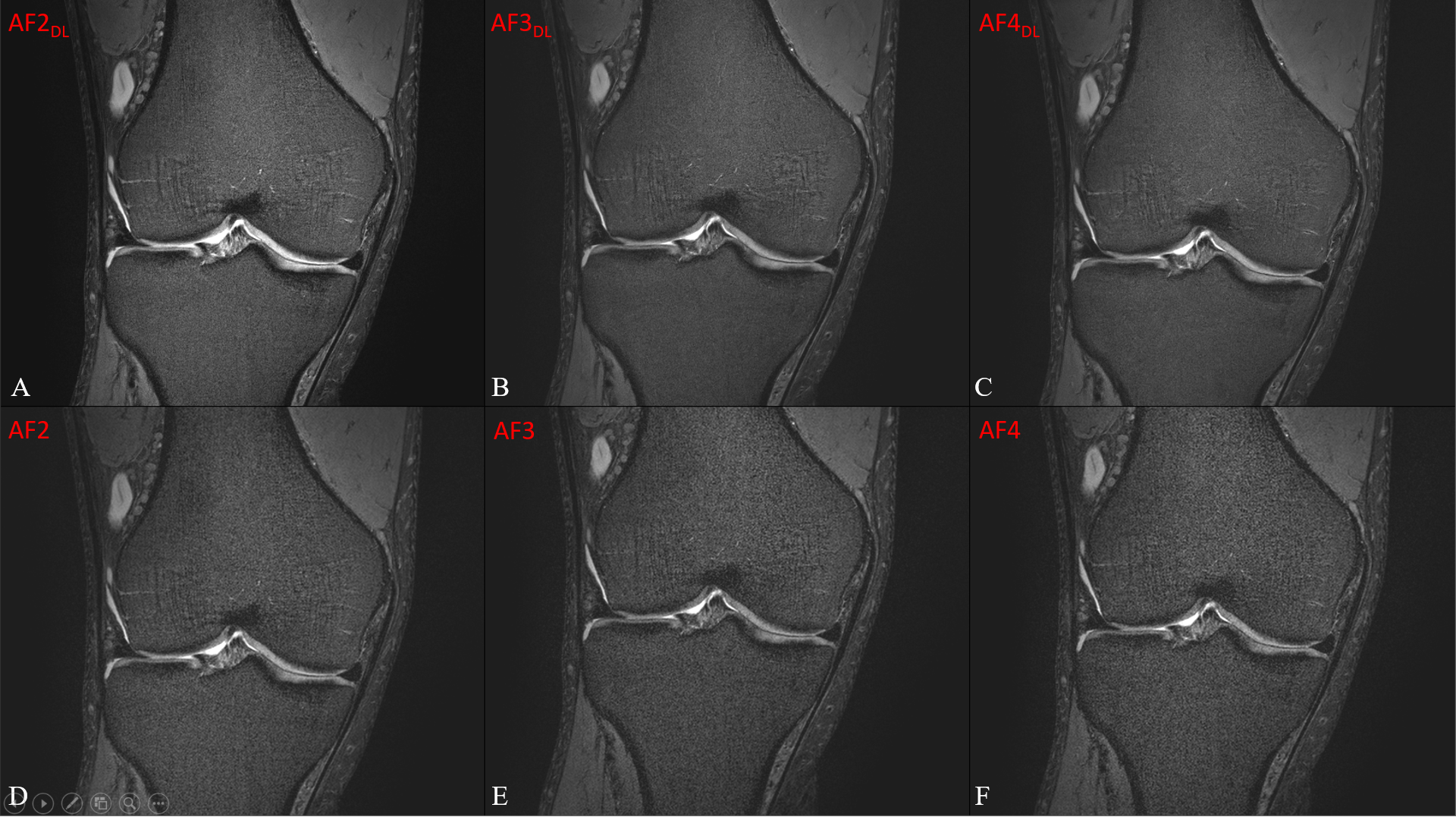
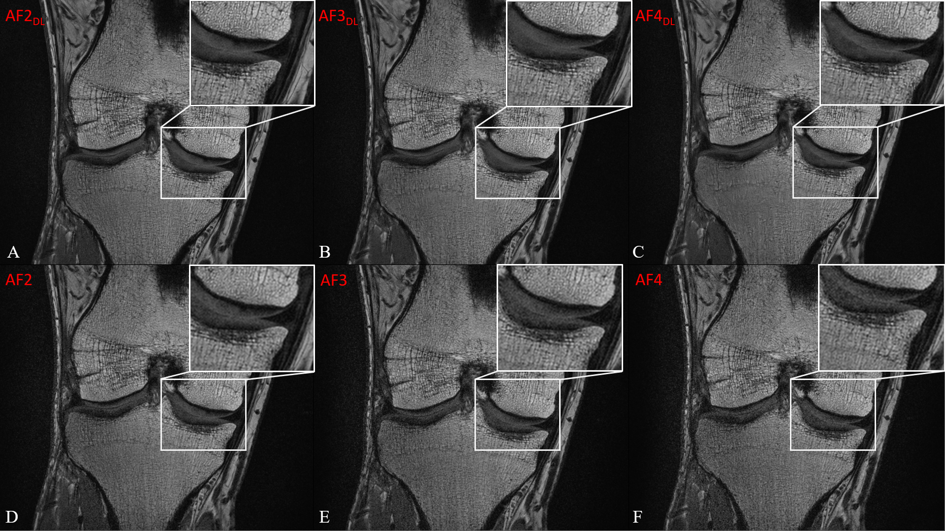
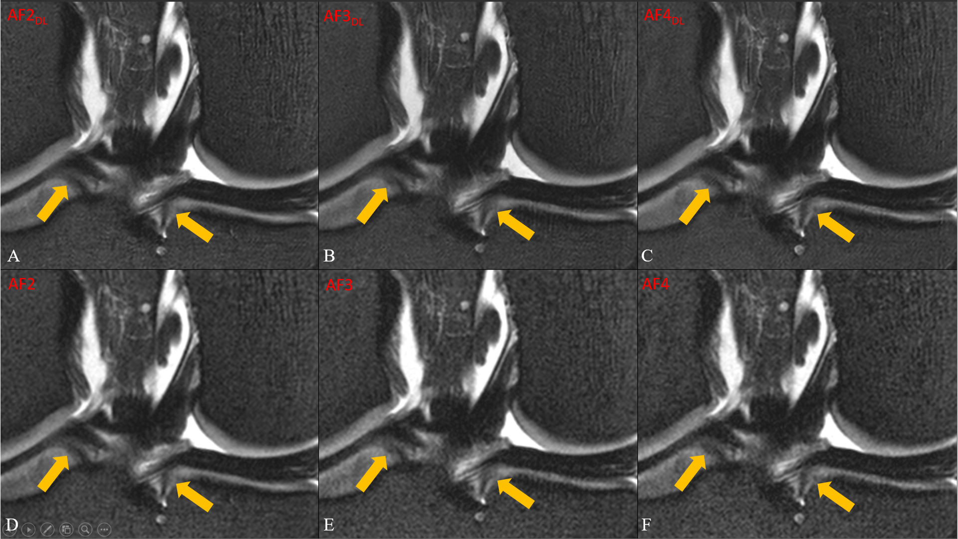
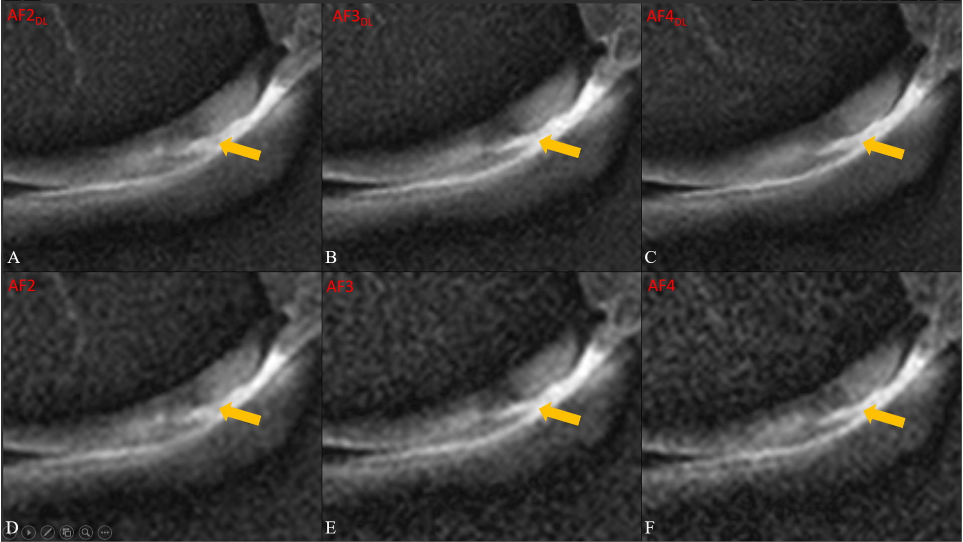
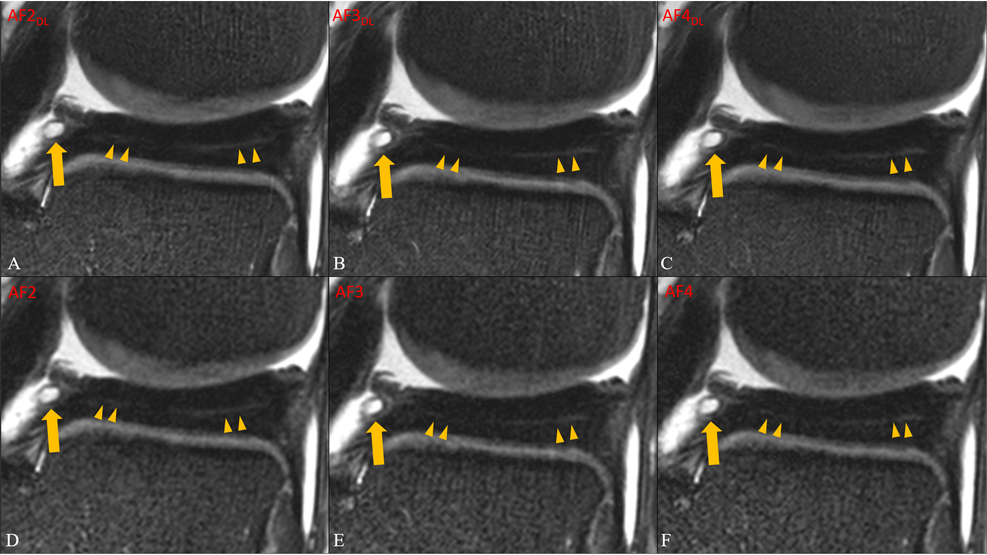
Acquisition times are summarized in Figure 1. Figure 2 and 3 shows coronal fat-suppressed PD- and T1-weighted knee images with different acceleration factors (AF=2-4) and reconstruction methods (DL and GRAPPA). Bone marrow and cartilage signal is significantly denoised with the DL reconstruction, particularly for AF=4. Figure 4 shows an excellent visualization of the insertion of the meniscal roots, while two incidental findings are shown in Figure 5 and 6: A cartilage delamination located at the medial femoral condyle and a parameniscal cyst as well as a subtle meniscal degeneration. All aspects of image quality were rated significantly higher in fourfold accelerated and DL reconstructed images compared to those without DL reconstruction (p < 0.001 for both, PD-fs and for T1-weighted images). There was no significant difference in all image quality aspects between two-, three- and fourfold accelerated DL-reconstructed images for both sequences (p ≥ 0.082).Discussion & Conclusion
This is the first study to report about DL-reconstructed TSE images of the knee at ultra-high field. The main finding of the present study was that high-resolution (voxel volume: 0.16 x 0.16 x 1.5 mm3), fourfold accelerated and DL reconstructed images exhibited excellent image quality which was significantly superior compared to conventional reconstructed images. This finding is consistent with numerous studies comparing DL- and non-DL-reconstructed images at field strengths of 1.5-T and 3-T 13-17. Additionally, image quality was comparable between DL reconstructed two- and fourfold acceleration which allows an effective reduction of acquisition time of up to 50.4%. Parallel imaging with high acceleration factors is necessary due to scan time and SAR constraints at ultra-high fields; it is, however, challenging given the SNR penalty by data subsampling. The advent of DL algorithms could push the boundaries of data under-sampling at 7T, as DL reconstructions are less prone to SNR loss compared to conventional reconstruction methods 6,7. This study is limited by the small sample size and no conclusions can be drawn about the diagnostic impact of our findings, as only healthy volunteers were included in this study. In conclusion, DL reconstructions have the potential to elevate the benchmark for reducing acquisition time, while still delivering high-resolution images of excellent quality.Acknowledgements
No acknowledgement found.References
1 Heiss, R. et al. Clinical Application of Ultrahigh-Field-Strength Wrist MRI: A Multireader 3-T and 7-T Comparison Study. Radiology 307, e220753, doi:10.1148/radiol.220753 (2023).
2 Pazahr, S., Nanz, D. & Sutter, R. 7 T Musculoskeletal MRI: Fundamentals and Clinical Implementation. Investigative Radiology 58, 88-98, doi:10.1097/rli.0000000000000896 (2023).
3 Welsch, G. H. et al. Magnetic resonance imaging of the knee at 3 and 7 tesla: a comparison using dedicated multi-channel coils and optimised 2D and 3D protocols. Eur Radiol 22, 1852-1859, doi:10.1007/s00330-012-2450-1 (2012).
4 von Deuster, C. et al. Controlling Through-Slice Chemical-Shift Artifacts for Improved Non-Fat-Suppressed Musculoskeletal Turbo-Spin-Echo Magnetic Resonance Imaging at 7 T. Invest Radiol 56, 545-552, doi:10.1097/rli.0000000000000778 (2021).
5 Recht, M. P. et al. Using Deep Learning to Accelerate Knee MRI at 3 T: Results of an Interchangeability Study. AJR Am J Roentgenol 215, 1421-1429, doi:10.2214/ajr.20.23313 (2020).
6 Schlemper, J., Caballero, J., Hajnal, J. V., Price, A. N. & Rueckert, D. A Deep Cascade of Convolutional Neural Networks for Dynamic MR Image Reconstruction. IEEE Trans Med Imaging 37, 491-503, doi:10.1109/tmi.2017.2760978 (2018).
7 Aggarwal, H. K., Mani, M. P. & Jacob, M. MoDL: Model-Based Deep Learning Architecture for Inverse Problems. IEEE Trans Med Imaging 38, 394-405, doi:10.1109/tmi.2018.2865356 (2019).
8 Koonjoo, N., Zhu, B., Bagnall, G. C., Bhutto, D. & Rosen, M. S. Boosting the signal-to-noise of low-field MRI with deep learning image reconstruction. Scientific Reports 11, 8248, doi:10.1038/s41598-021-87482-7 (2021).
9 Hammernik, K. et al. Learning a variational network for reconstruction of accelerated MRI data. Magn Reson Med 79, 3055-3071, doi:10.1002/mrm.26977 (2018).
10 Pruessmann, K. P., Weiger, M., Scheidegger, M. B. & Boesiger, P. SENSE: sensitivity encoding for fast MRI. Magn Reson Med 42, 952-962 (1999).
11 Pruessmann, K. P., Weiger, M., Börnert, P. & Boesiger, P. Advances in sensitivity encoding with arbitrary k-space trajectories. Magn Reson Med 46, 638-651, doi:10.1002/mrm.1241 (2001).
12 Griswold, M. A. et al. Generalized autocalibrating partially parallel acquisitions (GRAPPA). Magn Reson Med 47, 1202-1210, doi:10.1002/mrm.10171 (2002).
13 Johnson, P. M. et al. Deep Learning Reconstruction Enables Prospectively Accelerated Clinical Knee MRI. Radiology 307, e220425, doi:10.1148/radiol.220425 (2023).
14 Almansour, H. et al. Deep Learning Reconstruction for Accelerated Spine MRI: Prospective Analysis of Interchangeability. Radiology 306, e212922, doi:10.1148/radiol.212922 (2023).
15 Herrmann, J. et al. Deep Learning MRI Reconstruction for Accelerating Turbo Spin Echo Hand and Wrist Imaging: A Comparison of Image Quality, Visualization of Anatomy, and Detection of Common Pathologies with Standard Imaging. Acad Radiol, doi:10.1016/j.acra.2022.12.042 (2023).
16 Herrmann, J. et al. Feasibility of an accelerated 2D-multi-contrast knee MRI protocol using deep-learning image reconstruction: a prospective intraindividual comparison with a standard MRI protocol. Eur Radiol 32, 6215-6229, doi:10.1007/s00330-022-08753-z (2022).
17 Feuerriegel, G. C. et al. Evaluation of a deep learning-based reconstruction method for denoising and image enhancement of shoulder MRI in patients with shoulder pain. European Radiology 33, 4875-4884, doi:10.1007/s00330-023-09472-9 (2023).
Figures