0491
3D Turbo-Spin-Echo with VERSE Excitation Improves SNR for Brachial Plexus Magnetic Resonance Neurography1Siemens Medical Solutions USA, Inc., New York, NY, United States, 2Siemens Healthineers AG, Erlangen, Germany, 3Siemens Medical Solutions USA, Inc., Rochester, MN, United States, 4Department of Radiology and Imaging, Hospital for Special Surgery, New York, NY, United States
Synopsis
Keywords: Neurography, MSK, pulse sequence design, neurography, peripheral nerves
Motivation: The conventional slab-selective 3D TSE approach for brachial plexus magnetic resonance neurography (MRN) has a longer echo spacing for the first refocusing pulse and hence violates CPMG conditions. The signal available can decrease significantly due to field inhomogeneities.
Goal(s): We aimed to improve slab-selective 3D TSE for brachial plexus MRN.
Approach: We incorporated the variable-rate selective excitation (VERSE) method to shorten the excitation pulse duration and eliminate the need for the first long-echo-spacing refocusing module.
Results: The 3D TSE with VERSE excitation achieved higher SNR in both phantom and in-vivo experiments.
Impact: The proposed 3D TSE using VERSE excitation improved slab-selective imaging in brachial plexus MRN with higher SNR when compared to the conventional approach.
Background
Magnetic resonance neurography (MRN) of the brachial plexus employs a fluid-sensitive 3D turbo-spin-echo (TSE) short-tau inversion recovery (STIR) sequence for its excellent tissue contrast and high through-plane resolution1-3. Slab-selective acquisition is preferred over non-selective acquisition for unilateral brachial plexus MRN to mitigate against aliasing wrap artifact, as it provides excellent proximal to distal coverage of the nerve branches using an oblique coronal plane4. Conventionally, slab-selective 3D TSE employs a selective 90° excitation pulse and subsequent non-selective refocusing pulses5 to provide a high-quality slab profile with short echo spacing during the echo train, at the expense of a longer echo spacing at the first refocusing pulse6. This approach violates CPMG conditions and significantly diminishes the signal available in the echo train, especially in the presence of B0- and B1-field inhomogeneities7. In this study, we aimed to improve slab-selective 3D TSE for MRN by utilizing an excitation pulse designed with a variable-rate selective excitation (VERSE) method8 that shortens the slab-selective pulse duration with a time-varying gradient waveform.Methods
We implemented the research 3D TSE sequence on a 3T system (MAGNETOM Vida, Siemens Healthineers AG, Erlangen, Germany). Non-selective imaging uses hard pulses for both excitation and refocusing with durations of 0.6 and 0.74 ms, respectively (Fig.1A). Conventional slab-selective imaging uses an excitation pulse with a time-bandwidth product of 22 and duration of 10.24 ms5 (Fig.1B). We implemented the VERSE algorithm, similar to a prior study8, to calculate a VERSE version of the excitation pulse in real time minimizing the duration based on a 24 mT/m maximum gradient strength and a 144 mT/m/ms maximum gradient slew rate. This algorithm resulted in a much shorter duration of 0.96 ms, similar to that of non-selective excitation (Fig.1C). The slab profile remains the same for on-resonance spins and may degrade with off-resonance as shown by Bloch simulation (Fig.2).We scanned phantoms with a 16-channel transmit/receive knee coil and acquired each echo without phase encoding. We then compared the signal at each echo center among the 3 excitation methods – non-selective, conventional slab-selective, and VERSE slab-selective. Scan parameters included: TR/TE 3000/173 ms, receiver bandwidth 382 Hz/Pixel, echo spacing 4.8 ms, turbo factor 130, constant flip angle 120°, and slab thickness 160 mm covering the phantom entirely. We separately scanned 2 agar compartments with T1/T2 mimicking skeletal muscle (1337/39 ms) and peripheral nerve (1400/72 ms) tissue. We then repeated measurements with variable flip angles calculated with 4 control points (100-20-80-120)9.
We scanned the unilateral brachial plexus of 5 healthy subjects (mean age 34.4 years old, 2 females) with IRB approval and written consent using a combination of a 20-channel head/neck, 18-channel Ultraflex small coil, and 5-6 elements of a posterior spine coil with elements selected depending on subject positioning and size. Imaging parameters included: TR/TE/TI 3000/161/250 ms, field-of-view 304x168x120 mm, slice oversampling 20%, resolution 1.0x1.0x1.0 mm, 2 averages, receiver bandwidth 358 Hz/Pixel, echo spacing 4.88 ms, turbo factor 130, variable flip angles (100-20-80-120)9, 4-fold acceleration with CAIPIRINHA10 with a total acquisition time of 6 min. A C-FOCI adiabatic pulse was used for STIR11,12. We calculated the signal-to-noise (SNR) map using the pseudo multiple replica approach13 and measured SNR for the anterior scalene muscle, extraforaminal C7 nerve root, and subcutaneous fat. SNR was compared using a paired t-test with significance set at p<0.05.
Results
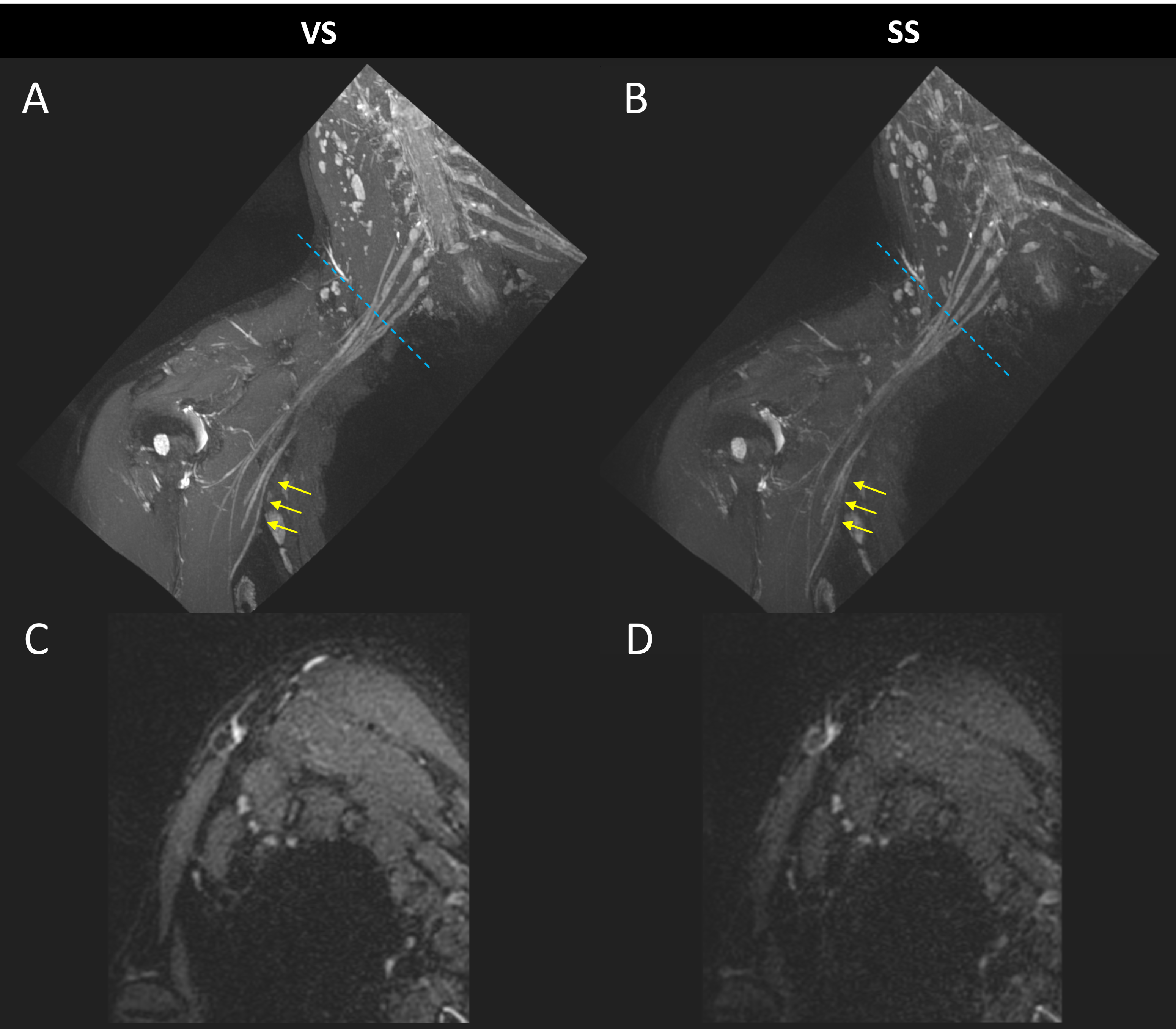
Fig. 3 shows the results of phantom experiments. The measured echo train signals following VERSE excitation were equivalent to those from non-selective excitation, and much higher than those from conventional slab-selective excitation. The differences in echo signal were more prominent with variable flip (red plots) than with constant flip angles. The same observations apply to both muscle (Fig.3A) and nerve (Fig.3B) vials. In-vivo comparison confirmed that the VERSE excitation produced higher SNR than the conventional slab-selective method (Fig.4) for both muscle (16.95±7.49 vs. 11.62±5.19, p < 0.05) and nerve (23.74±6.98 vs. 18.60±7.06, p<0.05). SNR of fat tissue was marginally superior with VERSE excitation (6.61±3.77 vs. 4.93±3.86, p=0.086), amidst the application of fat suppression.Discussion
Slab-selective 3D TSE with VERSE excitation eliminates the need for the first long-echo-spacing refocusing module. This design achieves higher SNR than conventional slab-selective imaging as demonstrated in phantoms and a small sample of human subjects. Although the slab profile with VERSE is expected to degrade in the presence of off-resonance, we observed no fold-over artifacts in this application. Future work includes applying the VERSE pulse for imaging patient subjects undergoing clinical brachial plexus scans and investigating its utility for other anatomical regions.Acknowledgements
No acknowledgement found.References
- Sneag, Darryl B. and Sophie Queler. "Technological advancements in magnetic resonance neurography." Current Neurology and Neuroscience Reports 19 (2019): 1-6.
- Chhabra, A., et al. "High-resolution 3T MR neurography of the brachial plexus and its branches, with emphasis on 3D imaging." American Journal of Neuroradiology 34.3 (2013): 486-497.
- Vargas, M. I., et al. "New approaches in imaging of the brachial plexus." European journal of radiology 74.2 (2010): 403-410.
- Davidson EJ, Tan ET, Pedrick EG, Sneag DB. Brachial Plexus Magnetic Resonance Neurography: Technical Challenges and Solutions. Invest Radiol. 2023 Jan 1;58(1):14-27. doi: 10.1097/RLI.0000000000000906. Epub 2022 Aug 2. PMID: 35926072.
- JP, MUGLER. "Efficient spatially-selective single-slab 3D turbo-spin-echo imaging." Proc Intl Soc Mag Reson Med. Vol. 11. 2004.
- Mugler III, John P. "Optimized three‐dimensional fast‐spin‐echo MRI." Journal of magnetic resonance imaging 39.4 (2014): 745-767.
- Park, Jaeseok, John P. Mugler III, and Timothy Hughes. "Reduction of B1 sensitivity in selective single‐slab 3D turbo spin echo imaging with very long echo trains." Magnetic Resonance in Medicine: An Official Journal of the International Society for Magnetic Resonance in Medicine 62.4 (2009): 1060-1066.
- Conolly, Steven, et al. "Variable-rate selective excitation." Journal of Magnetic Resonance (1969) 78.3 (1988): 440-458.
- Busse, Reed F., et al. "Effects of refocusing flip angle modulation and view ordering in 3D fast spin echo." Magnetic resonance in medicine 60.3 (2008): 640-649.
- Breuer, Felix A., et al. "Controlled aliasing in parallel imaging results in higher acceleration (CAIPIRINHA) for multi‐slice imaging." Magnetic Resonance in Medicine: An Official Journal of the International Society for Magnetic Resonance in Medicine 53.3 (2005): 684-691.
- Ordidge RJ, Wylezinska M, Hugg JW, Butterworth E, Franconi F. Frequency offset corrected inversion (FOCI) pulses for use in localized spectroscopy. Magn Reson Med. 1996;36(4):562-566.
- Wang, Xinzeng, et al. "Frequency offset corrected inversion pulse for B0 and B1 insensitive fat suppression at 3T: application to MR neurography of brachial plexus." Journal of Magnetic Resonance Imaging 48.4 (2018): 1104-1111.
- Robson, Philip M., et al. "Comprehensive quantification of signal‐to‐noise ratio and g‐factor for image‐based and k‐space‐based parallel imaging reconstructions." Magnetic Resonance in Medicine: An Official Journal of the International Society for Magnetic Resonance in Medicine 60.4 (2008): 895-907.
Figures
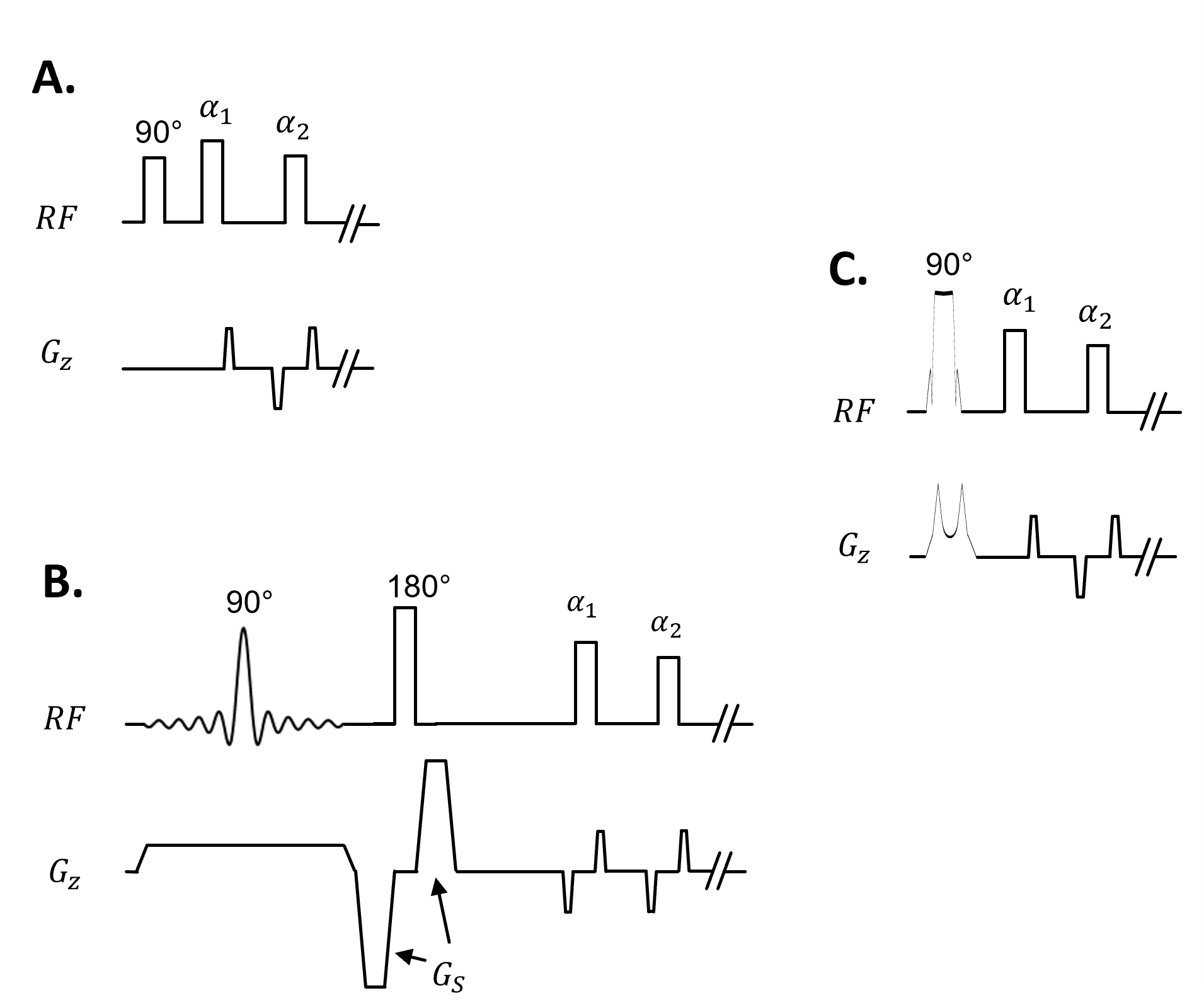
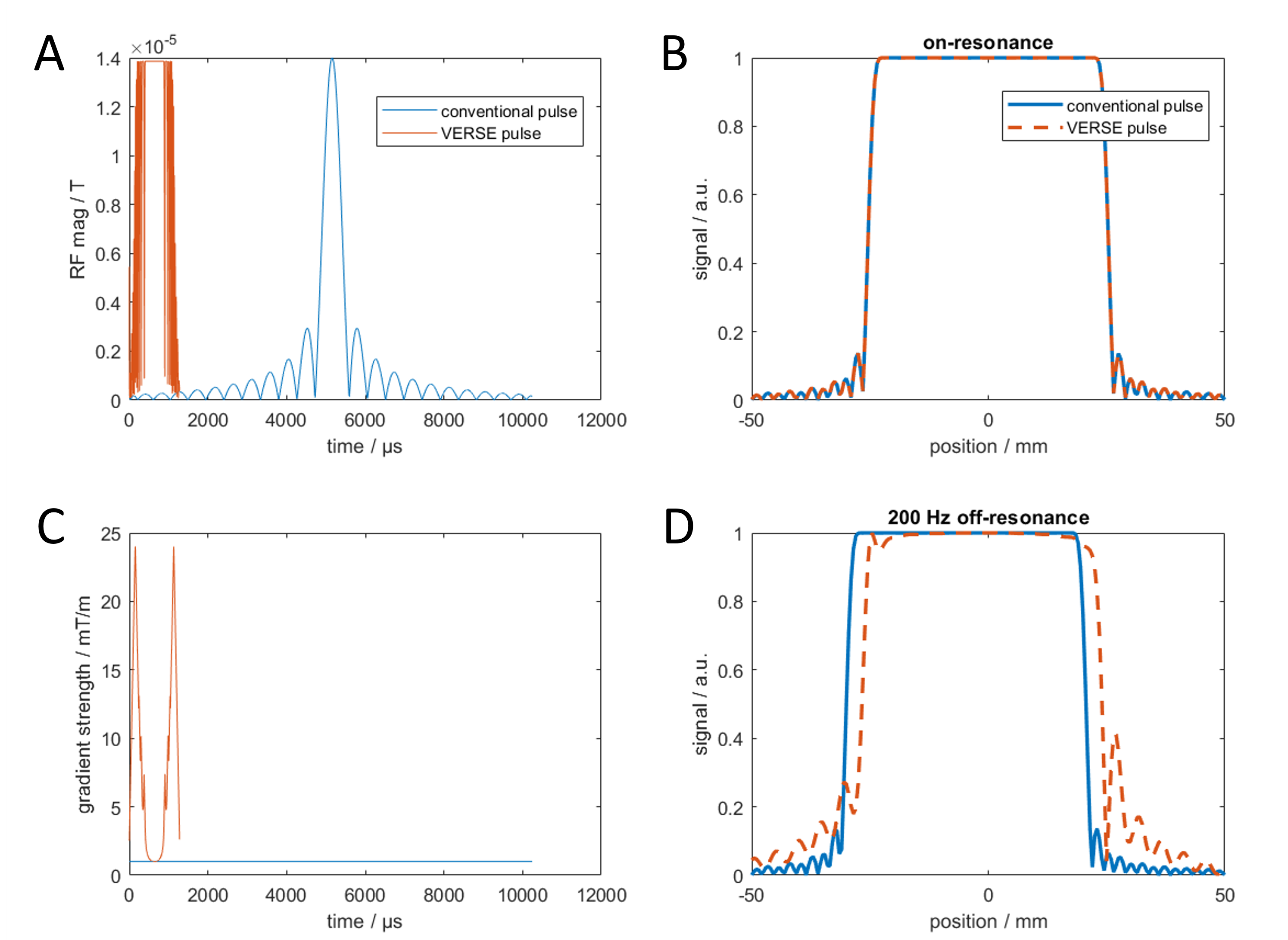
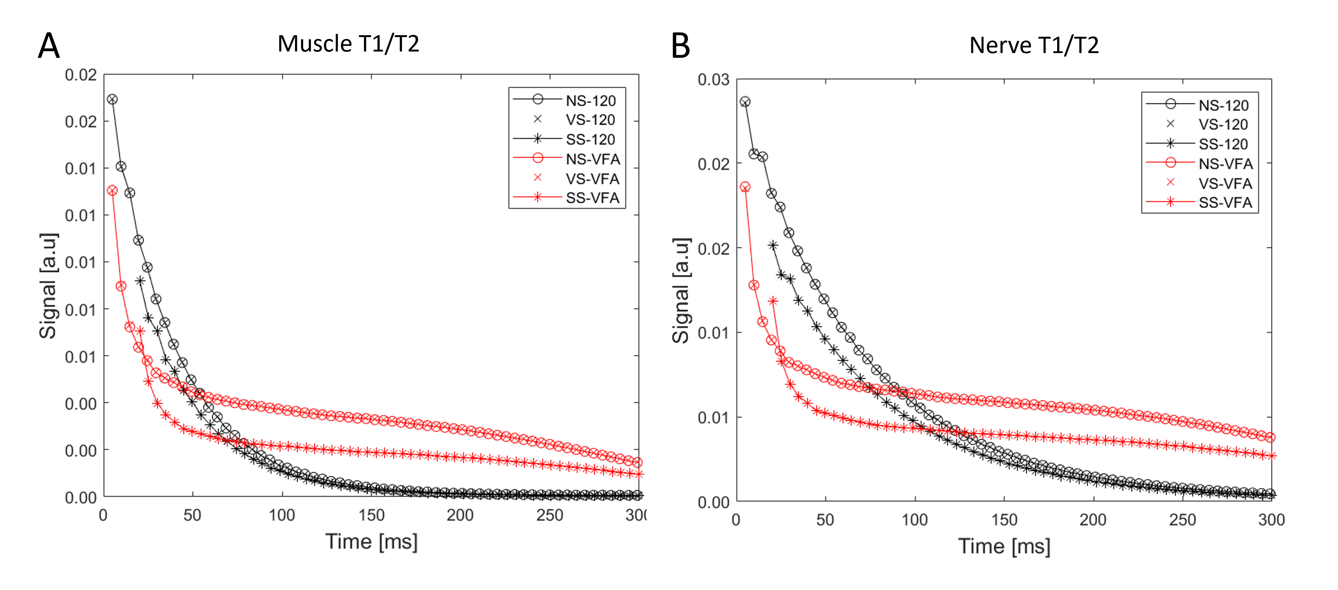
Fig.3. 3D TSE signal comparison in phantom experiments. VERSE-selective (VS) acquisitions are virtually identical to non-selective (NS) acquisitions with higher signals than those from conventional slab-selective (SS). These results are applicable for both constant 120-degree flip angle (black plots) and variable flip angle (VFA) (red plots) acquisitions, and for both muscle (A) and nerve (B) phantoms. The first 300 ms of the echo train is shown.