0489
Feasibility of Bone Porosity Assessment Using Dual-Echo uTE-MR Fingerprinting1Department of Radiology, Stanford University, Stanford, CA, United States, 2Department of Electrical Engineering, Stanford University, Stanford, CA, United States, 3Department of Radiology and Biomedical Imaging, University of California San Francisco, San Francisco, CA, United States
Synopsis
Keywords: Bone, Bone, MSK, Quantitative Imaging, MR Fingerprinting, Data Processing
Motivation: Bone porosity is crucial for bone strength, yet standard multi-echo uTE-GRE techniques are too time-consuming for clinical use. uTE MR Fingerprinting (MRF) has not been tested for porosity assessment. If feasible it may offer faster porosity mapping for clinical applications.
Goal(s): Assessing the feasibility of using dual-echo 3D-uTE-MRF to measure porosity through simulations and preliminary in vivo testing.
Approach: A dual-echo 3D-uTE-MRF sequence was tested for porosity accuracy and precision against standard multi-echo uTE-GRE via simulations. In-vivo, a volunteer's tibia was imaged to demonstrate the technique's preliminary viability.
Results: In simulations, dual-echo uTE-MRF outperformed uTE-GRE, but in-vivo applications, despite feasibility, need further development.
Impact: We demonstrated the feasibility of a dual-echo uTE MRF approach for measuring bone porosity trough simulation and a preliminary in-vivo acquisition.
Introduction
Trabecular and cortical porosity deterioration plays a key role in determining bone strength. Multi-echo uTE gradient echo (GRE) acquisitions with tri-exponential modeling allow effective measurement of cortical bone porosity in proximal bone sites in vivo1. However, their long scan time impairs their use in clinical settings. Porosity Index2 and Suppression Ratio3 have been proposed to overcome long scan times, but they do not allow full quantification of water compartments. Magnetic Resonance Fingerprinting (MRF) is a fast quantitative MRI methodology for multi-parametric mapping4. While ultra-short Echo-Time (uTE) MRF has been recently proposed for myelin water mapping5,6, its feasibility for porosity mapping in bone has not been investigated. If feasible, the approach may circumvent long acquisition times required by multi-echo uTE-GRE acquisitions and allow porosity measurement in clinically viable times. In this work, we investigated the feasibility of using a dual-echo 3D uTE-MRF approach to measure bone porosity using simulations and show a prelaminar acquisition in vivo.Methods
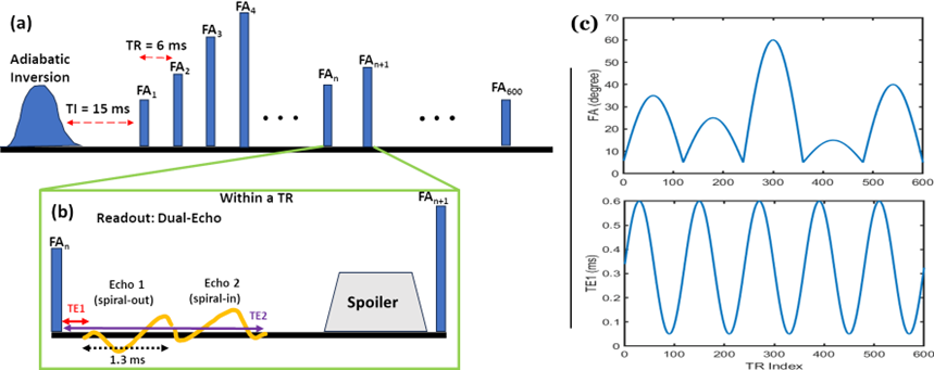
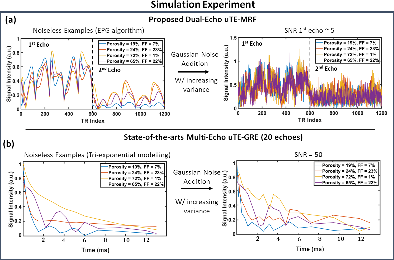
Sequence: A 3D dual-echo uTE-MRF sequence inspired by the recently developed sequence applied for Myelin mapping in the brain5 was implemented (Figure 1). For each repetition time (TR) between consecutive pulses, two echoes are acquired using a spiral-out spiral-in readout with an echo spacing of 2.6 ms (see Fig. 1(a)). The first echo is varied among successive pulses according to a sinusoidal pattern within the range [0.1 - 0.6] ms (Fig. 1(c)).Simulation experiments: The performance of the dual-echo uTE-MRF sequence in estimating bone porosity as a function of noise compared with using the standard multi-echo uTE-GRE sequence was tested using simulations. 1000 bone voxel signals with porosity ranging from 100% (bone marrow) to 10% (very compact cortical bone) were simulated. Each signal was a mixture of 3 components: fat, pore water, and bound water. For each mixture, T1, T2, and T2* of each component were randomly sampled within physiological ranges (estimated from literature(1,7–9)). In addition, for each signal, B0 inhomogeneity and B1 inefficiency were randomly sampled in the ranges (-100, 100) Hz and (0.5, 1.2) respectively. Fat was modeled as one peak at +3.4ppm. For the multi-echo uTE-MRF, the signals were simulated with an extended phase graph (EPG) formalism10, while a tri-exponential model was used for simulating the multi-echo uTE-GRE signals1. White Gaussian noise was added with increasing variance to simulate different levels of noise corruption (see Figure 2). The SNR was defined as the power of the signal to the power of the noise. A non-linear least square (NLSQ) optimization procedure with the VARPRO technique was used to fit the data11 using a 3-component model: tri-exponential and tri-component EPG for uTE-GRE and uTE-MRF, respectively. The mean error and the mean absolute error were used as measures of accuracy and precision, respectively.
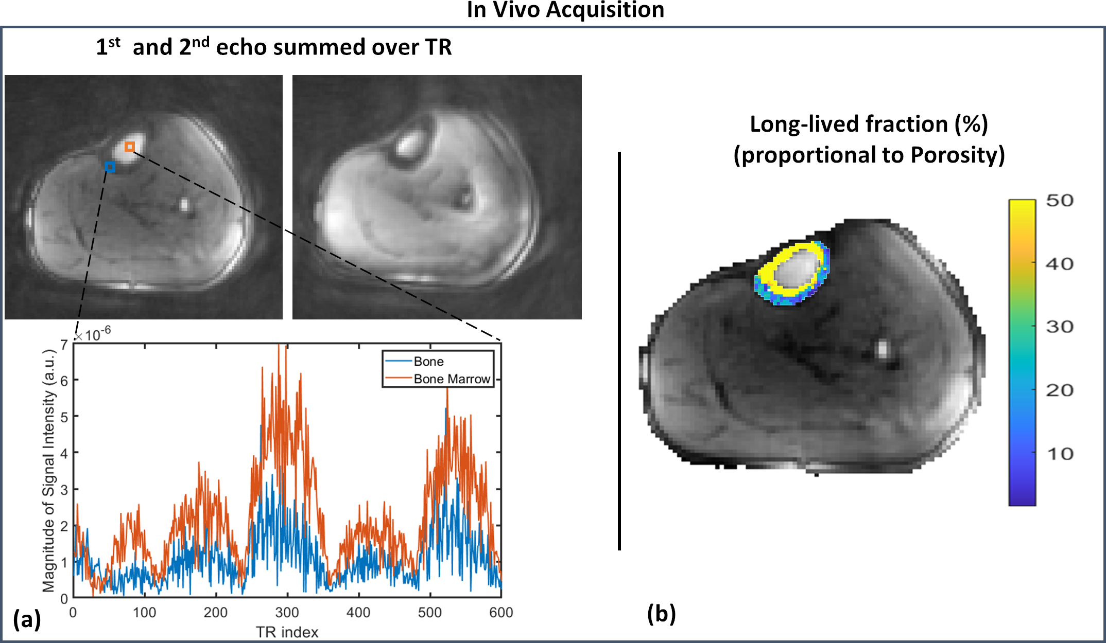
In-vivo acquisition: The tibia of a healthy volunteer was scanned a 3T (MAGNETOM Vida, Siemens Healthineers, Erlangen, Germany), after obtaining informed consent, using the 3D uTE-MRF sequence with isotropic resolution (FOV = 20 cm3 , matrix size of 150 x 150 x 150). For this preliminary proof of concept, an undersampling factor R=64 for each spiral readout was used with 289 sequence repetitions (tacq = 806 s). The data were reconstructed using filtered back-projection with NUFFT. NLSQ fitting was applied voxel-wise in the tibia.
Results
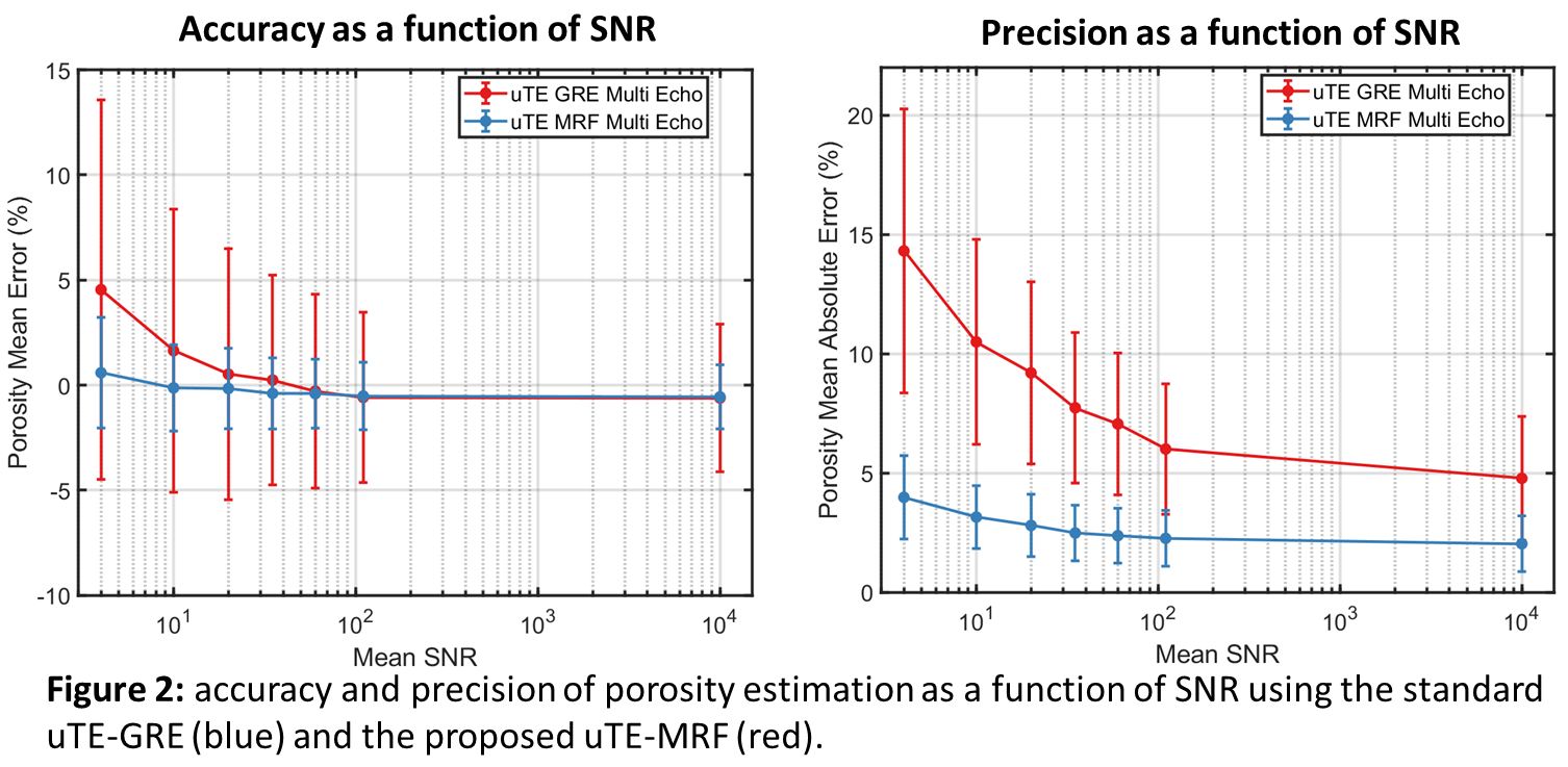
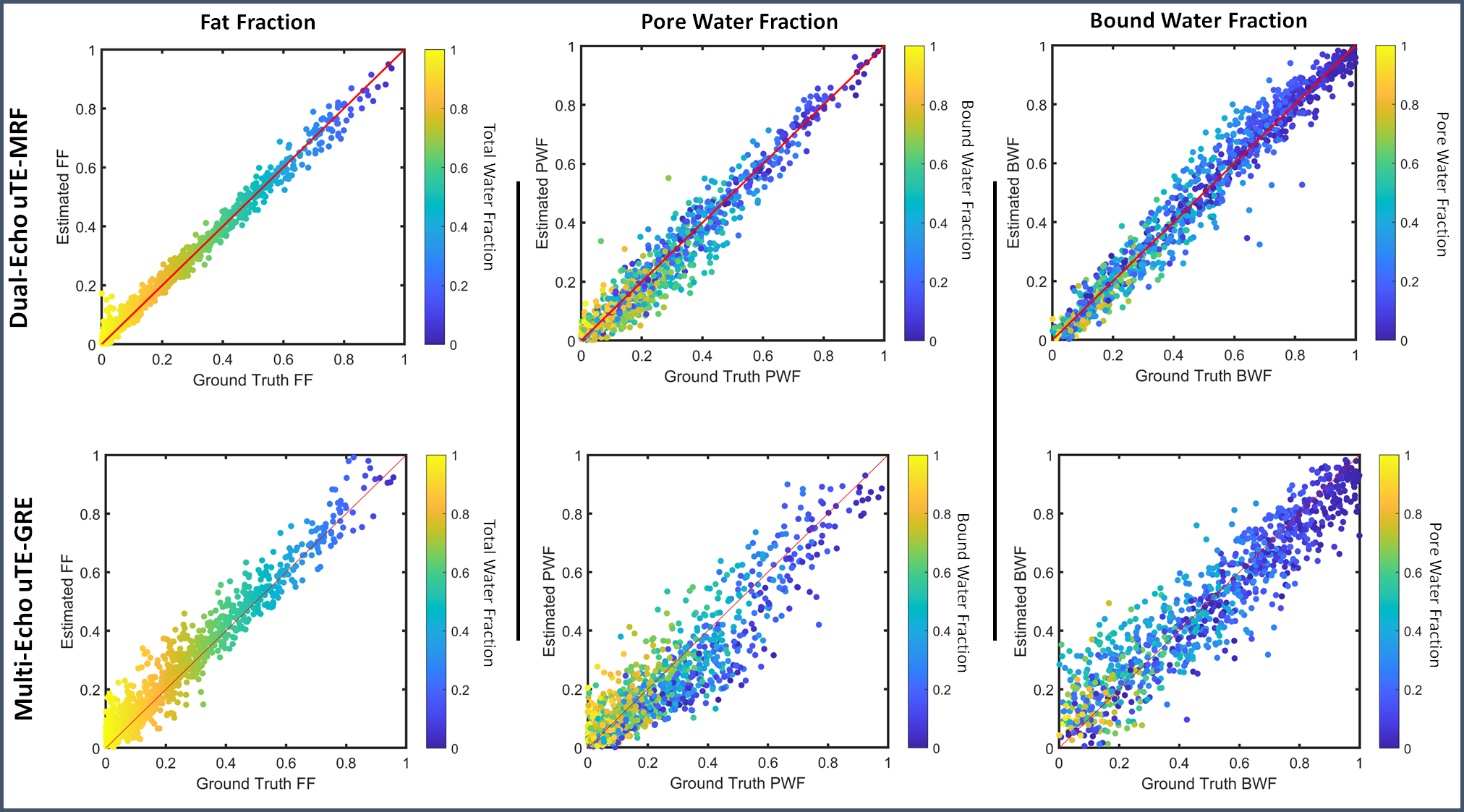
Simulation results are shown in Figures 3 and 4. Dual-echo uTE-MRF outperformed the reference uTE-GRE method, providing more accurate and consistent porosity estimates across different SNRs (Figure 3). Figure 4 shows a higher level of agreement between ground-truth component fractions and estimated fractions according to uTE-MRF and uTE-GRE for an SNR of 5 (first echo) and 50, respectively. The long-lived fraction map, which combines fat and pore-water and is proportional to porosity, from the preliminary in-vivo acquisition is reported in Figure 5b. It is possible to observe some artifacts in the external portion of the tibia, where the long-lived component fraction is much higher than the internal part of the tibia.Discussion and Conclusion
Our simulation experiments indicate that dual-echo uTE-MRF is capable of accurately estimating bone porosity, demonstrating robustness at low SNRs and better sensitivity to tissue properties than traditional uTE-GRE. While initial in vivo results showed differentiation of different compartments in the tibia, they also highlighted the need for further refinement due to observed artefacts. Optimizations, possibly using Cramér–Rao Lower Bound principles for improved SNR efficiency, along with thorough in vivo and phantom validations, are necessary steps forward.In summary, our findings suggest that with further development, dual-echo uTE-MRF may enable precise and clinically timely bone porosity measurements.
Acknowledgements
NIH (R01AR079431, R21EB030180, R01AR077604, R01EB002524), Wu Tsai Human Performance Alliance, CIHR Postdoctoral FellowshipReferences
1. Lu X, Jerban S, Wan L, Ma Y, Jang H, Le N, et al. Three-dimensional ultrashort echo time imaging with tricomponent analysis for human cortical bone. Magn Reson Med. 2019 Jul 1;82(1):348–55.
2. Jones BC, Jia S, Lee H, Feng A, Shetye SS, Batzdorf A, et al. MRI-derived porosity index is associated with whole-bone stiffness and mineral density in human cadaveric femora. Bone. 2021;143:115774.
3. Jerban S, Ma Y, Moazamian D, Athertya J, Dwek S, Jang H, et al. MRI-based porosity index (PI) and suppression ratio (SR) in the tibial cortex show significant differences between normal, osteopenic, and osteoporotic female subjects. Front Endocrinol. 2023;14:1148345.
4. Ma D, Gulani V, Seiberlich N, Liu K, Sunshine JL, Duerk JL, et al. Magnetic resonance fingerprinting. Nature. 2013;495(7440):187–92.
5. Li Q, Cao X, Ye H, Liao C, He H, Zhong J. Ultrashort echo time magnetic resonance fingerprinting (UTE-MRF) for simultaneous quantification of long and ultrashort T 2 tissues. Magn Reson Med. 2019 Oct 1;82(4):1359–72.
6. Zihan Zhou, Qing Li, Congyu Liao, Xiaozhi Cao, Ting Gong1, Qiuping Ding, Hongjian He and JZ. 3D Ultrashort Echo Time MR Fingerprinting (3D UTE-MRF) for Whole Brain Myelin Imaging. In: Annual meeting of ISMRM. 2021.
7. Chen J, Chang EY, Carl M, Ma Y, Shao H, Chen B, et al. Measurement of bound and pore water T1 relaxation times in cortical bone using three-dimensional ultrashort echo time cones sequences. Magn Reson Med. 2017;77(6):2136–45.
8. Horch RA, Nyman JS, Gochberg DF, Dortch RD, Does MD. Characterization of1H NMR signal in human cortical bone for magnetic resonance imaging. Magn Reson Med. 2010 Sep;64(3):680–7.
9. Ren J, Dimitrov I, Sherry AD, Malloy CR. Composition of adipose tissue and marrow fat in humans by 1H NMR at 7 Tesla. J Lipid Res. 2008 Sep;49(9):2055.
10. Weigel M. Extended phase graphs: Dephasing, RF pulses, and echoes - Pure and simple. J Magn Reson Imaging. 2015;41(2).
11. Golub G, Pereyra V. Separable nonlinear least squares: the variable projection method and its applications. Inverse Probl. 2003 Feb 14;19(2):R1.
Figures