0488
Robust single-point UTE Dixon imaging.1Department of Diagnostic and Interventional Radiology, Technical University of Munich, Munich, Germany, 2Philips GmbH Market DACH, Munich, Germany
Synopsis
Keywords: Bone, Bone, Fat & Fat/Water Separation
Motivation: A recent approach solved the unwanted phase estimation in single point Dixon imaging using an iterative optimization method.
Goal(s): The goal of this work is to properly initialize the previously proposed iterative optimization approach for a more robust water-fat separation.
Approach: The iterative optimization method is initialized with the result of the polynomial fit of the signal phase and its derived water and fat signals.
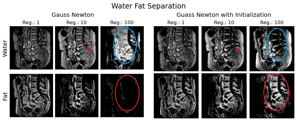
Results: By initializing the Gauss Newton method with the result of a polynomial fit of the signal phase, better and more robust water fat separation can be achieved in sUTE Dixon imaging.
Impact: The proposed polynomial fit initialization has improved water-fat separation quality and its robustness against regularization parameters while reducing the number of optimization iterations.
Introduction
Single-point ultra-shot echo time Dixon imaging techniques (sUTE-Dixon) enabled the visualization of short T2* components of the musculoskeletal anatomies and the spine [1-8]. Unlike multi-point Dixon approaches where the unwanted phase terms coming from sources such as B1 can be reliably calibrated by echo referencing, sUTE-Dixon must rely on the single complex image to both estimate and remove the unwanted phase terms and perform water-fat separation. With the assumption that the unwanted phase varies smoothly across the field of view (FOV), a recently proposed iterative optimization sUTE-Dixon approach -hereinafter referred to as Gauss Newton- has been applied for the simultaneous detection and assessment of acute vertebral fractures as well as bone marrow edema of the thoracolumbar spine [4, 5] and has also been shown to yield good water-fat separation across various musculoskeletal anatomies [6]. However, due to the non-convexity of the optimization problem and its ill-posed nature, the obtained results can vary significantly with the regularization factor. In this work, we show that by initializing the Gauss Newton method with the result of the polynomial fitting of the signal phase and its derived water and fat signals, the optimization problem solved robustly and converges faster.Methods
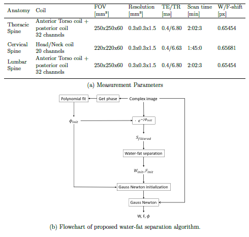
Data Acquisition:3D-UTE measurements were performed with a stack-of-stars center-out radial acquisition and phase-encoding in the third cartesian dimension on a clinical 3T system (Elition X, Philips Healthcare). Sequence parameters are shown in Fig. 1a.
Post Processing:
A simplified flow chart describing the proposed algorithm is depicted in Fig. 1b. The phase of the image was fitted to a 4th degree polynomial in 3D. The limited memory Broyden–Fletcher–Goldfarb–Shanno algorithm (L-BFGS) method was used for the fitting to account for speed as well as memory problems with high dimensional images without risking accuracy. The result of the polynomial fit was the first estimation of the unwanted background phase $$$(\phi_{init})$$$ and was removed from the complex UTE image, resulting in a filtered image $$$(S_{filtered})$$$. The filtered image was used for first estimations of the water and fat: \begin{align*} F_{init} &= \frac{\mathrm{Im}(S_{filtered})}{\mathrm{sin}(\Theta(TE))}, &W_{init} &= \mathrm{Re}(S_{filtered}) - F_{init}\cdot \mathrm{cos}(\Theta(TE)) \end{align*} where $$$\mathrm{Re}(\cdot)$$$ and $$$\mathrm{Im}(\cdot)$$$ are the real part and imaginary part operator of a number respectively. $$$\Theta(TE)$$$ is the phase between water and fat components, and was computed using a nine-peak fat model [9]. For the current acquisitions $$$\Theta(0.4ms)=-0.93048$$$.
$$$\phi_{init}, F_{init}, W_{init}$$$ were used as initialization for the Gauss Newton method, which solves the cost function: \begin{align*} \phi &= \underset{\phi}{\mathrm{argmin}} \psi(\phi)\\ &= \underset{\phi}{\mathrm{argmin}} \frac{1}{2} \lVert \left( W_{init} + F_{init} \exp^{i\Theta(TE)} \right)\exp^{i\phi} - S_{measured}\rVert _{2}^{2} + \lambda \rVert M\nabla\phi \lVert _{2} ^{2}, \end{align*} where $$$\lambda$$$ is the regularization parameter and $$$M$$$ is the used mask. This optimization yields $$$\phi$$$ with $$$\phi_0 = \phi_{init}$$$, which generates the final estimation of water and fat.
The two variants of the Gauss Newton method with and without the polynomial fit initialization were compared in terms of water-fat separation results and the number of iteration till convergence at regularization factors of 1, 10 and 100. The convergence criteria included when the relative phase update $$$\frac{\lVert d\phi \rVert _2}{\lVert \phi \rVert _2}$$$ is less than 0.01 or the number of iterations exceeds a 100. [4]
Results
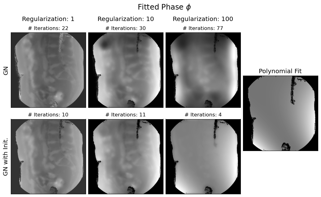
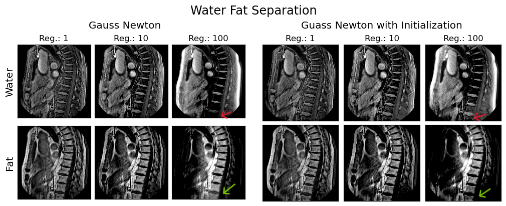
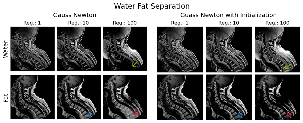
The estimated background phase images for different regularization parameters and the number of iterations needed for the computation are depicted in Fig. 2. The Gauss Newton method with initialization takes less iterations to converge at all the investigated regularization parameters. As expected the fitted phase image becomes increasingly similar to the phase generated by the polynomial fit when increasing the regularization parameter.The water-fat separation results for the three scanned anatomies are in Figs. 3, 4, 5. In the case of the thoracic spine there are no observable differences in stability or in quality of the water fat separation (Fig. 3). Fig. 4 shows that for mid-high regularization the swapping of the water in the lower region of the lumbar spine pointed out by the arrows are not present when initialization was used; there is however, a more severe fat swap at regularization factor of 100 in the case with initialization. Fig. 5 shows improvements in quality and stability of water-fat separation after using the initialization.
Conclusion
By initializing the Gauss Newton method with the result of a polynomial fitting of the signal phase, better and more robust water fat separation can be achieved in sUTE Dixon imaging.Acknowledgements
The present work was supported by the German Research Foundation (project number 455422993, FOR5298-iMAGO-P1). The authors also acknowledge research support from Philips Healthcare.
References
- Sophia Kronthaler, Christof Boehm, Georg Feuerriegel, Peter Boernert, Ulrich Katscher, Kilian Weiss, Marcus R. Makowski, Benedikt J. Schwaiger, Alexandra S. Gersing, and Dimitrios C. Karampinos. Assessment of vertebral fractures and edema of the thoracolumbar spine based on water-fat and susceptibility-weighted images derived from a single ultra-short echo time scan. Magnetic Resonance in Medicine, 87(4):1771–1783, 2022.
- Michael Carl, Graeme M. Bydder, and Jiang Du. Ute imaging with simultaneous water and fat signal suppression using a time-efficient multispoke inversion recovery pulse sequence. Magnetic Resonance in Medicine, 76(2):577–582, 2016.
- Hyungseok Jang, Michael Carl, Yajun Ma, Saeed Jerban, Tan Guo, Wei Zhao, Eric Y. Chang, and Jiang Du. Fat suppression for ultrashort echo time imaging using a single point Dixon method. NMR in Biomedicine, 32(5):e4069, 2019.
- Jingfei Ma. A single-point Dixon technique for fat-suppressed fast 3d gradient-echo imaging with a flexible echo time. Journal of Magnetic Resonance Imaging, 27(4):881–890, 2008.
- Georg C. Feuerriegel, Sophia Kronthaler, Christof Boehm, Martin Renz, Yannik Leonhardt, Florian Gassert, Sarah C. Foreman, Kilian Weiss, Markus Wurm, Thomas Liebig, Marcus R. Makowski, Benedikt J. Schwaiger, Dimitrios C. Karampinos, and Alexandra S. Gersing. Diagnostic value of water-fat-separated images and CT-like susceptibility-weighted images extracted from a single ultrashort echo time sequence for the evaluation of vertebral fractures and degenerative changes of the spine. European Radiology, 33(2): 1445–1455, Feb 2023.
- Kronthaler S, Feuerriegel GC, Boehm C, Gersing AS, Schwaiger BJ, Makowski MR, Weiss K, and Karampinos DC. On the robustness of single ute-dixon for simultaneous short T2* , water and fat imaging across skeletal anatomies. Proc ISMRM 2022, page 536, 2022.
- Anh T. Van, Kilian Weiss, Georg C. Feuerriegel, Philipp Braun, Guruprasad Krishnamoorthy, Alexandra S. Gersing, James G. Pipe, and Dimitrios C. Karampinos.Accelerated single UTE-Dixon for simultaneous short T2* water and fat imaging using a floret trajectory. Proc ISMRM 2023, 2023.
- Nathan Newbury, Sam Sedaghat, Jiyo Athertya1, Michael Carl, Jiang Du, and Hyungseok Jang. Ultrashort echo time single point Dixon using 3d phase modeling. Proc ISMRM 2023, 2023.
- Jimin Ren, Ivan Dimitrov, A. Dean Sherry, and Craig R. Malloy. Composition ofadipose tissue and marrow fat in humans by 1H NMR at 7 tesla. Journal of LipidResearch, 49(9):2055–2062, Sep 2008.
Figures