0481
Safety of Implanted Auditory Prostheses at 7T1Cochlear Technology Centre Belgium, Mechelen, Belgium, 2Laboratory of Soft Matter and Biophysics, Department of Physics and Astronomy, KU Leuven, Leuven, Belgium, 3Research group Experimental Otorhinolaryngology, Department of Neurosciences, KU Leuven, Leuven, Belgium, 4Philips Healthcare N. V., Best, Netherlands, 5Department of Otolaryngology, Head and Neck Surgery, University Hospitals Leuven, Leuven, Belgium, 6Sir Peter Mansfield Imaging Centre, School of Physics and Astronomy, University of Nottingham, Nottingham, United Kingdom, 7National Institute for Health Research (NIHR) Nottingham Biomedical Research Centre, Nottingham, United Kingdom, 8Hearing Sciences, Division of Mental Health and Clinical Neurosciences, School of Medicine, University of Nottingham, Nottingham, United Kingdom
Synopsis
Keywords: Safety, Safety, Prostheses, active auditory implants
Motivation: While scanning individuals with active hearing implants (e.g., cochlear implants) at 1.5 and 3 T has become commonplace, scanning these patients at ultra-high field strengths (e.g., 7 T) is likely to be associated with an increased risks of damaging the device or causing soft-tissue damage, pain, and/or discomfort.
Goal(s): To evaluate the safety of three different active hearing implants in the 7 T MR environment.
Approach: Five potential interactions between the devices and the MR environment were assessed using industry-recommended practices for 1.5/3 T.
Results: Preliminary findings show no adverse effects within the predefined test conditions.
Impact: While scanning individuals with active hearing implants at 1.5/3 T has become commonplace, 7 T is associated with significantly greater safety risks. This study is the first to provide some confidence in the safety of such implants at 7 T.
Introduction
The use of magnetic resonance (MR) imaging and spectroscopy in patients with active implanted auditory prostheses has become daily practice for diagnostic medical assessment. Many such prostheses contain implanted retaining magnets that align the internal and external components of the device. Recently developed implants feature rotatable magnets, significantly improving the safety of MR scanning individuals using these implants at field strengths up to 3T. Such scanning is conditionally approved by international regulators, whereby the conditions imposed range from software restrictions of the radiofrequency specific absorption rate (SAR) and gradient strength during scanning, to the removal of an implanted retaining magnet, if present. Failure to comply with these safety conditions carries the risk of damaging the implanted prosthesis, and/or causing pain and/or tissue damage. Scanners using field strengths of 7T are becoming more widely available. Scanning implanted patients at ultra-high field strengths is likely to be associated with even greater risks for the patient. This study aims to evaluate the safety of multiple active hearing implants in the 7T MR environment by assessing the mutual interactions occurring between the implantable device and the MR scanner and its environment.Methods
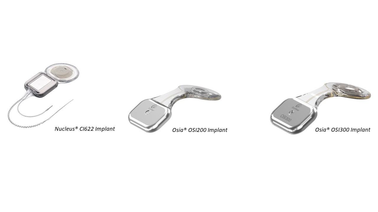
Several potential interactions were investigated using a 7T Philips Achieva MR scanner (Philips, Best, Netherlands), adhering to the relevant active industry standards. Measurements included: the magnetically induced force experienced by the implant housing and the implantable retaining magnet, demagnetisation of the implantable retaining magnet, electrical function of the implant following scanning, scanning-induced heating of the implant as caused by radiofrequency pulses and gradients, and quantification of image artefacts. Experiments were performed on three active hearing implants: Nucleus cochlear implant CI622 and Osia bone conduction implants OSI200 and OSI300 (Cochlear Ltd., Sydney, AU).Results and discussion
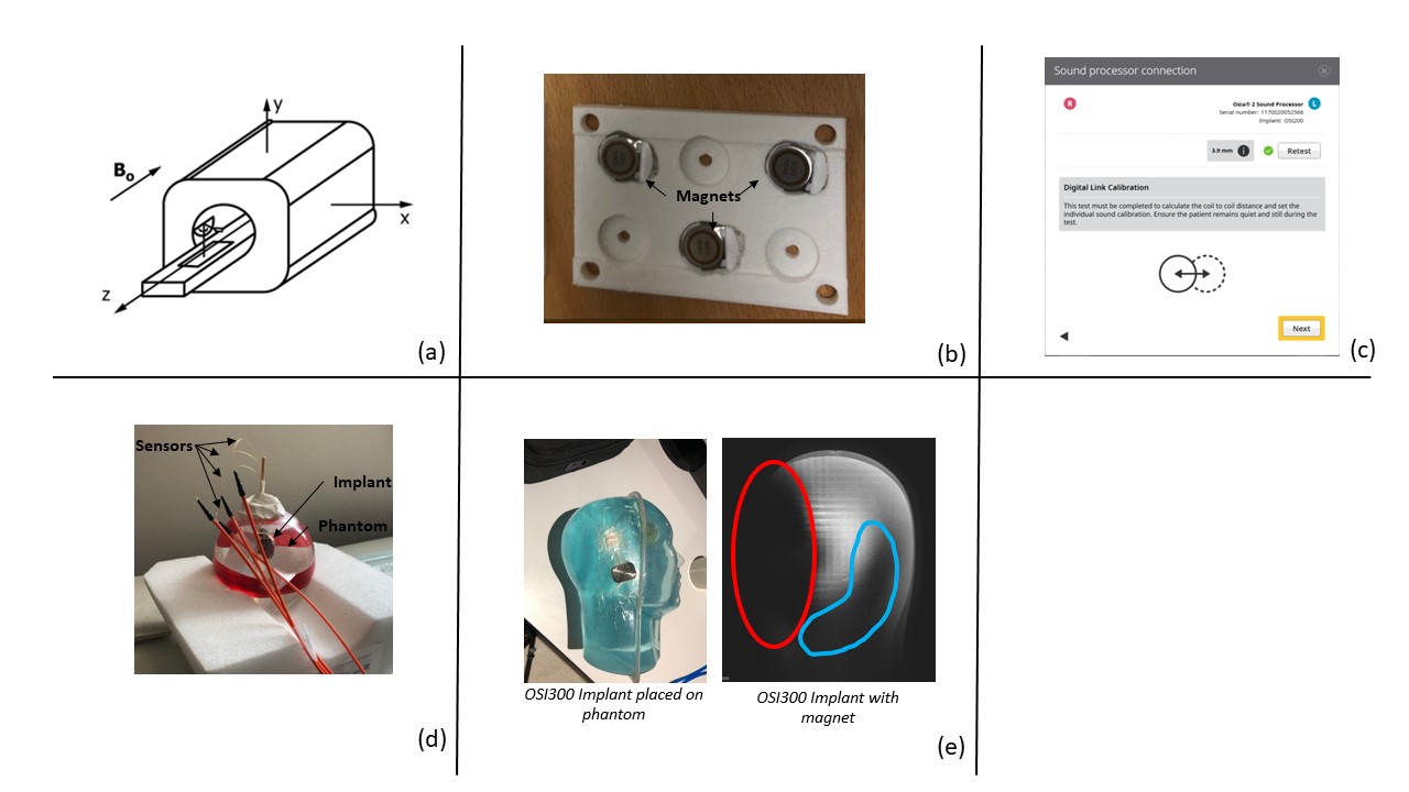
Magnetically induced forces were measured by quantifying the deflection force when the device was suspended on a string close to the bore entry, where the spatial gradient field was maximal [1] (Fig. 2a). When no magnet was present, the force ratio (defined as the magnetically-induced force divided by the force induced by gravity) remained below 0.3 for all devices. With the magnet in place, the force ratio increased up to 11 for the CI622 device.Several implant magnets were exposed to the scanner’s B0 field at body temperature, by moving them into the scanner's isocentre, either with their main axis aligned with the field (N=12) or perpendicular to it (N=6).
Retaining magnet magnetization was measured before and after exposure using a magnetic field camera (Fig. 2b). In line with industry standards, a total of 10 exposures were performed [2]. Changes in magnetization were quantified per magnet and averaged for the population. Average magnetization changes of ±1% were measured in both orientations, which was similar to the average population spread at baseline.
Device functionality after scanning was verified using the device fitting software (Fig. 2c). A dedicated experiment was performed for all devices where functionality was verified after a total of ten exposures, which according to industry standards [2] covers the needs of 99.9% of the population. The same devices were subsequently used during all other tests, and intermediate functionality tests were performed.
Device heating was measured using fiber-optic sensors attached around the devices when placed in a liquid phantom at isocentre (for RF-induced heating) or 20 cm from isocentre (gradient-induced heating) [2] (Fig. 2d). For all devices, heating remained limited to 0.15°C compared to background heating after 15 minutes consecutive scanning at 1 W/kg (RF-induced heating) or with a gradient field strength of 41.8 T/s (gradient-induced heating).
Image datasets were acquired with the device attached to the right side of the head of a commercial head phantom (Fig. 2e). Pulse sequences were selected based on the commonly used clinical examinations. Images were acquired in a coronal direction to limit scan time. When scanning with the magnet in place, the artefact covered almost the entire ipsilateral side of the head. When the magnet was replaced with the non-magnetic cassette, the extent of the artefact was notably reduced.
Conclusion
This study documents the first results of a feasibility trial that investigated several interactions between active hearing implants and the ultra-high field 7T MR environment. Five interactions that may result in patient harm were assessed. These preliminary findings show no adverse effects on the implant within the predefined test conditions, and image artefacts similar to those seen at 3T.Preliminary outcomes of this feasibility study are positive, yet do not imply implant safety in the 7T MR environment. Formal verification will be required to label a device for use within the 7T MR environment.
Acknowledgements
N Verhaert was supported by Fonds Wetenschappelijk Onderzoek (FWO) Senior Clinical Investigator Fellowship 1804816N. This work was funded by Cochlear Belgium and supported by the National Institute for Health Research (NIHR) Nottingham Biomedical Research Centre.References
[1] American Society for Testing and Materials, “Standard Test Method for Measurement of Magnetically Induced Displacement Force on Medical Devices in the Magnetic Resonance Environment (F2052-21),” Annual Book of ASTM Standards, vol. i, pp. 1–6, 2021.
[2] ISO, “Assessment of the safety of magnetic resonance imaging for patients with an active implantable medical device. ISO/TS 10974:2018,”International Organization for Standardisation, vol. 2018, 2018.
Figures