0479
Evaluation of a Numerical Approach Alternative to MR Thermometry in the Safety Validation of Multi-Channel RF Coils1Center for Magnetic Resonance Research (CMRR), University of Minnesota, Minneapolis, MN, United States
Synopsis
Keywords: Safety, Safety
Motivation: The existing safety validation process for multi-channel RF coils requires MR thermometry tests, which are difficult to carry out.
Goal(s): To propose an alternative approach to MRT in the validation process of multi-channel RF coils.
Approach: The simulated B1+ error was measured and propagated to the SAR error using Monte-Carlo simulations. To validate this approach MRT experiments were conducted with a 16-channel body coil at 10.5T, and the measured SAR errors were compared against the predicted ones.
Results: For two different excitation patterns, measured SAR errors were 36% and 40%, while predicted SAR errors were 49% and 46%, respectively.
Impact: The new strategy presented replaces MR Thermometry in the safety validation process of multi-channel RF coils. This approach can simplify and accelerate the development and testing of innovative multi-channel RF coils which are essential for UHF MRI applications.
Introduction
According to International guidelines1, to minimize patient safety risks related to multi-channel RF transmit coils, peak local SAR (pSAR) is one of several limits that must not be exceeded. Due to the lack of accurate pSAR measurement methods for in-vivo applications, the prediction of the pSAR relies on experimentally validated EM simulation models2,3. Despite the clear workflow that exists for the experimental validation of the EM model3, error in the prediction of pSAR is inevitable due to different sources of uncertainties. If quantified, these uncertainties—listed as inter-subject variability ($$${e_{ISV}}$$$), power monitoring uncertainty ($$${e_{PM}}$$$), and EM modeling error ($$${e_{EMM}}$$$)—can be combined in a sum-of-squares fashion to calculate a safety factor4 (SF) and used to scale the predicted peak local SAR:$$SF = 1 + \sqrt {{e_{PM}}^2 + {e_{ISV}}^2 + {e_{EMM}}^2}$$
To quantify the $$${e_{EMM}}$$$, it was previously proposed4 to calculate the deviation between SAR maps determined by simulation and experiment using MR Thermometry5 (MRT). The main hurdles of this approach are related to the technical challenges of conducting MRT experiments including the need to perform several different excitation patterns, phase drift errors, system instability, specialized phantoms, and the need for high transmit-power—which can be harmful to sensitive electronics, especially in the presence of a dedicated receiver coil inside the transmitter. To overcome these challenges, we previously proposed6 a new method to quantify $$${e_{EMM}}$$$ with respect to peak 10g-averaged SAR (pSAR10g) without the need for conducting MRT experiments. Our approach propagates the quantitative differences between measured and simulated B1+-maps to find an upper bound for pSAR10g error. In the current study, we evaluate the performance of our approach against experimental MRT measurement using a 16-channel dipole transceiver body coil7 with two different excitation patterns at 10.5T.
Methods
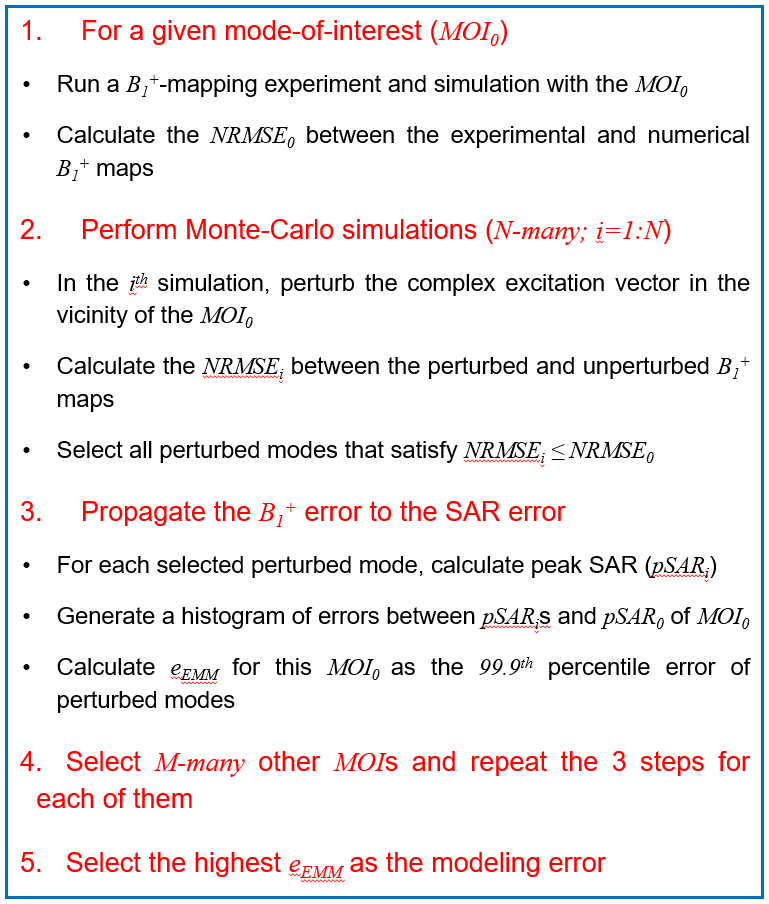
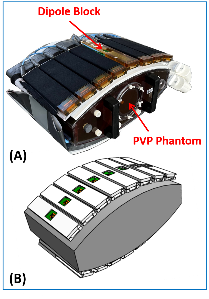
Figure1 summarizes the workflow we previously proposed6 for quantifying the $$${e_{EMM}}$$$ corresponding to any particular mode of excitation.To evaluate the capability of this approach in predicting the SAR error, CP and alternating-phase modes of interest (MOI0) of a 16-channel dipole transceiver body coil7 (Figure2) at 10.5T were investigated (i.e., the first step in Figure1). In the MRT-based validation technique proposed by Steensma et al4, the voxel-wise comparison between simulation and experimental B1+ and SAR maps was used for the $$${e_{EMM}}$$$ quantification, whereas in our proposed technique, the normalized root-mean-square error (NRMSE) between the measured and simulated B1+-maps of any mode of excitation was propagated to the pSAR10g-error space of that particular mode of excitation. The error propagation was carried out using 105 Monte-Carlo simulations following the second step in Figure1. The perturbed modes around the MOI0 were then used to generate the pSAR10g-error space for that MOI0. Eventually, the 99.9th percentile of the pSAR10g-error space was calculated as the $$${e_{EMM}}$$$ of that particular mode of interest (the third step in Figure1). The $$${e_{EMM}}$$$ calculated using this numerical technique were compared to those calculated from the experimentally measured SAR in Schmidt et al’s work7.
Results
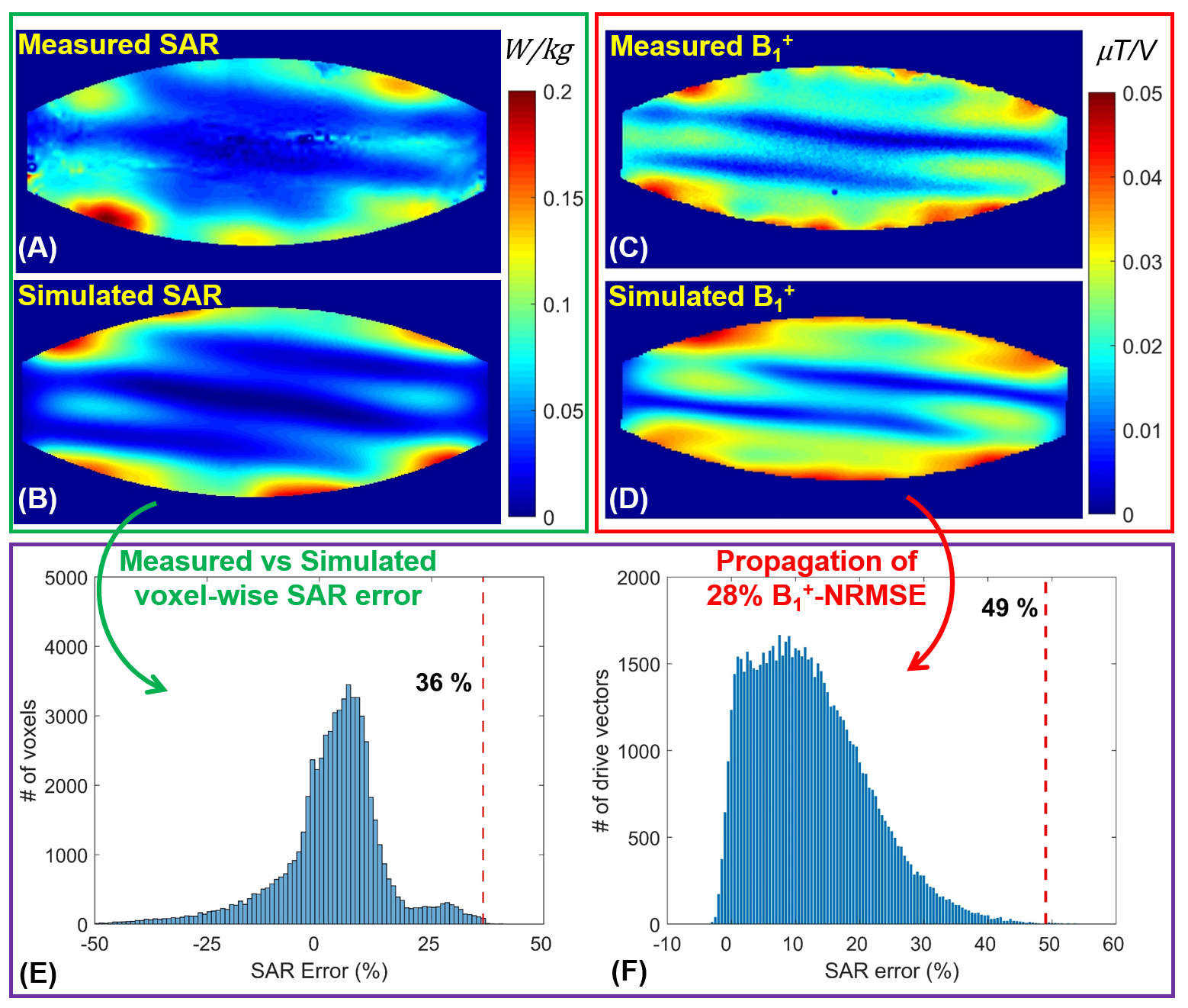
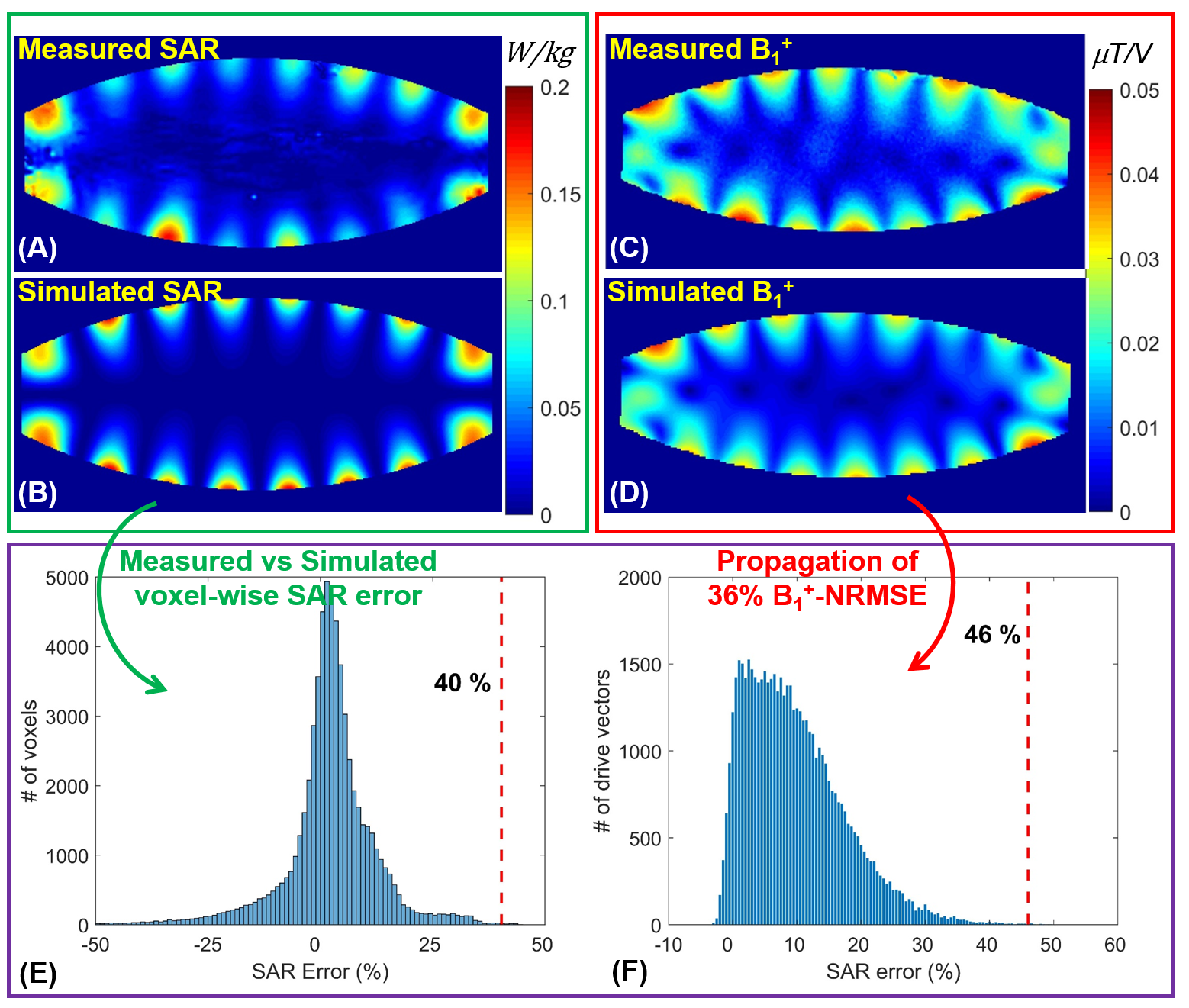
Figure3A-D shows the measured and simulated B1+ and SAR maps corresponding to the CP mode of excitation. For this mode, Figure3E shows the voxel-wise local SAR error assessment proposed by Steensma et al4, where the 99.9th percentile (red dashed line at 36%) of voxel-wise difference between simulated and measured SAR maps is introduced as the $$${e_{EMM}}$$$. Whereas Figure3F shows the pSAR10g-error space generated by propagating a 28% NRMSE between measured and simulated B1+ maps to the SAR error. The 99.9th percentile (red dashed line at 49%) of this pSAR10g-error space is defined as the $$${e_{EMM}}$$$.Figure4 is a demonstration of a similar comparison between two techniques for the alternating-phase mode of excitation where adjacent channels are shifted by 180 degrees.
Discussions and Conclusion
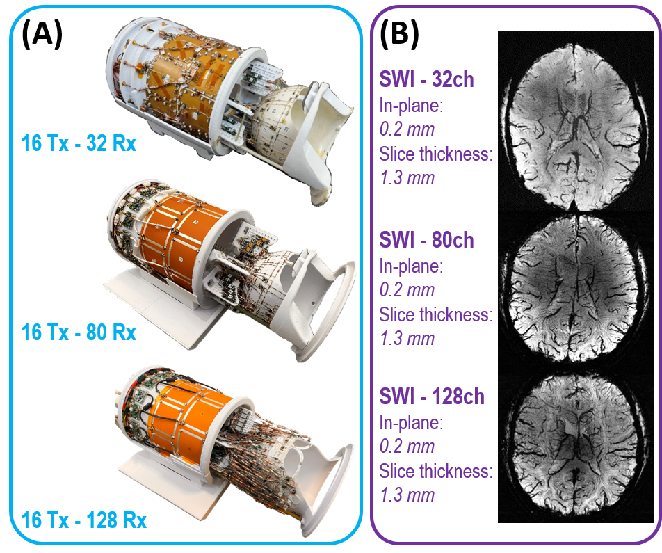
We developed a strategy to handle EM modeling uncertainties ($$${e_{EMM}}$$$) with a simulation-based analysis. In this study, the reliability of our proposed technique was investigated and demonstrated by comparing the numerically predicted $$${e_{EMM}}$$$ to that of measured SAR data. While the MRT-based approach resulted in a 36% for the CP mode of excitation, our simulation-only-based technique conservatively predicts a 49% $$${e_{EMM}}$$$. MRT- and simulation-based $$${e_{EMM}}$$$ for alternating-phase mode of excitation are 40% and 46%, respectively. Note that only two excitation modes were experimentally investigated and validated. In future studies, more modes will be analyzed.The proposed technique, which offers an alternative approach to MRT experiments in the safety validation process of RF coils, is free from the aforementioned challenges associated with MRT experiments. It enabled the validation of three head coils with dedicated high-channel receivers8-10 (16-channel transmitters with 32-, 80-, and 128-channel receivers). The corresponding preliminary susceptibility-weighted imaging results are presented (Figure5).
Acknowledgements
This work was supported by the following grants: NIBIB P41 EB027061, NINDS R01NS115180, NIH U01 EB025144, and NIH R01 EB029985.References
1. International Electrotechnical Commission. IEC 60601-2-33-Edition 4.0: Medical Electrical Equipment—Particular Requirements for the basic safety and essential performance of magnetic resonance equipment for medical diagnosis. 2022.
2. Restivo M, Raaijmakers A, van den Berg C, Luijten P, Hoogduin H. Improving peak local SAR prediction in parallel transmit using in situ S‐matrix measurements. Magnetic resonance in medicine. 2017 May;77(5):2040-7.
3. Sadeghi‐Tarakameh A, DelaBarre L, Lagore RL, Torrado‐Carvajal A, Wu X, Grant A, Adriany G, Metzger GJ, Van de Moortele PF, Ugurbil K, Atalar E. In vivo human head MRI at 10.5 T: A radiofrequency safety study and preliminary imaging results. Magnetic resonance in medicine. 2020 Jul;84(1):484-96.
4. Steensma BR, Sadeghi‐Tarakameh A, Meliadò EF, van den Berg CA, Klomp DW, Luijten PR, Metzger GJ, Eryaman Y, Raaijmakers AJ. Tier‐based formalism for safety assessment of custom‐built radio‐frequency transmit coils. NMR in Biomedicine. 2023 May;36(5):e4874.
5. Kuroda K, Oshio K, Chung AH, Hynynen K, Jolesz FA. Temperature mapping using the water proton chemical shift: A chemical shift selective phase mapping method. Magn Reson Med. 1997; 38: 845-851.
6. Sadeghi-Tarakameh A, Radder J, Lagore RL, et al. Safety Assessment of Custom-Built Multi-Channel RF Coils: EM Modeling Uncertainties: Proceedings of the International Society of Magnetic Resonance for Medicine 30, London, UK. April 2022. ISMRM; 2022:538.
7. Schmidt S, Ertürk MA, He X, Haluptzok T, Eryaman Y, Metzger GJ. Improved 1H body imaging at 10.5 T: Validation and VOP‐enabled imaging in vivo with a 16‐channel transceiver dipole array. Magnetic Resonance in Medicine. 2023 Sep 13.
8. Tavaf N, Lagore RL, Jungst S, Gunamony S, Radder J, Grant A, Moeller S, Auerbach E, Ugurbil K, Adriany G, Van de Moortele PF. A self‐decoupled 32‐channel receive array for human‐brain MRI at 10.5 T. Magnetic resonance in medicine. 2021 Sep;86(3):1759-72.
9. Waks M, Lagore R, Auerbach E, et al. A Self-Decoupled 16-Channel Transmit, 80-Channel Receive Array For 10.5 Tesla Human Head Imaging. Proceedings of the International Society of Magnetic Resonance for Medicine 31, Toronto, ON, Canada. June 2023. ISMRM; 2023:02
10. Lagore RL, Grant A, DelaBarre L, et al. 128-Channel Brain Imaging Array With Improved Acceleration At 10.5 Tesla. Proceedings of the International Society of Magnetic Resonance for Medicine 31, Toronto, ON, Canada. June 2023. ISMRM; 2023:1059.
Figures