0477
Efficient RF Shimming Strategies for Cardiac MRI at 5T1Paul C. Lauterbur Imaging Research Center, Shenzhen Institutes of Advanced Technology, Chinese Academy of Sciences, Shenzhen, China, 2Key Laboratory for Magnetic Resonance and Multimodality lmaging of Guangdong Province, Shenzhen, China, 3Chongqing University of Technology, Chongqing, China, 4United lmaging Healthcare, Shanghai, China, 5Department of Biomedical Engineering, State University of New York at Buffalo, New York, NY, United States
Synopsis
Keywords: Parallel Transmit & Multiband, Parallel Transmit & Multiband, SAR;RF Shimming;CMR;
Motivation: Enhance RF shim efficiency, reduce power, and provide uniformity for ultra-high field cardiac imaging.
Goal(s): To significantly enhance excitation efficiency, reduce excitation power, and mitigate the risk of SAR exceeding safe levels, while minimizing loss of uniformity.
Approach: Optimizing excitation intensity and uniformity through joint loss function and validating the approach with simulation models and in vivo imaging.
Results: The research results indicate that, when compared to MLS, the proposed Eff-MLS leads to an average 3.3% reduction in CV, while improving safety excitation efficiency by 442.26% in the simulation model. Similar effects were also observed in in-vivo experiments.
Impact: By enhancing RF shim efficiency and reducing the required excitation power while maintaining uniformity, this approach paves the way for more precise, high-resolution cardiac imaging at ultra-high field strengths. The potential impact includes broader applications of ultra-high field CMR.
Introduction
In the context of cardiac magnetic resonance imaging (CMR), ultra-high field (>3T) improves myocardial-blood pool contrast and anatomical resolution, enhancing cardiac functional assessment [1]. However, higher field strengths exacerbate radiofrequency (RF) field inhomogeneity in the heart, resulting in dark band artifacts due to dielectric effects. This diminishes contrast and hinders clinical diagnosis. RF shimming, a common solution, necessitates increased RF excitation power due to factors like heart position and volume [2].This study presents an efficient RF shimming for 5T CMR, aimed at improving excitation efficiency while minimizing uniformity loss, reducing RF power requirements, and mitigating the risks associated with high specific absorption rate (SAR) and excessive power. The effectiveness and robustness of the proposed algorithm are validated through electromagnetic simulations and in vivo experiments.
Method
RF shimming is commonly regarded as a magnitude least squares (MLS) optimization problem. To enhance excitation efficiency within the target region while optimizing uniformity, a constraint on the excitation strength is introduced on top of the uniformity optimization loss function. Additionally, less weight is assigned to uniformity in areas outside the heart in the optimization equation to reduce the risk of elevated local SAR. This approach is referred to as Eff-MLS and is represented in Equation.$$min\left\{|||Ax|-\theta||^{2}+\beta_{1}||x||^2+\beta_2\frac{1}{||Ax||^2}\right\}$$
In this equation, matrix A represents the sensitivity maps obtained from a prescan for each transmit channel. The vector x denotes the complex weighting factors for each transmit channel relative to a default channel, and vector θ represents the normalized target flip angle.The coefficient of variation (CV) is employed to assess the uniformity of the transmit field, while the rayleigh quotient is utilized to evaluate the excitation efficiency[3].
The simulation dataset, sourced from Sim4life, comprises 15 human models[4]. Simulations focus on a 5mm thick transverse section at the heart's center for single-layer RF shimming, facilitating thorough parameter analysis and comparison. Standardization of the average magnetic field within the cardiac region across all simulation results ensures comparability with real system calibration requirements.Validation used cardiac data from a healthy male volunteer scanned on a 5T MRI system (United Imaging Healthcare, Shanghai, China). Transmit channel sensitivity maps were acquired using the self-developed DREAM sequence with specific parameters: 'STE first' echo ordering, a 54.7°STEAM preparation pulse, 1.2ms shot-to-shot delay, and a 450×450×10mm³FOV. Multi-channel transmit encoding with encoding coefficients of -1 was employed to improve signal-to-noise ratio and sensitivity map reliability[5].
Result
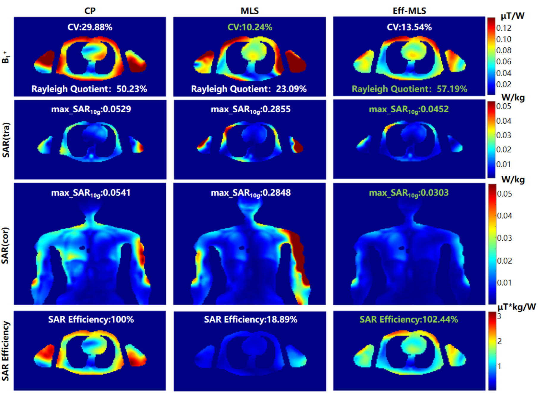
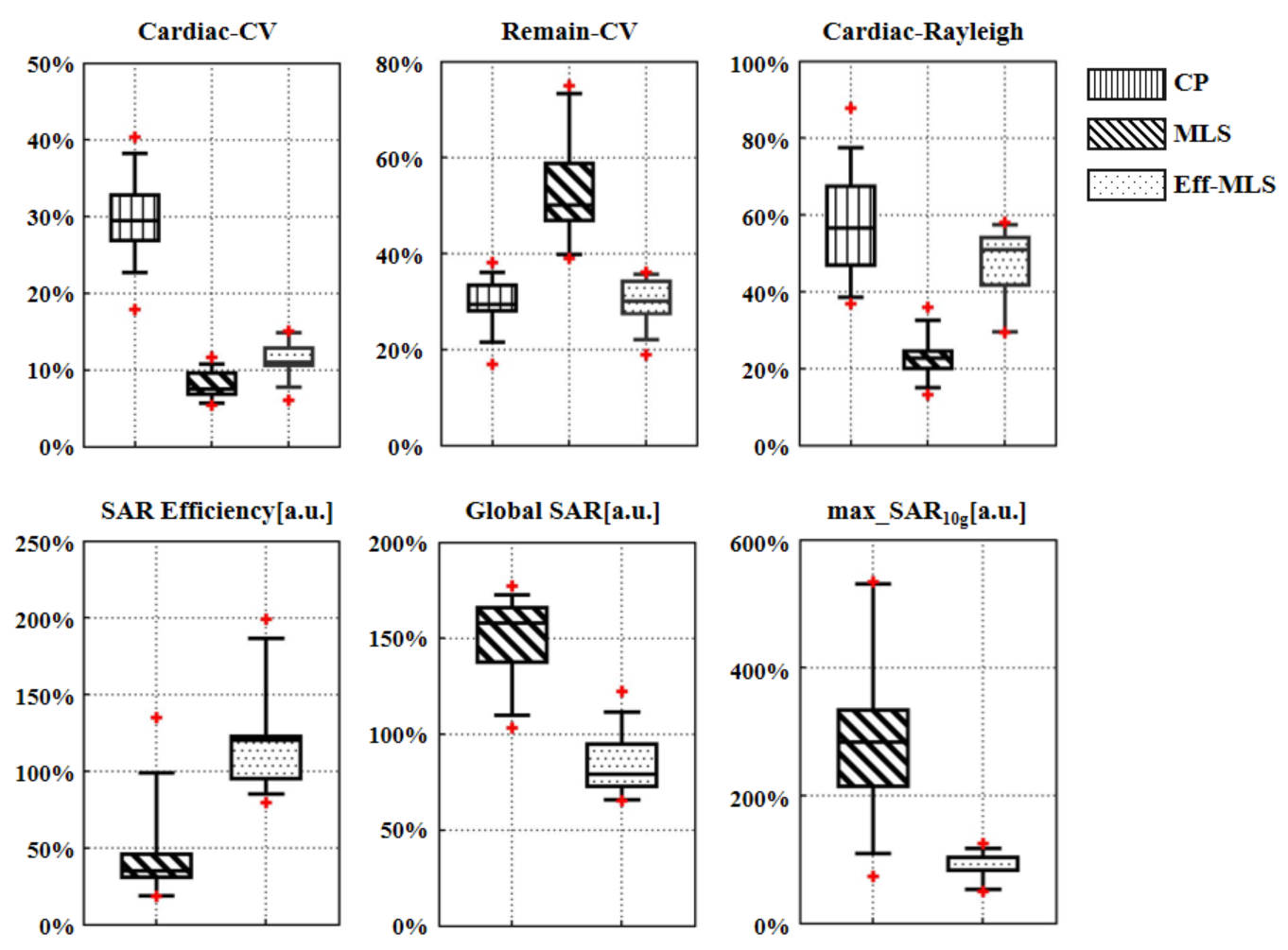
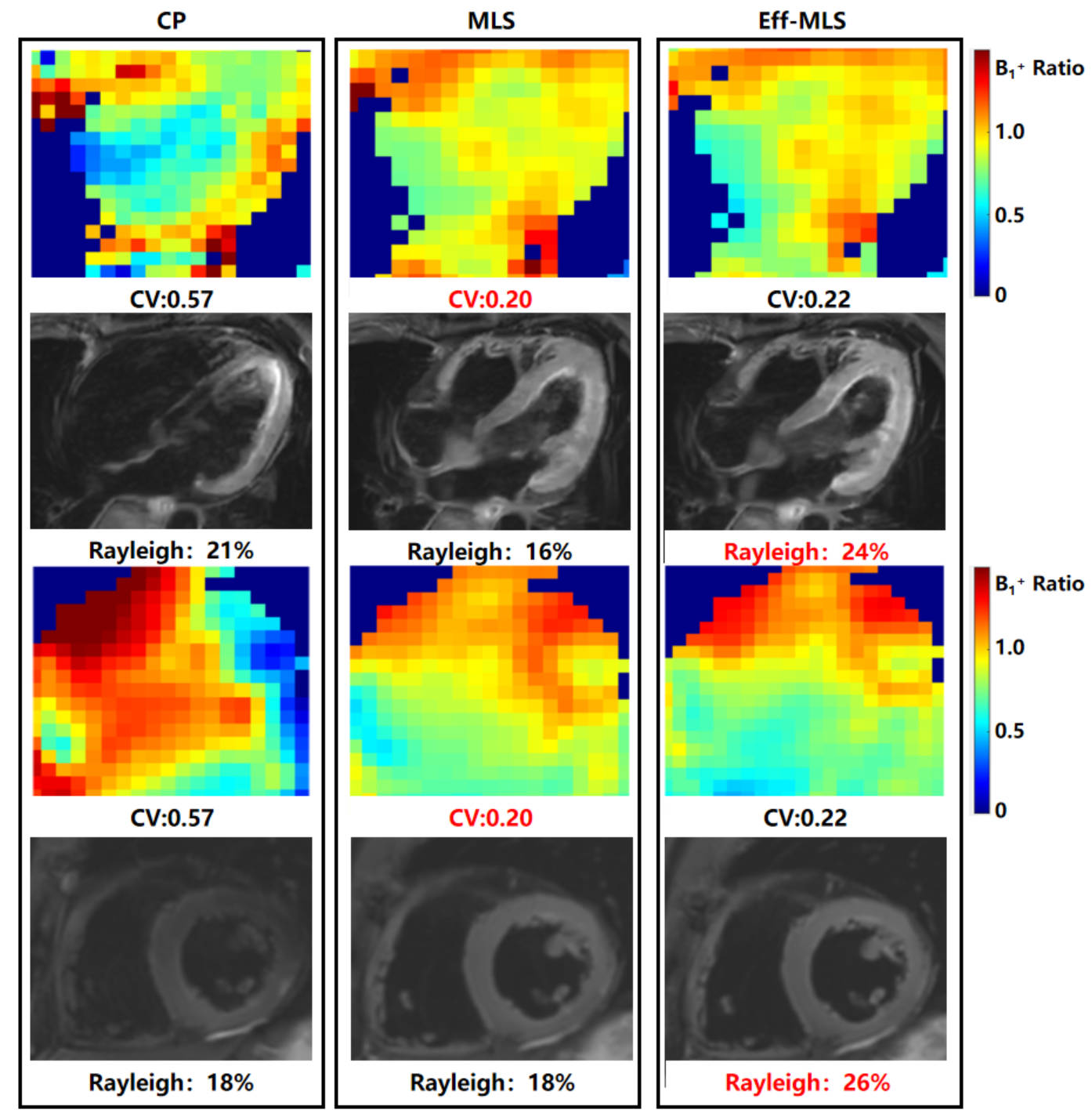
The results in Figure 1 display shim outcomes for the Duke simulation model. They reveal that CP excitation without RF shimming leads to magnetic field distribution non-uniformity. Application of the conventional RF shimming MLS algorithm enhances uniformity in the cardiac region but at the expense of excitation efficiency. In contrast, Eff-MLS results indicate a slight reduction in cardiac uniformity compared to MLS, with a 3.3% increase in the CV. Notably, hotspots' number and intensity in the arm region decrease. Enhanced excitation efficiency is observed with a 34.10% increase in local efficiency, a 59.32% reduction in overall RF power, an 81.56% reduction in maximum local SAR10g, a 48.92% reduction in global SAR, and a 442.26% increase in average safety excitation efficiency.In Figure 2, Eff-MLS results across all samples show an average CV reduction of 3.38%, a 24.32% increase in average excitation efficiency, and a 64.13% reduction in global SAR when compared to MLS.Figure 3 shows T2-FSE images and corresponding B1+ maps from a healthy male volunteer under various RF shimming parameters. CP excitation results in a noticeable loss of contrast in the right cardiac region.After applying MLS, there is an improvement in cardiac uniformity. Eff-MLS maintains cardiac uniformity with a 2.8% increase in B1+ non-uniformity in the four-chamber view. Eff-MLS reduces excitation power by 36.67% compared to MLS, and the predicted global SAR decreases by 29.41%. In the short-axis view, excitation power is reduced by 39.29% while maintaining consistent myocardium-blood pool contrast.Discussion
Both electromagnetic simulations and in-vivo imaging confirm that Eff-MLS, in comparison to MLS, significantly improves excitation efficiency while slightly reducing RF excitation field uniformity. This reduction mitigates the risk of SAR exceeding safe levels, closely resembling CP excitation SAR values. Future experiments will expand the sample size for further validation.Conclusion
This study presents an efficient RF shimming strategy for cardiac imaging, which enhances RF excitation efficiency while preserving uniformity and reducing excitation power. Validation was performed on a 5T MRI system. Simulation results show significant excitation power reduction across 15 different models, while maintaining excitation performance. In vivo imaging results confirm reduced SAR risk in both the short-axis and four-chamber viewsAcknowledgements
This work was supported in part by, the NSFC grant(U22A20344),the Project on Global Common Challenges of Chinese Academy of Sciences(No. 321GJHZ2022081GC), the Key Laboratory for Magnetic Resonance and Multimodality Imaging of Guangdong Province (2023B1212060052), the Funding Program of Shenzhen ,China (RCYX20200714114735123),the Chinese Academy of Sciences Youth Innovation Promotion Association funded project (Y2021098)References
1. Noeske R, Seifert F, Rhein KH, Rinneberg H. Human cardiac imaging at 3T using phased array coils. Magn Reson Med. 2000;44:978-982.
2. Setsompop K, Wald L, Alagappan V, Gagoski B, Adalsteinsson E. Magnitude least squares optimization for parallel radio frequency excitation design demonstrated at 7 Tesla with eight channels. Magn Reson Med. 2008;59:908-915.
3. He X, Schmidt S, et al. Improved TSE imaging at ultrahigh field using nonlocalized efficiency RF shimming and acquisition modes optimized for refocused echoes (AMORE). Magn Reson Med. 2022;88(4):1702-1719.
4. Gosselin M C , Neufeld E , Moser H , et al. Development of a new generation of high-resolution anatomical models for medical device evaluation: the Virtual Population 3.0[J]. Physics in Medicine & Biology, 2014, 59(18):5287-5303.
5. Tse DH, Poole MS, Magill AW, Felder J, Brenner D, Jon Shah N. Encoding methods for B1+ mapping in parallel transmit systems at ultra high field. J Magn Reson. 2014;245:125-32.
Figures