0471
Rapid Specific Absorption Rate Estimation of High-Field MRI via 3D U-net Architectures for MRI Safety1School of Electrical and Electronic Engineering, Nanyang Technological University, Singapore, Singapore, 2Engineering Product and Development, Singapore University of Technology and Design, Singapore, Singapore
Synopsis
Keywords: Safety, Safety
Motivation: Advancements in MRI technology towards high fields demand rapid and accurate SAR estimation tools for enhancing MRI safety, currently hindered by the computational cost of conventional physics-based simulators.
Goal(s): The goal is to develop an efficient machine learning framework capable of estimating subject-specific SAR values rapidly.
Approach: The study employs 3D U-net deep learning models with their variants to achieve rapid and accurate SAR estimations.
Results: The proposed neural network model provides SAR estimations within 183ms, achieving approximately 10,000x acceleration over traditional physics-based simulators, with a mean relative error of 7.6%.
Impact: The near real-time accurate SAR estimation achieved by proposed machine learning framework will allow (i) checking patient-specific SAR while patient is lying in the MRI machine and (ii) performing ultra-fast optimization and uncertainty quantification studies while designing new high-field coils.
Introduction
As a non-invasive diagnostic technique, MRI is widely used for visualizing internal body structures. Among medical imaging modalities, it is distinguished by its capability to provide high soft tissue contrasts without ionizing radiation. It is nowadays indispensable for detecting and localizing tumors and cancerous tissues. The demand for enhanced image resolution in MRI has driven extensive research efforts on high-field MRI systems with magnetic fields exceeding 3 Tesla (T). While improving resolution, these systems introduce challenges regarding the non-uniform electric field (E-field) distributions and the corresponding thermal effects on human tissues, characterized by the Specific Absorption Rate (SAR). Compliance with SAR regulations 1,2 is essential to ensure the MRI safety. Nonetheless, the task of predicting the local SAR distributions prior to the actual scanning is challenging due to the unique anatomical structures and electrical properties of individual subjects, specifically tissues’ relative permittivities ($$$\epsilon_r$$$) and conductivities ($$$\sigma$$$). Currently, the task can be accomplished by physics-based simulators through iterative methods 3,4, which take anatomical models derived from MRI data with corresponding electrical properties of human tissues to calculate the induced E-fields and SAR distributions within the human body. However, these simulators are resource-intensive and unable to deliver real-time computations, underscoring the necessity for innovative frameworks to accelerate the estimation of subject-specific SAR. So far, a deep learning-based model utilizing a 2D U-Net architecture has been proposed to estimate the MRI-induced SAR in real time 5. Although the proposed architecture allows real-time SAR estimation, its capabilities are limited to two-dimensional estimations, and it does not incorporate the variability in electrical properties. To this end, our study aims to develop a framework capable of processing three-dimensional human head models with varying electrical properties and accurately estimating subject-specific SAR values.Methods
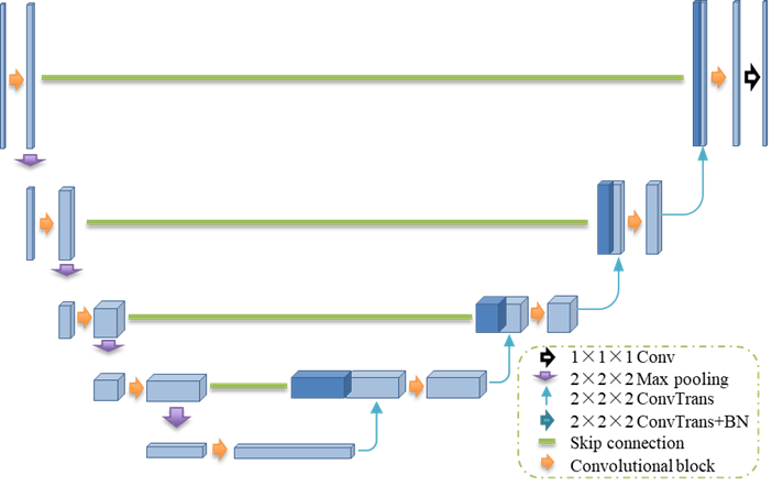
This study employs a convolutional neural network (CNN) approach based on a 3D U-net architecture and its variant for estimating subject-specific SAR in human head models at 7T (300 MHz). The U-net architecture, as shown in Figure 1, excels in detailed feature mapping and complex pattern recognition, with its encoder-decoder structure and skip connections. The networks are trained on a large dataset generated by the physics-based simulator MARIE 3. Inputs to the networks include permittivity and conductivity maps of human head models and the transmitted E-fields from a high-pass birdcage head coil. Then, the models provide the estimations of the three-dimensional E-field and the corresponding 10-g SAR value distributions through post-processing.Results and Discussion
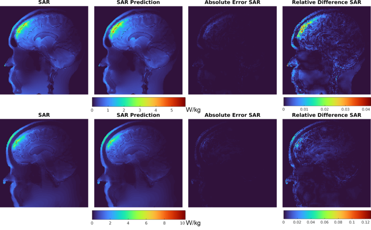
This dataset was first derived from different human head models from open-source datasets 6,7, with each model consisting of 500 data samples with varying permittivity and conductivity maps at a resolution of 1.4 millimeters. Subsequently, the dataset was separated into training, validation, and testing sets, where the division was performed to ensure that the head models within each set were unique, with no repetition between sets. Such partitioning guaranteed that the head models used for testing remain entirely unseen by the neural network during training. The dataset has been deployed for training on standard U-net and attention U-net. Among the network structures employed, the standard U-net provided better results, yielding a mean relative error (MRE) of 7.6% on the testing dataset. This level of accuracy is complemented by its computational efficiency, where the network performs a single SAR estimation within 183ms, which corresponds to approximately 10,000x acceleration compared to the traditional physics-based simulator, MARIE. To visually demonstrate the accuracy of the proposed method, selected slices from two head models are presented in Figure 2.Conclusion
This study presents an innovative framework by employing 3D CNNs to estimate subject-specific SAR distributions in near real-time. The results indicate that the framework yields promising results in terms of accuracy and computational speed. By utilizing deep-learning methods, this study provides new possibilities on optimizing SAR safety and understanding of non-uniform SAR distribution within human. In future work, additional architectures will be explored to improve the accuracy of SAR estimation. Furthermore, the proposed framework will be extended for subject-specific field optimization in MRI coils and real-time SAR checks or temperature checks working with real-time-acquired anatomical information.Acknowledgements
No acknowledgement found.References
1. Medical electrical equipment-Part 2-33: Particular requirements for the basic safety and essential performance of magnetic resonance equipment for medical diagnosis, IEC 60601-2-33, Geneva, Switzerland, 2010.
2. IEEE Standard for Safety Levels with Respect to Human Exposure to Electric, Magnetic, and Electromagnetic Fields, 0 Hz to 300 GHz, IEEE Std C95.1-2019, 2019.
3. A. G. Polimeridis, J. F. Villena, L. Daniel, and J. K. White, "Stable FFT-JVIE solvers for fast analysis of highly inhomogeneous dielectric objects," J. Comput. Phys., vol. 269, pp. 280-296, 2014.
4. M. Kozlov and R. Turner, "A comparison of Ansoft HFSS and CST microwave studio simulation software for multi-channel coil design and SAR estimation at 7T MRI," PIERS Online, vol. 6, no. 4, pp. 395-399, 2010.
5. S. Gokyar, F. J. Robb, W. Kainz, A. Chaudhari, and S. A. Winkler, "MRSaiFE: an AI-based approach towards the real-time prediction of specific absorption rate," IEEE Access, vol. 9, pp. 140824-140834, 2021.
6. S. Marcus, et al. "Open access series of imaging studies: longitudinal MRI data in nondemented and demented older adults," J. Cogn. Neurosci., vol. 22, no. 12, pp. 2677-2684, 2010.
7. R. Bilder, et al. ”UCLA Consortium for Neuropsychiatric Phenomics LA5c Study,” OpenNeuro, 2018.
Figures