0468
Estimating Variations in SAR Calculations due to Within-Scan Patient Motion Using cGANs for Parallel RF Transmission at Ultrahigh Field MRI1Cardiff University Brain Research Imaging Centre, Cardiff University, Cardiff, United Kingdom, 2Life Sciences & Medicine, Biomedical Engineering & Imaging Sciences, Department of Biomedical Engineering, King's College London, London, United Kingdom
Synopsis
Keywords: Safety, Safety, UHF-MRI, SAR, pTx design
Motivation: Specific absorption rate (SAR), a proxy measure for tissue heating, is affected by patient motion. SAR safety factors during MRI scanning are intentionally overconservative.
Goal(s): While designed to ensure patient safety, overconservativeness impedes the utility of scanning with parallel-transmit (pTx) 7T MRI. We aim to relieve pTx MRI from overconservativeness while maintaining patient safety.
Approach: We used deep learning to predict the location of hot-spots during head motion and applied them to a pTx design method which considers patient motion.
Results: We report that hot-spots are overcalculated almost 1.5-fold when the degree of motion is not included compared to when it is.
Impact: Deep learning-estimated local specific absorption rate (SAR) variations caused by patient motion may be combined with within-scan motion tracking and subject-specific SAR models to create personalized SAR supervision for patients who cannot remain still for high-performance scanning while ensuring patient-safety.
Introduction
The shorter radio frequency (RF) wavelengths for ultra-high field (UHF) magnetic resonance imaging (MRI) cause inhomogenous excitations and cause an increase in the prevalence of tissue heating, or specific absorption rate (SAR)1. RF inhomogeneity can be mitigated by parallel RF transmission (pTx) which can lead to localized SAR increases in unexpected locations. The literature shows that these effects worsen with unplanned patient motion 2–6. Ref7 used conditional Generative Adversarial Networks (cGANs) to predict variations in B1+ from head motion. The current work applies a similar approach to predict variations in local SAR from head motion using practical pulses designed using a near-real-time pTx pulse design method 8.Ref9 proposed using cGANs to predict the effect of rigid head motion on the magnitudes of Q-matrices . However, predicting the phases of Q-matrices was unsuccessful. An algorithm10 was applied to recover phase information by creating real-valued 64-channel intermediary local SAR matrices (ilSAR). Last year’s preliminary pipeline was rewritten, completed, and is presented here.
Methods
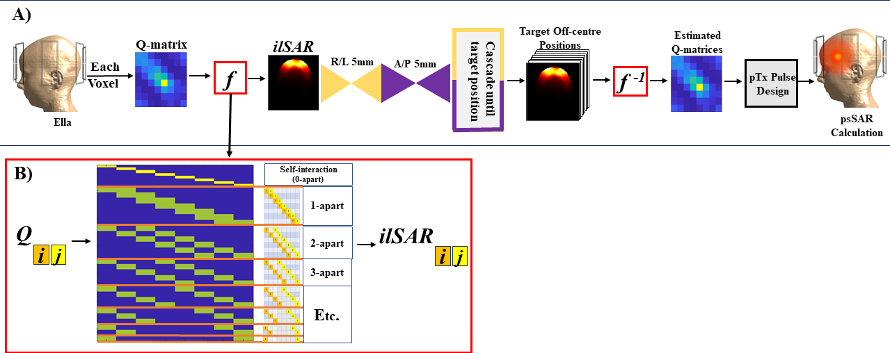
The dataset used was the same as the one generated for 2, which consists of Sim4Life simulations (ZMT, Zurich, Switzerland) of body models (BMs) Billie, Duke, Fats, Ella and Glenn from the Virtual Population (IT’IS, Zurich,Switzerland)11 at 35 positions (combinations of rightward-leftward (R-L):-20/-10/-5/0/5/10/20 and anterior-posterior (A-P):-10/-5/0/5/10). Q-matrices for the 8-channel parallel-transmit array were derived using the approach in Ref2. Q-matrices were converted to ilSAR with an algorithm10 designed to predict the power absorption consequence of 64 linearly-independent combinations of the coil elements (figure 1).The cGAN was implemented in TensorFlow 2.4.1 and Python 3.7.11 on a NVIDIA DGX GPU in Linux. The architecture was identical to pix2pix12, excluding these parameters: epochs = 60; filter size = 1; strides = 1; a ReLu replaced with a leaky ReLu and no batch normalization in the generator network during downsampling. The data was normalized channel-wise, labeled with the degree of motion, and paired as given input and output ilSAR distribution. A leave-one-out approach was used to create the training, validation,and testing data at the body-model level to prevent cross-talk (ie. TRAINING: Fats, Duke, and Billie; VALIDATION: Glenn; TESTING: Ella). To create the training pairs, the degrees of motion were paired both center-out and with displacement given an off-center reference (eg. R0 mm → R5 mm and R5 mm → R10 mm both represent R5 mm displacements). The training data contained 5,040 pairs for the P-A and 4,200 for the R-L networks. Testing was conducted using for all simulated positions by cascading trained 5mm displacement networks as required (figure 1). Network estimation error (NEerr) of ilSAR was evaluated with an L1-norm of the difference with the ground truth (GT) voxel-wise and averaged across slices per channel at each position. This was compared to motion-induced (MI) error (Mierr; L1-norm) (displaced GT simulations vs. centered ilSAR). R-P ilSAR estimations were remapped to Q-matrices13 and applied to a pTx pulse design method8 (figure 1).
Results and Discussion
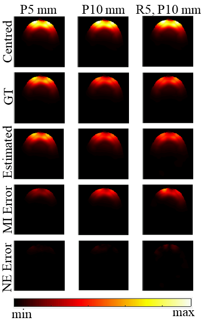
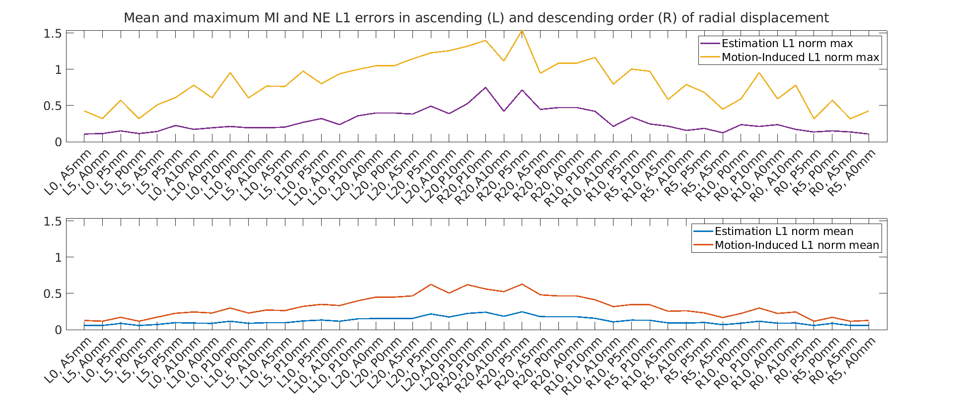
Figure 2 shows that the NE ilSAR distributions resemble the GT across slices and movement types. 1-3 cascades are displayed from channel combinations yielding the worst error. Compared to MIerr maps (GT-centred), NE error (NEerr) maps (GT-NE) were considerably less pronounced.Figure 3 plots the mean and maximum L1 errors across all pTx channel combinations and slices, per degree and direction of motion. The network-estimations reduced mean and maximum error in all cases, and mean NEerr remained low throughout. While mean NEerr was 0.1%, never exceeding 0.7%, mean MIerr was 0.3%, reaching 1.5%.
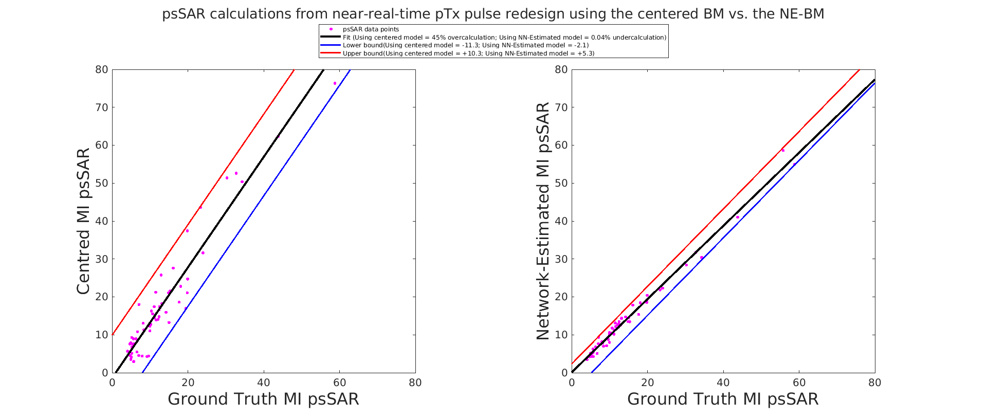
Figure 4 displays the improvement in the estimated Q-matrices derived from the NE ilSAR distributions when used as a constraint in a pTx design protocol. The resulting worst-case underestimation is 2.5-fold when calculating psSAR with the centered body-model (cBM), and 1.3-fold with the NE-BM, which reduces the required safety margin. Overall, using the cBM leads to 45% overcalculation, while using the NE-BM leads to 0.04% undercalculation. When worst-case underestimation factors are used as safety margin factors, the cBM yields an overall trend of 3.6-fold overestimation whereas for NE-BM the overall overestimation is 1.25-fold.
Conclusion
We have established a pipeline to estimate local SAR variations arising from head motion during 8-channel pTx at UHF-MRI. Since our NE and GT ilSAR distributions are compatible, this approach can be developed for near-real-time pTx with revised, less overconservative safety factors. Future work will expand the pipeline to include yaw and 2 mm degrees of motion.Acknowledgements
This project was supported in part by the Wellcome Trust [204824/Z/16/Z], Welsh Government [Wales Data Nation Accelerator project], and EPSRC [Doctoral Training Program].References
1. Frank Seifert, Gerd W ̈ubbeler, Sven Junge, Bernd Ittermann, and Herbert Rinneberg. Patient safety concept for multichannel transmit coils. Journal of Magnetic Resonance Imaging, 26:1315–1321, 11 2007.
2. Emre Kopanoglu, Cem M. Deniz, M. Arcan Erturk, and Richard G. Wise.Specific absorption rate implications of within-scan patient head motion for ultra-high field mri. Magnetic Resonance in Medicine, 84:2724–2738, 112020.
3. Emre Kopanoglu. Actual patient position versus safety models: Specific absorption rate implications of initial head position for ultrahigh field magnetic resonance imaging. NMR in Biomedicine, 5 2022.
4. Morgane Le Garrec, Vincent Gras, Marie France Hang, Guillaume Ferrand,Michel Luong, and Nicolas Boulant. Probabilistic analysis of the specific absorption rate intersubject variability safety factor in parallel transmission mri. Magnetic Resonance in Medicine, 78:1217–1223, 9 2017.
5. Amer Ajanovic, Joseph V Hajnal, and Shaihan Malik. Positional sensitivity of specific absorption rate in head at 7t. volume 4251, ISMRM Annual Proceedings, 2020.
6. Amer Ajanovic, Joseph V Hajnal, Raphael Tomi-Tricot, and Shaihan Malik.Motion and pose variability of sar estimation with parallel transmission at7t. volume 2487, ISMRM Annual Proceedings, 2021.
7. Alix Plumley, Luke Watkins, Matthias Treder, Patrick Liebig, Kevin Murphy, and Emre Kopanoglu. Rigid motion-resolved prediction using deep learning for real-time parallel-transmission pulse design. Magnetic Resonance in Medicine, 87:2254–2270, 5 2022.
8. Emre Kopanoglu. Near real-time parallel-transmit pulse design. ISMRM Annual Proceedings, 2018.
9. Katherine Blanter, Alix Plumley. Shaihan Malik, Emre Kopanoglu. Towards applying deep learning to predict rigid motion-induced changes in q-matrices from uhf-mri ptx simulations. ISMRM Annual Proceedings, 2023.
10. Yudong Zhu, Leeor Alon, Cem M. Deniz, Ryan Brown, and Daniel K. Sodickson. System and sar characterization in parallel rf transmission. Magnetic Resonance in Medicine, 67:1367–1378, 5 2012.
11. Honegger Katharina, Zefferer Marcel, Neufeld Esra, Oberle Michael, Szczerba Dominik, Kuster Niels, Kainz Wolfgang, Guag Joshua W, Hahn Eckhart, G Rascher Wolfgang, Janka Rolf, Bautz Werner, Chen Ji, Shen Jianxiang, Kiefer Berthold, Schmitt Peter, Hollenbach Hans-Peter, Christ Andreas and Anthony Kam. The virtual family-development of surface-based anatomicalmodels of two adults and two children for dosimetric simulations, Jan 2010.
12. Phillip Isola, Jun-Yan Zhu, Tinghui Zhou, Alexei A Efros, and Berkeley Ai Research. Image-to-image translation with conditional adversarial networks,2017.5
13. Arian Beqiri, JV Hajnal, and SJ Malik. Local q-matrix computation for parallel transmit mri using optimal channel combinations. In Proceedings of the 24th Annual Meeting of ISMRM, Singapore, page 3658, 2016.
Figures