0466
Local SAR and Uncertainty Estimation for Brain Imaging by Bayesian Deep Learning1Department of Radiology, University Medical Center Utrecht, Utrecht, Netherlands, 2Computational Imaging Group for MR diagnostics & therapy, Center for Image Sciences, University Medical Center Utrecht, Utrecht, Netherlands, 3Tesla Dynamic Coils BV, Zaltbommel, Netherlands, 4Department of Radiotherapy, University Medical Center Utrecht, Utrecht, Netherlands, 5Biomedical Image Analysis, Dept. Biomedical Engineering, Eindhoven University of Technology, Eindhoven, Netherlands
Synopsis
Keywords: Safety, Safety, specific absorption rate; local SAR; deep learning; Bayesian deep learning; uncertainty estimation
Motivation: Local SAR cannot be measured during an MRI examination. Deep learning approaches are proving to be a solution for on-line subject-specific SAR assessment.
Goal(s): The brain is the region of greatest clinical interest for ultra-high field MRI. Therefore, we apply for brain imaging a deep learning approach presented for local SAR assessment for body imaging.
Approach: The Bayesian deep-learning approach maps the relation between subject-specific complex B1+-maps and the corresponding local SAR distribution, and predicts the spatial distribution of uncertainty at the same time
Results: The Bayesian deep-learning approach for local SAR assessment in brain outperforms the previous application for prostate imaging.
Impact: The application of Bayesian deep-learning can allow the reduction of overly conservative RF safety constraints that limit the performance of UHF-MRI. Furthermore, the joint estimation of uncertainty can help the acceptance of such methods in clinical contexts.
PURPOSE
Since local SAR cannot be measured during an MRI examination, it is usually estimated by off-line numerical simulations using generic body models1. Software tools and deep-learning methods3 to perform on-line predictions2 are being developed.Deep-learning methods have become very popular because they have achieved impressive results. In particular, Bayesian deep-learning approach also allows to predict subject-specific local SAR and uncertainty distributions at the same time4. Recently a new deep-learning approach based on B1+-maps and anatomical MRI image was presented for local SAR assessment in brain5.
Indeed, brain is the region of greatest clinical interest for ultra-high field MRI. However, we believe in importance of mapping the underlying physical relationships to enhance robustness and generalizability, as well as the importance of evaluating uncertainty for the acceptance of deep learning methods in clinical contexts. In this study, we apply our Bayesian deep-learning approach4 to map the relation between subject-specific complex B1+-maps and the corresponding 10g-averaged SAR (SAR10g) distribution in brain.
METHODS
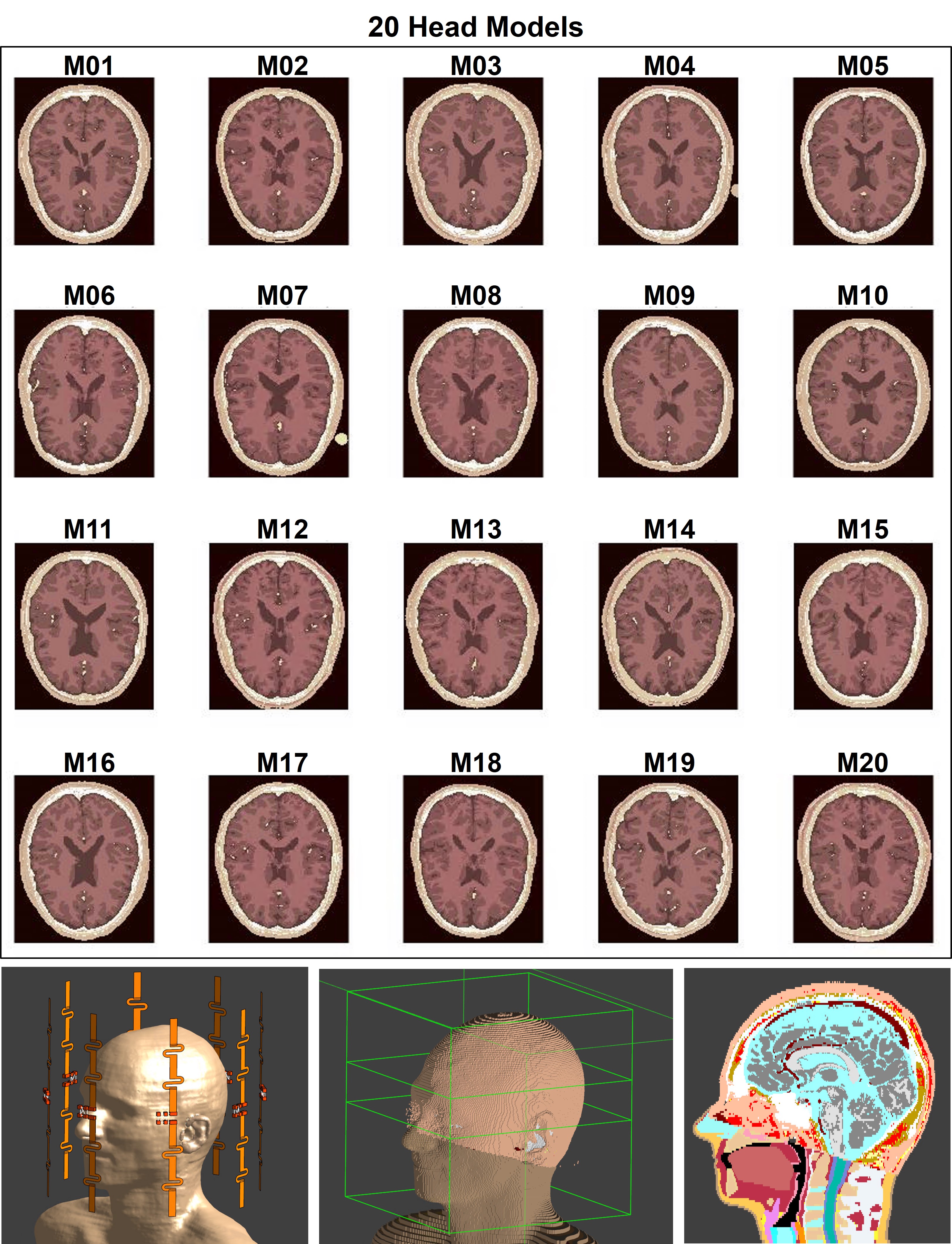
We used a database of 20 subject-specific head models6,7 with an 8-fractionated dipole array8,9 for brain imaging at 7T (Figure 1) to build a synthetic dataset consisting of complex B1+-maps and corresponding SAR10g distributions3,4 (20×1000=20000 data samples), Sim4Life (ZMT, Zürich, Switzerland).This dataset was partitioned into 5 sub-datasets according to the models that have generated the data samples and a 5-Fold Cross-Validation was performed.
To capture the aleatory uncertainty $$$\widehat{\sigma}_{Data_i}$$$ (e.g. due to noisy data), we trained a convolutional neural network (U-Net)10 by minimizing the Gaussian log-likelihood loss function (NLL)4,11.
$$\mathcal{L}_{NLL} = \frac{1}{N_{voxel}}\sum_{i=1}^{N_{voxel}}\frac{1}{2}\frac{|Ground\texttt{-}Truth_i-Output_i|^2}{\widehat{\sigma}_{Data_i}^2}+\frac{1}{2}\texttt{log}\widehat{ \sigma}_{Data_i}^2$$
The epistemic uncertainty $$$\widehat{\sigma}_{Model_i}$$$ (e.g., uncertainty in the model parameters due to a limited amount of training data) was estimated by Monte Carlo (MC) dropout5,12, performing T=50 stochastic predictions for each complex B1+-map.
MC dropout consists of randomly removing a number of nodes from the network during training and inference time. For each prediction, the architecture of the network is slightly different and can be considered as a MC sample from the space of all plausible networks given the data (i.e. a kind of ensembles of networks).
Then, by performing multiple (T=50) stochastic forward passes through the network, SAR10g ($$$\widehat{\mu}_i$$$ ) and uncertainties ($$$\widehat{\sigma}_{Data_i}$$$ and $$$\widehat{\sigma}_{Model_i}$$$) were calculated for each voxel i.
$$\widehat{\mu}_i=\frac{1}{2}\sum_{t=1}^{T}Output_{i_t}$$
$$\widehat{\sigma}_{Model_i}=\sqrt{\frac{1}{T}\sum_{t=1}^{T}(Output_{i_t})^2-\left(\frac{1}{T}\sum_{t=1}^{T}Output_{i_t}\right)^2}$$
$$\widehat{\sigma}_{Data_i}=\sqrt{\frac{1}{T}\sum_{t=1}^{T}\widehat{\sigma}_{Data_{i_t}}}$$
$$\widehat{\sigma}_i=\sqrt{\widehat{\sigma}_{Model_{i}}^2+\widehat{\sigma}_{Data_{i_t}}^2}$$
To assess the performance of the proposed approach, a 5-Fold Cross-Validation was performed.
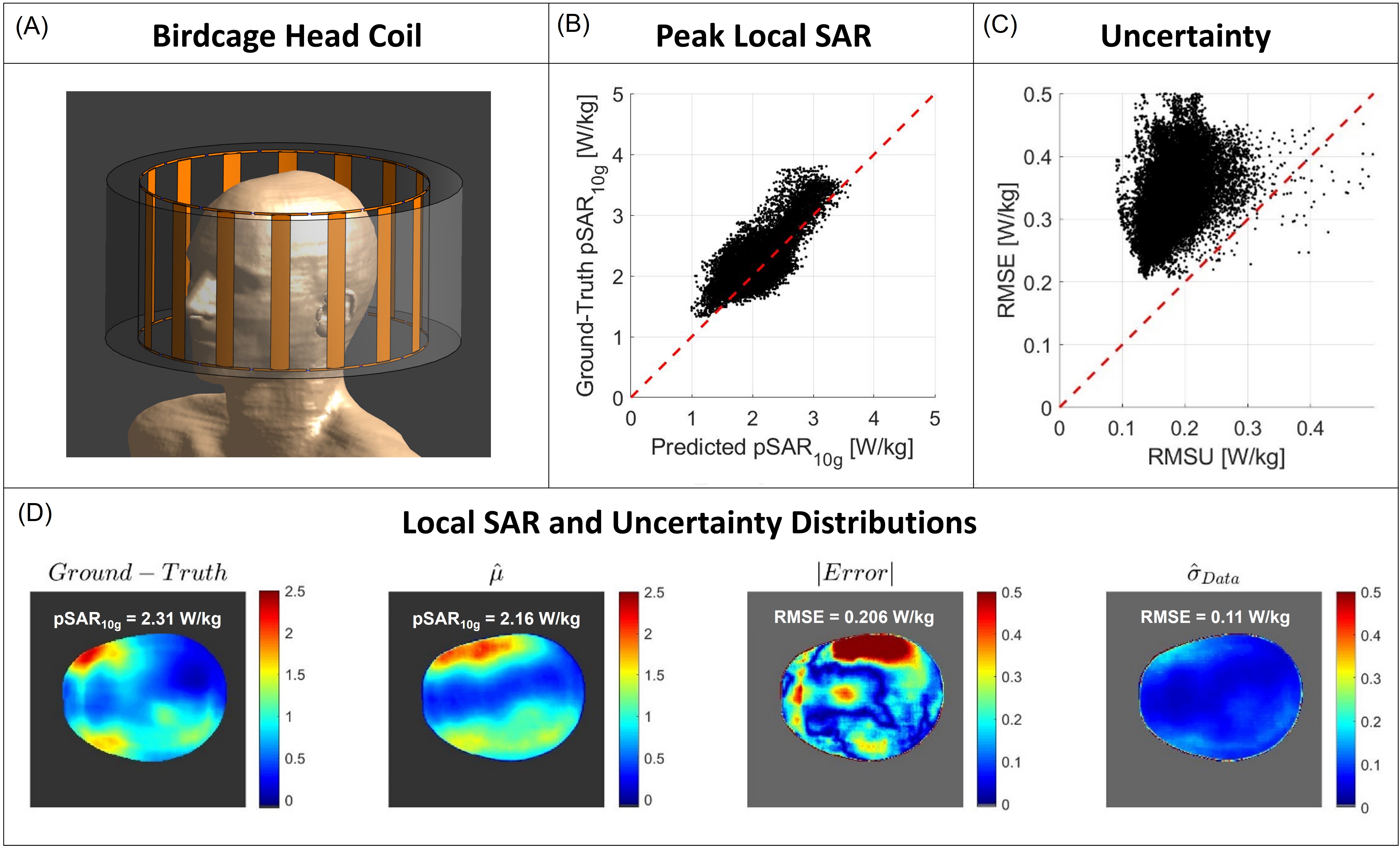
In theory, with Bayesian deep-learning approaches there is no guarantee that predictions on samples out of the training distribution will be considered uncertain13. However, the results of our previous study14 suggested that the network might be able to predict high uncertainty levels for array setups that it was not trained for. Therefore, to verify this hypothesis, we tested the trained network with a similar test set (20×1000=20000 data samples) obtained by simulating the head models with a birdcage head coil with 16 rungs (Diameter: 310mm – Length: 170mm).
We qualitatively assessed the spatial distribution of the predicted local SAR compared to the ground-truth and the predicted uncertainty with the absolute error. Then, we quantitatively evaluated the peak local SAR error, the RMS of uncertainty (RMSU), and the RMS of absolute error (RMSE).
RESULTS AND DISCUSSION
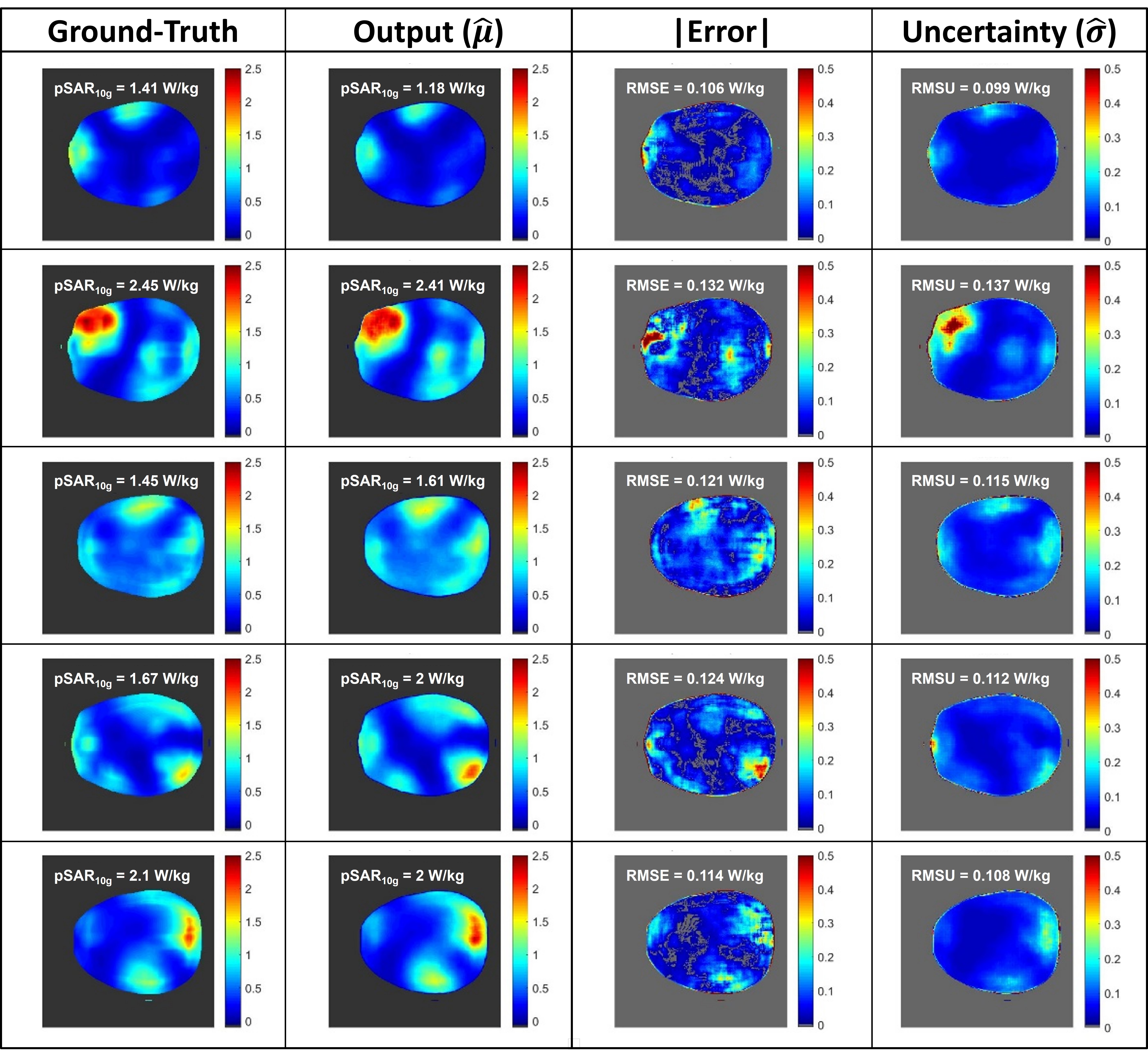
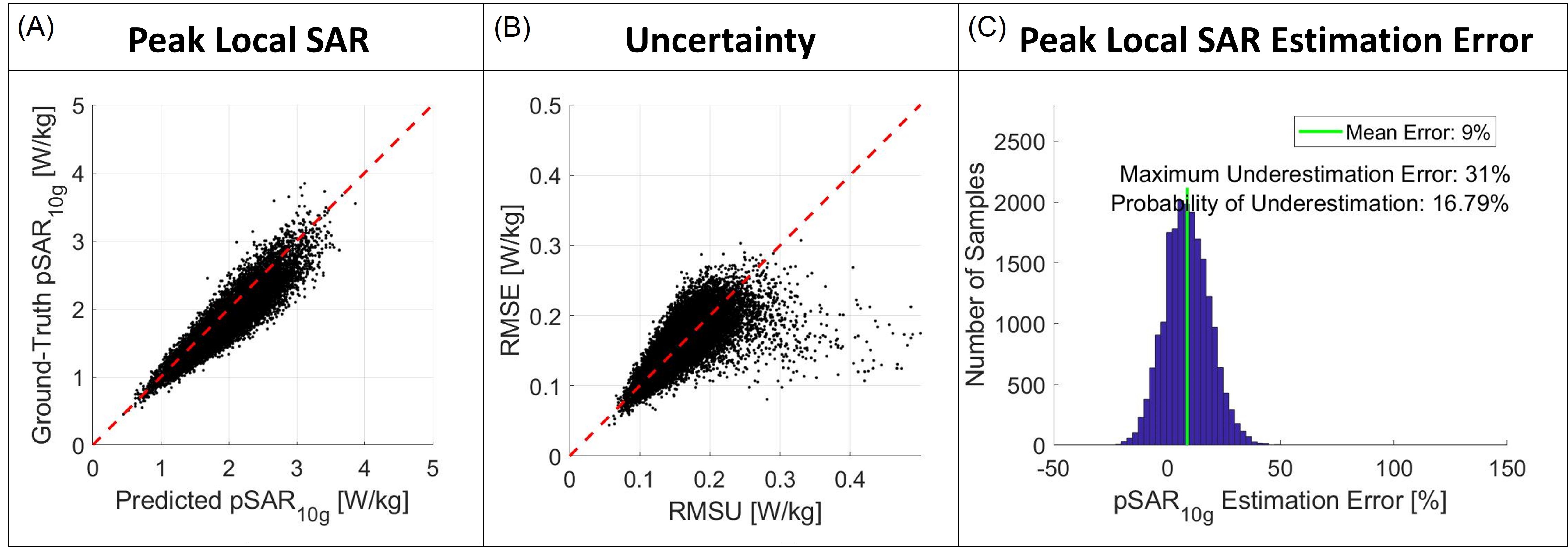
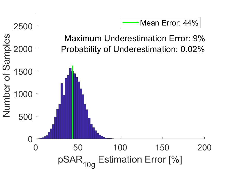
The distributions in Figure 2 show a good qualitative and quantitative match of both SAR10g and uncertainty distributions for five cross-validations. The scatter plots in Figure 3 show a good correlation in all cross-validations (A-C). The histogram of the pSAR10g estimation error in Figure 4 shows a mean overestimation error of 44% with 0.1% probability of underestimation (outperforming the Bayesian deep learning approach for body imaging4, mean overestimation error of 51%).Although a quite good qualitative match between the ground-truth and predicted SAR10g distributions can also be observed in the test set with the birdcage head coil in Figure 5, contrary to our previous study, the lower quantitative performances are not accompanied by appropriately large uncertainty predictions. This means that the network is not always able to correctly predict high uncertainty levels for array setups that it was not trained for.
CONCLUSION
The Bayesian deep-learning approach for SAR10g prediction in brain outperforms our previous application for prostate imaging (7% less mean overestimation). The predicted uncertainty closely matches quite well to the errors between predicted and actual SAR10g predictions. However, although the proposed approach is able to predict SAR10g distributions for array setups that were not used in training, confirming its capability to effectively learning the mathematical model that can simulate the underlying physics, the uncertainty underestimation that occurs highlights the need for methods to detect/reject these potentially dangerous situations (not included in the training set).Acknowledgements
The second author receives funding from the Netherlands Organization for Scientific Research (NWO; VENI grant no. 18078). This work was also financially supported by the Artificial Intelligence working group of the EWUU allianceReferences
[1] Christ A, Kainz W, Hahn EG, et al. The Virtual Family—development of surface‐based anatomical models of two adults and two children for dosimetric simulations. Phys Med Biol. 2010;55:N23–N38.
[2] Villena JF, Polimeridis AG, Eryaman Y, et al. Fast Electromagnetic Analysis of MRI Transmit RF Coils Based on Accelerated Integral Equation Methods. IEEE Trans Biomed Eng. 2016; 63(11):2250-2261.
[3] Meliadò EF, Raaijmakers AJE, Sbrizzi A, et al. A deep learning method for image‐based subject‐specific local SAR assessment. Magn Reson Med. 2019;00:1–17.
[4] Meliadò EF, Raaijmakers AJE, Maspero M, et al. Subject-specific Local SAR Assessment with corresponding estimated uncertainty based on Bayesian Deep Learning. Proceedings of the ISMRM 29th Annual Meeting, 8-14 August 2020. p. 4195.
[5] Gokyar S, Zhao C, Ma SJ,Wang DJJ. Deep learning-based local SARprediction usingB1maps and structural MRI of thehead for parallel transmission at 7T.Magn ResonMed. 2023;90:2524-2538. doi: 10.1002/mrm.29797 6. B. Aubert-Broche, D.L. Collins, A.C. Evans: "A new improved version of the realistic digital brain phantom" NeuroImage, in review - 2006.
[7] B. Aubert-Broche, M. Griffin, G.B. Pike, A.C. Evans and D.L. Collins: "20 new digital brain phantoms for creation of validation image data bases"
[8] Steensma BR, Luttje M, Voogt IJ, et al. Comparing Signal-to-Noise Ratio for Prostate Imaging at 7T and 3T. J Magn Reson Imaging. 2019;49(5):1446-1455.
[9] Raaijmakers AJE, Italiaander M, Voogt IJ, Luijten PR, Hoogduin JM, et al. The fractionated dipole antenna: A new antenna for body imaging at 7 Tesla. Magn Reson Med. 2016;75:1366–1374.
[10] O. Ronneberger, P. Fischer, T. Brox, U-Net: Convolutional Networks for Biomedical Image Segmentation, Medical Image Computing and Computer-Assisted Intervention – MICCAI 2015 pp 234-241.
[11] Kendall A, Gal Y. What Uncertainties Do We Need in Bayesian Deep Learning for Computer Vision?. In: Advances in Neural Information Processing Systems. 2017;5580–5590.
[12] Gal Y, Ghahramani Z. Dropout as a bayesian approximation: representing model uncertainty in deep learning. In: Proceedings of the 33rd International Conference on International Conference on Machine Learning, ICML. 2016;1050-1059.
[13] Ståhl N, Falkman G, Karlsson A, Mathiason G. Evaluation of Uncertainty Quantification in Deep Learning. In: Lesot MJ. et al. (eds) Information Processing and Management of Uncertainty in Knowledge-Based Systems. IPMU 2020. Communications in Computer and Information Science, vol 1237. Springer, Cham. https://doi.org/10.1007/978-3-030-50146-4_41.
[14] Meliadò EF, Raaijmakers AJE, Luijten PR, et al. Detection of Samples Out of Training Distribution: Rejection of Potential Erroneous Local SAR Predictions, Proceedings of the ISMRM 31st Annual Meeting, 7-12 May 2022. p. 2545.
Figures