0463
7 Tesla 31P Birdcage / 16-channel 1H Loop and Dipole Array integrated with Parallel Transmit System1Advanced Imaging Research Center, UTSouthwestern Medical Center, Dallas, TX, United States, 2Philips Healthcare, Best, Netherlands
Synopsis
Keywords: RF Arrays & Systems, Parallel Transmit & Multiband, 31P/1H imaging, Dual-tuned array design, ultra-high field, parallel transmit system
Motivation: Integrating multinuclear applications into parallel transmit system (PTx) at ultra-high field MRI systems will provide uniform transmit field and good SNR for both proton and the x-nuclei.
Goal(s): Design, build and integrate a 7T 31P/1H head array into a 7T PTx system that supports multinuclear applications.
Approach: Use 8 loops and 8 folded dipoles as 1H transceivers, and a birdcage as 31P transceiver, for 31P/1H MRS and MRI in a 7T PTx system
Results: Phantom and in vivo experimental results show that the coil can provide homogeneous and good SNR signals for both 31P and 1H in ROI
Impact: Elevating multinuclear studies with 7T parallel transmit system, providing uniform transmit fields and good receive sensitivity profile for both proton and x-nuclei in ROI. A potential game-changer for metabolic studies in body applications at ultra-high fields, advancing medical research.
INTRODUCTION
Phased array principle (1) has been widely used in dual-tuned array designs at ultra-high fields (≥7 Tesla (T)). So far, these dual-tuned arrays have been constrained to use in conventional 1-channel 1H transmit systems. However, 1H B1+ inhomogeneity is a well-known issue at ultra-high fields, and parallel transmit (PTx) system is a must to tackle it. It is a tendency that the 7T PTx system will be available for clinical applications. Therefore, it is essential to support multinuclear functionality in 7T PTx systems. In this work, we present the first 7T 31P/1H head array, consisting of 8 loops and 8 folded dipoles as 1H transceivers, as well as a birdcage as 31P transceiver, for 31P/1H MRS and MRI in a 7T PTx system (Philips Healthcare, 7T dSync). By leveraging RF shimming capability of the PTx, the array provides homogenous proton B1+ and good SNR for both 1H and 31P.METHOD
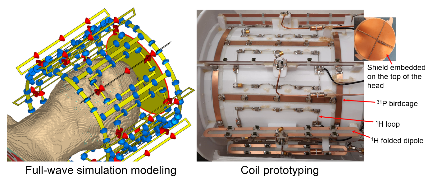
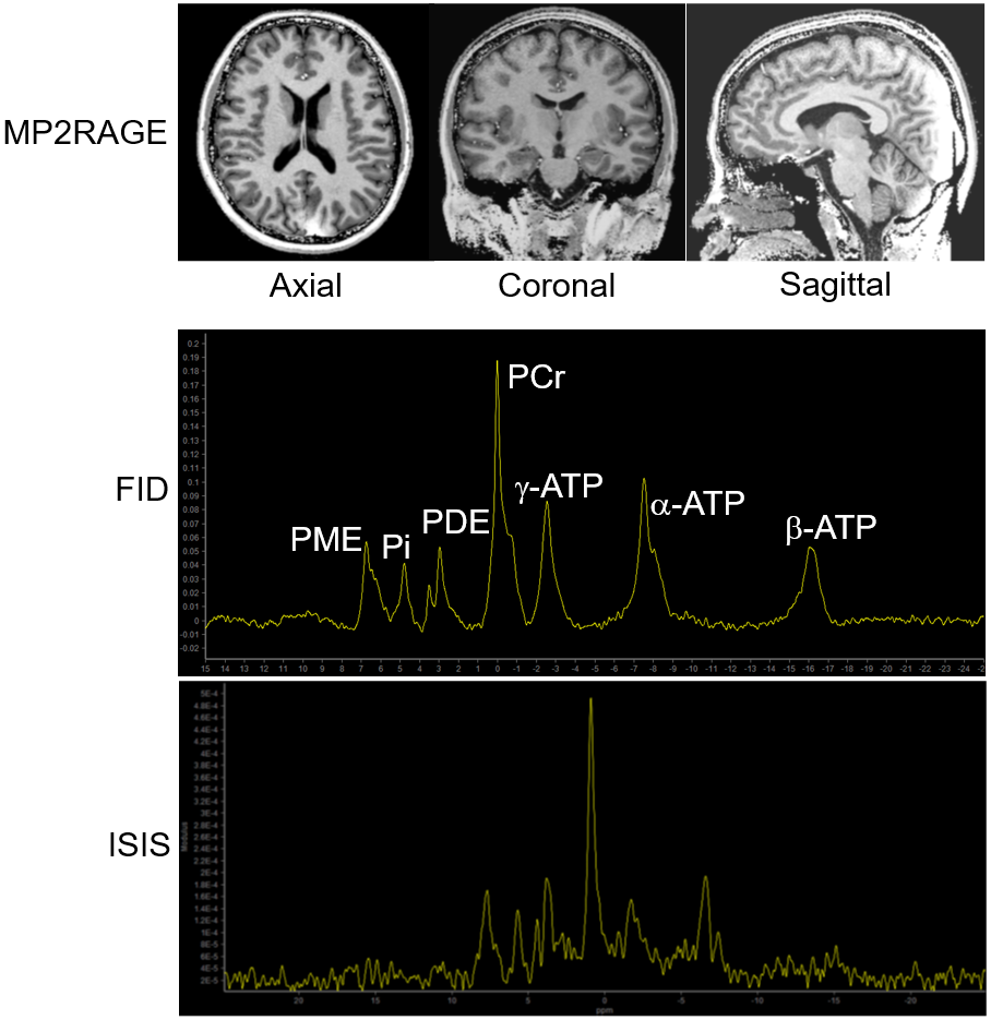
Figure 1 shows the full-wave simulation modeling of the array and the coil prototype. The coil layout, capacitor distribution, and capacitance values were the same in both simulation and prototype. All the coil elements were sitting on a 276mm-diameter cylinder. A shield was placed on the top of the head to enhance the B1+ in the dome. Coil layout: The 31P birdcage is a 22cm-long 8-rung bandpass birdcage, whose copper width is 1cm. Five capacitors (22.7pF) were evenly distributed on each rung, and eight capacitors (68pF) on each end-ring. Eight 1H 14cm×7.5cm loops, made with 14-awg copper wire, were nested inside the 31P birdcage. 11 capacitors were used for tuning, and matching accomplished with a lattice balun. Eight 1H 26cm-long 2-folded dipoles sit 1cm above the birdcage and the loops. The dipoles geometrically decouple with the loops. Eight inductors were used for tuning, and a lattice balun parallel to a variable capacitor for matching. Coil interface: Shielded cable traps were implemented on every ¼-λ coax cable. A 31P quadrature hybrid was used to create circularly polarized (CP) B1+ field, while the 0° and 90° ports receive individually via their own preamplifiers. Eight 1:2 1H Wilkinson power splitters were used to split 8 independent channels of 1H transmit power into eight pairs of loop and dipole. All loops and dipoles connected to their own preamplifiers to receive, thus creating a 16-channel receive array. Preamplifier decoupling of the 1H loops was achieved by adjusting the cable length. Experiments: All in vivo images were acquired using a 7T whole-body MRI scanner (Philips Healthcare, 7T dSync). Human subject studies adhered to an approved Institutional Review Board protocol with informed consent. Safety testing and SAR parameters were acquired in accordance with worst-case SAR guidelines (2). Phantom: 1H B1+ maps were acquired with a fast pre-saturated TurboFlash B1+ mapping method (3), and 31P spectra were acquired with non-selective, ISIS and 2D CSI sequences. In vivo: 1) 1H images were acquired with high-resolution 3D MP2RAGE, and 31P spectra with FID and ISIS.RESULT AND DISCUSSION
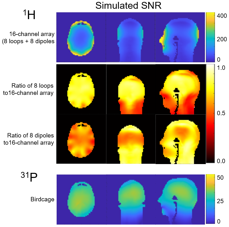
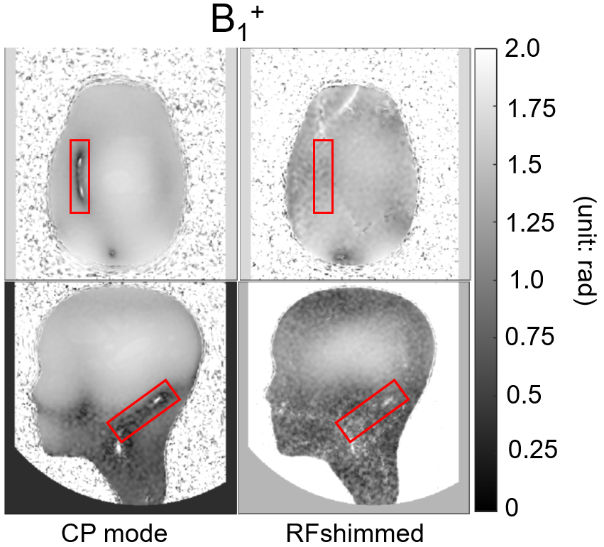
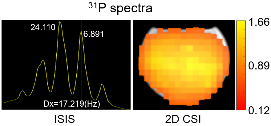
Simulation: Figure 1 shows that the 8 loops contribute most of SNR (82%) in the brain, while the 8 dipoles contribute to the longitudinal coverage. The loops have 54.7% higher SNR than the dipoles in the brain, and the SNR of the 16-channel array is dominated by the 8 loops; the 31P birdcage has homogenous signal in ROI. Workbench: Q ratio is 5.7 for the 31P birdcage, and 8.5 for the 1H loop, loaded with a head phantom (ɛr=62, σ=0.45 S/m at 7T). S21<-13dB among the coil elements. Phantom experiments: Figure 3 shows B1+ maps in CP and RF-shimmed modes in axial and sagittal planes respectively. The B1+ in the null band of the CP mode in axial plane as well as the B1+ in cortex in the sagittal plane were boosted with the RF shimming mechanism. Figure 4 demonstrated that the 31P birdcage has sufficient SNR and homogeneous field, by observing well resolved 17Hz J-coupling in a methyl phosphoric acid phantom acquired by the 31P ISIS spectra, as well as homogeneous 31P signal in the ROI over the same phantom by 2D CSI. The MP2RAGE images show nice contrast between grey and white matter (Figure 5, first row), consistent with the simulated SNR results. In vivo non-localized and ISIS 31P spectra shows sharp peaks of all typically 31P detected brain metabolites (Figure 5).CONCLUSION
We presented the first 31P/1H head array compatible and integrated with a 7T PTx system. Phantom and in vivo experimental results show that the coil can provide homogeneous B1+ and good SNR for both 31P and 1H in ROI.Acknowledgements
This work was fully funded by Cancer Prevention and Research Institute of Texas (CPRIT) RR180056 and was a collaborative project between Advanced Imaging Research Center, UTSW and Philips Healthcare.References
1. Roemer PB, Edelstein WA, Hayes CE, Souza SP, Mueller OM. The NMR phased array. Magn Reson Med 1990;16(2):192-225.
2. Hoffmann J, Henning A, Giapitzakis IA, Scheffler K, Shajan G, Pohmann R, Avdievich NI. Safety testing and operational procedures for self-developed radiofrequency coils. NMR Biomed 2016;29(9):1131-1144.
3. Chung S, Kim D, Breton E, Axel L. Rapid B1+ mapping using a preconditioning RF pulse with TurboFLASH readout. Magn Reson Med 2010;64(2):439-446.
Figures