0448
T1 Contrast-Augmented Single-Spoke Real-Time 4D MRI1Bernard and Irene Schwartz Center for Biomedical Imaging, Department of Radiology, New York University Grossman School of Medicine, New York, NY, USA, New York University Grossman School of Medicine, New York, NY, United States, 2Center for Advanced Imaging Innovation and Research (CAI2R), Department of Radiology, New York University Grossman School of Medicine, New York, NY, USA, New York University Grossman School of Medicine, New York, NY, United States, 3BioMedical Engineering and Imaging Institute (BMEII), Icahn School of Medicine at Mount Sinai, New York, NY, United States
Synopsis
Keywords: MR-Guided Radiotherapy, Radiotherapy, 4DMRI, Real-Time Imaging
Motivation: 4D MRI is a powerful technique for free-breathing volumetric imaging, holding great potential for application in MRI-guided radiotherapy. Traditional 4D MRI typically requires explicit motion detection and data binning for respiratory-resolved reconstruction and commonly employs fast steady-state MRI acquisition, which may not yield optimal contrast.
Goal(s): This work proposes a novel T contrast-augmented, free-breathing, real-time 4D MRI technique.
Approach: The proposed technique improves image contrast through highly-accelerated inversion recovery-prepared acquisition and reconstruction of real-time 4D images at a sub-second temporal resolution without requiring explicit motion compensation.
Results: T1 contrast-augmented real-time 4D MRI demonstrated improved image contrast over conventional 4D MRI with steady-state acquisition.
Impact: The contrast-augmented real-time 4D MRI technique proposed in this work can improve image contrast for free-breathing imaging without requiring explicit motion detection, data binning and respiratory motion compensation. It holds great potential for various applications, such as MRI-guided radiotherapy.
Introduction
4D MRI (3D plus a time dimension or a motion dimension) has proven to be a powerful technique for free-breathing volumetric imaging in many applications, such as MRI-guided radiation therapy1–3. Conventional 4D MRI typically involves an explicit step to extract a respiratory motion signal for guiding the binning of acquired data to generate respiratory motion-resolved 4D images4. However, this approach faces significant challenges when patients exhibit irregular breathing patterns or experience motion drift and/or bulk motion. Meanwhile, state-of-the-art 4D MRI techniques commonly employs fast steady-state acquisition (e.g., gradient echo imaging)1, which may not yield optimal contrast. This study proposes a novel T1 contrast-augmented, free-breathing, real-time 4D MRI technique, which combines inversion recovery-prepared golden-angle stack-of-stars sampling to improve image contrast and advanced iterative reconstruction to eliminate the need for motion detection and data binning by reconstructing each image frame from a single spoke to achieve a sub-second temporal resolution.Methods
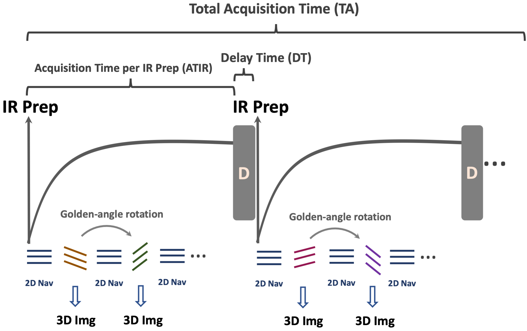
Data Acquisition and Image ReconstructionThe acquisition scheme for the proposed 4D MRI is illustrated in Figure 1. Inversion recovery (IR) preparation is periodically applied in golden-angle stack-of-stars sampling. Following each IR preparation, a series of radial stacks, rotated by a golden-angle, are continuously acquired, constituting one IR repetition. A 2D projection with a rotation angle of 0o is consistently acquired before each radial stack. A short delay is inserted between consecutive IR preparations to enhance T1 contrast. The acquisition time in each IR repetition, the delay time, and the total acquisition time are denoted as ATIR, DT, and TA, respectively, as shown in Figure 1.
Image reconstruction is performed using GRASP-Pro, which implements a combination of low-rank subspace constraint and spatiotemporal total variation (TV) constraints to reconstruct 4D dynamic images without explicit motion compensation, as detailed in previous works5,6. The 0o 2D navigators are employed to estimate a temporal basis for guiding image reconstruction and compressing the dynamic 4D images into a low-dimensional subspace for improved reconstruction performance. Images with desired contrast can be retrospectively selected at a specific TI.
In-Vivo Experiments
Imaging experiments were conducted in the liver of 4 subjects (3 healthy controls and 1 patient) using a 3T MRI scanner (Prisma, Siemens Healthineers). Relevant imaging parameters included: FOV=360x360x160mm², Matrix size= 256x256x32, TR/TE=2.6/1.2ms, flip angle=5o, ATIR of 2.2s, DT of 1.5s, and TA=8 min. A total of 130 IR repetitions were acquired, and 27 radial stacks were acquired after in each IR repetition. The 4D MRI reconstruction generated 13 3D images within each IR repetition with a temporal resolution of 0.16ms/3D volume, resulting in a total of 1690 images (from 130 IR repetitions) with inversion recovery and respiratory motion occurred simultaneously in real time. Images at different inversion times (TIs) and respiratory motion states can then be flexible selected from the reconstructed images.
Results
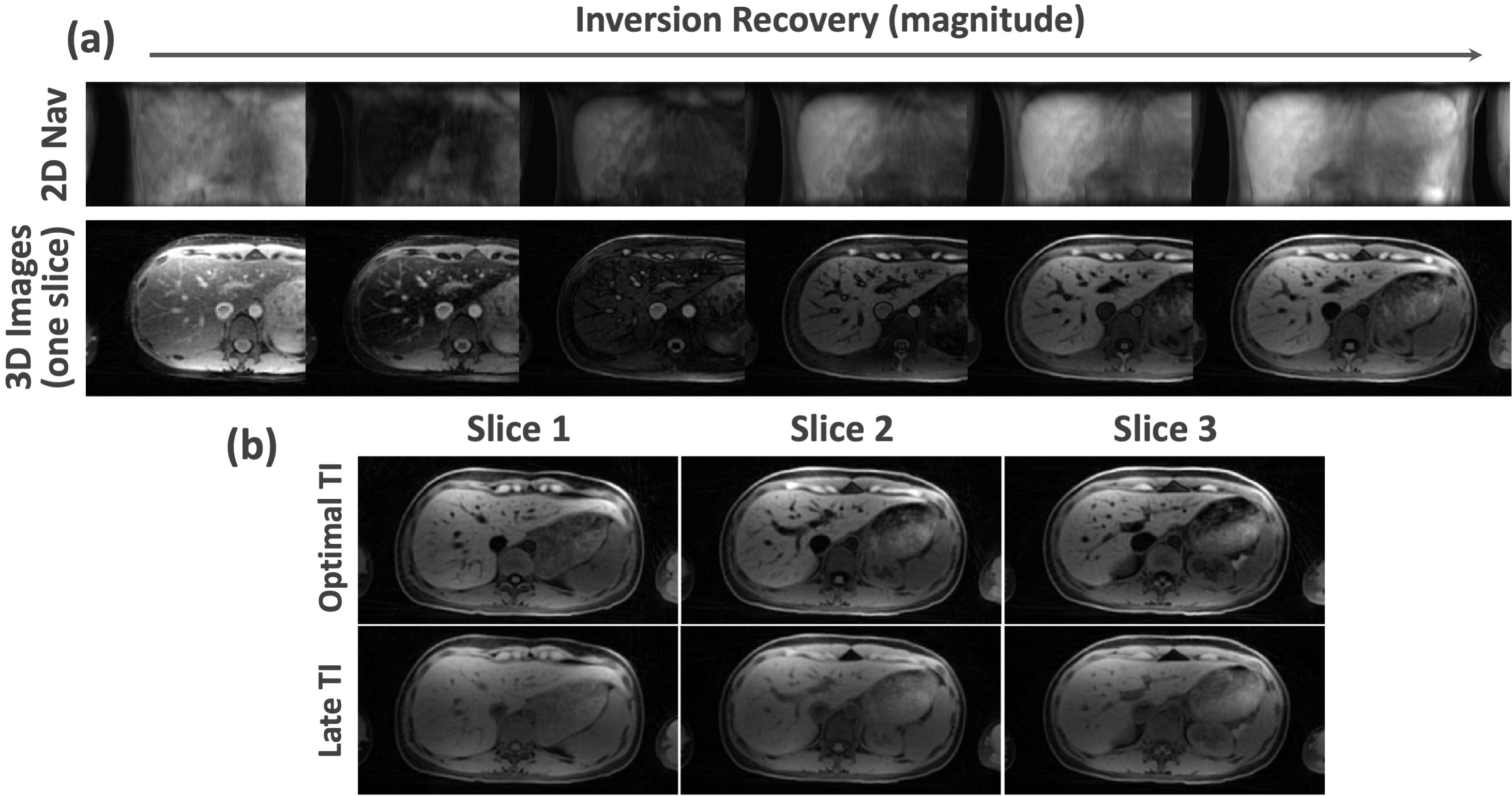
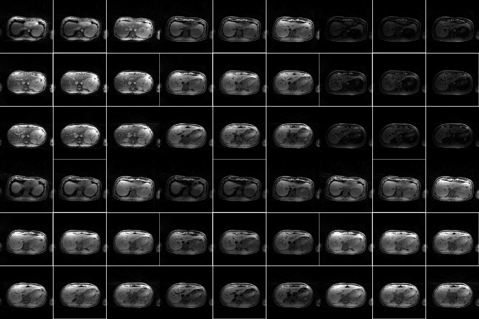
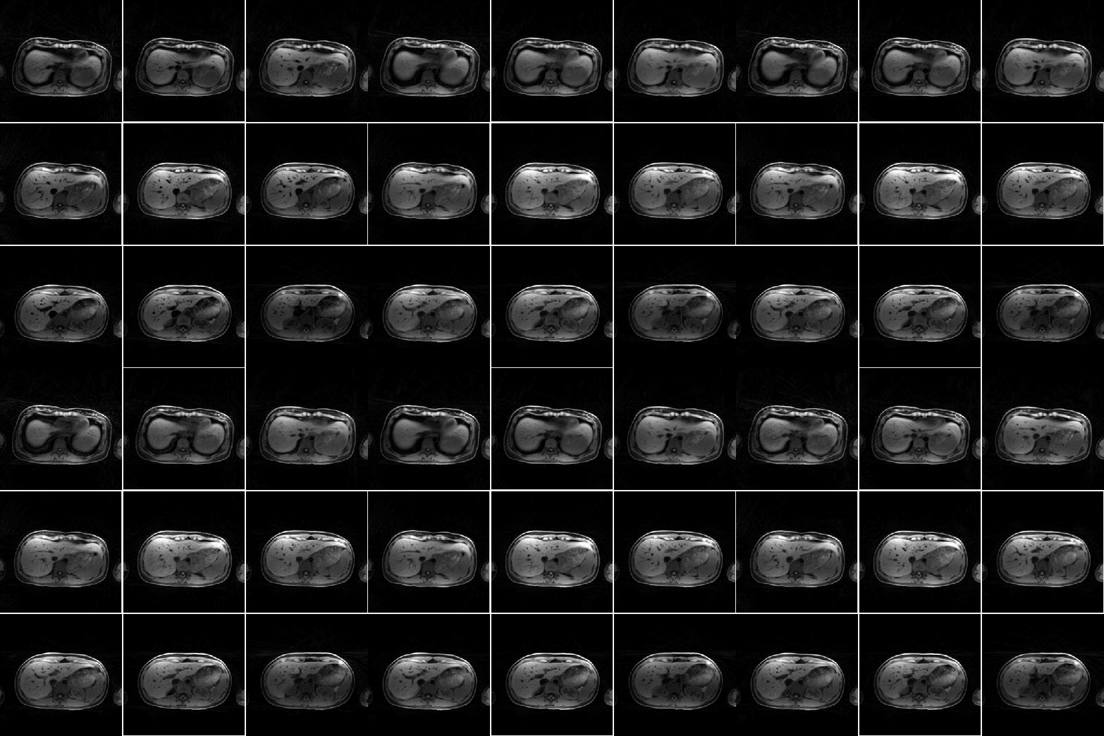
Figure 2a shows real-time 2D navigators and corresponding 3D images (single slice) in one IR repetition from one subject, which capture inversion recovery, underlying respiratory motion and potential body movement. Figure 2b compares real-time 3D images reconstructed at a TI with augmented T1 contrast and a late TI. Note that images at the late TI were acquired in a steady state, which exhibit similar image contrast as in conventional 4D MRI without IR preparation.Figure 3 shows a GIF movie including 9 slices of real-time images capturing simultaneous inversion recovery and respiratory motion. Corresponding images selected for optimal image contrast at a specific TI are displayed in the GIF movie presented in Figure 4.
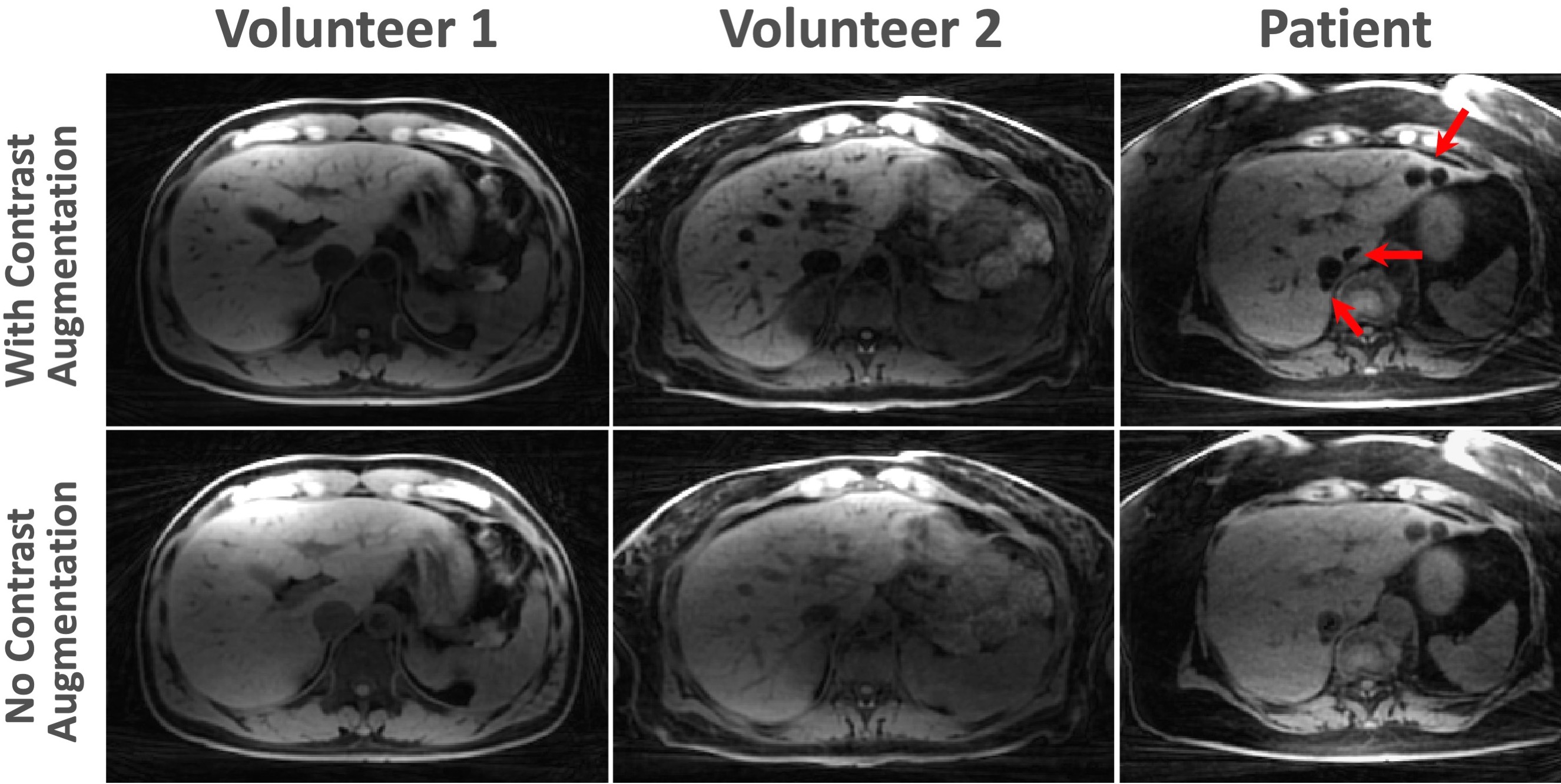
Figure 5 illustrates a similar comparison in the remaining subjects. Similar to the findings in previous figures, images with T1 contrast augmentation, selected from an optimal TI after IR preparation, demonstrate enhanced delineation of tissue structures and lesions (indicated by the red arrows in the patient images) in the liver.
Discussion
IR is known to provide the strongest T1 contrast. However, its integration into 4D MRI is limited due to substantial contrast variation from IR preparation and slow imaging speed. In this work, we combined IR preparation with advanced low-rank subspace reconstruction to enable free-breathing 4D MRI with augmented image contrast and high temporal resolution (~0.16 seconds per 3D volume). This effectively tackles the challenge associated with IR preparation and conventional 4D MRI, while eliminating the need for any explicit respiratory motion compensation. It is important to note that reconstructing each image frame from one radial spoke is essential in the presence of IR to minimize contrast blurring during the T1 recovery process. This technique is expected to be promising for improving the precision of treatment planning within the context of MRI-guided radiotherapy.Acknowledgements
This work was supported by the NIH (R01EB030549, R01EB031083 and P41EB017183) and was performed under the rubric of the Center for Advanced Imaging Innovation and Research (CAI2R), an NIBIB Biomedical Technology Resource Center.References
1. Stemkens, B., Paulson, E. S. & Tijssen, R. H. N. Nuts and bolts of 4D-MRI for radiotherapy. Phys Med Biol 63, (2018).
2. Feng, L. et al. Simultaneous Evaluation of Lung Anatomy and Ventilation Using 4D Respiratory-Motion-Resolved Ultrashort Echo Time Sparse MRI. Journal of Magnetic Resonance Imaging 49, 411–422 (2019).
3. Paulson, E. S. et al. 4D-MRI driven MR-guided online adaptive radiotherapy for abdominal stereotactic body radiation therapy on a high field MR-Linac: Implementation and initial clinical experience. Clin Transl Radiat Oncol 23, 72 (2020).
4. Feng, L. et al. XD-GRASP: Golden-angle radial MRI with reconstruction of extra motion-state dimensions using compressed sensing. Magn Reson Med 75, 775–788 (2016).
5. Feng, L. et al. GRASP-Pro: imProving GRASP DCE-MRI through self-calibrating subspace-modeling and contrast phase automation. Magn Reson Med 83, 94–108 (2020).
6. Feng, L. 4D Golden-Angle Radial MRI at Subsecond Temporal Resolution. NMR Biomed (2022) doi:10.1002/NBM.4844.
Figures