0447
Super-paramagnetic iron oxide nanoparticles improve liver tumor visualization throughout online MRI-guided liver stereotactic radiotherapy1Radiation Oncology, Allegheny Health Network, Pittsburgh, PA, United States
Synopsis
Keywords: MR-Guided Radiotherapy, Radiotherapy, MRI-guided radiotherapy;
Motivation: Can we provide superior liver tumor visualization for online adaptive planning? MRI enables direct visualization of tumor and organs-at-risk (OAR). However, MRI contrast agents are often required to differentiate primary and metastatic liver malignant lesions from functional hepatic parenchyma.
Goal(s): We employed super-paramagnetic iron oxide nanoparticles (SPION) as an MRI contrast agent.
Approach: SPION enhanced the liver-to-tumor contrast ration for rapid and accurate delineation of tumors and functional hepatic parenchyma throughout the entire treatment course.
Results: This study is the first to report the efficiency of a single SPION injection for multi-fractionated MRI-guided liver stereotactic body radiotherapy on a 1.5T Elekta MR-Linac.
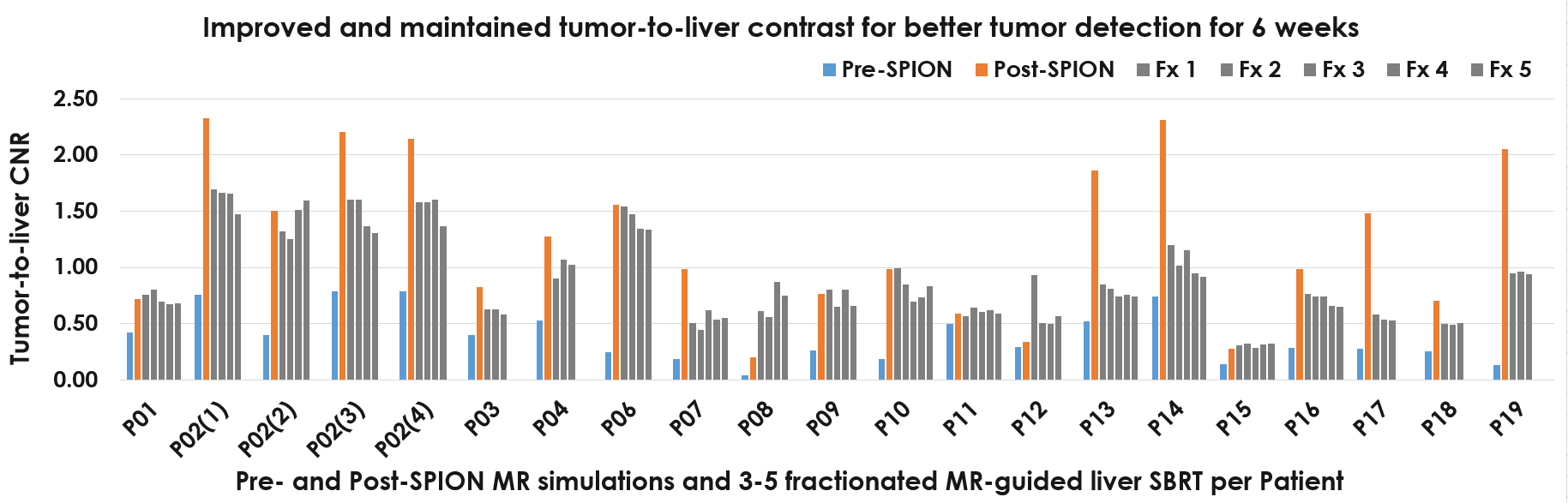
Impact: A single SPION injection significantly improved the tumor-to-liver contrast, and it was maintained throughout multi-fraction MRI-guided liver SBRT to provide rapid and accurate contouring tumor lesions from functional liver parenchyma for online adaptive planning.
INTRODUCTION:
Magnetic resonance imaging (MRI) can enable direct visualization of tumor and organs-at-risk (OAR).1–4 However, MRI contrast agents are often required to further enhance liver tumor visualization for the detection of primary and metastatic liver malignant lesions in respect to functional liver parenchyma during online adaptive planning.5–10 Super-paramagnetic iron oxide nanoparticles (SPION) are increasingly used as an MRI contrast agent for superior tumor visualization to account for tumor changes in volume and shape, and sparing surrounding organs-at-risk (OAR) including functional liver parenchyma volume (FLPV). Once IV injected, SPION stays within hepatic liver parenchyma for several weeks allowing precise delineation of hepatic tumors for conformal avoidance during MR-guided liver stereotactic body radiotherapy (SBRT). We proposed a novel approach of SPION-aided online adaptive planning on Elekta Unity® MR-Linac (Elekta; Stockholm, Sweden).METHODS:
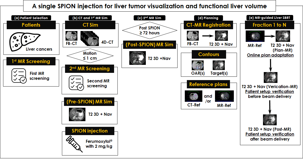
The workflow of this study was comprised of 5 steps (Figure 1). All patients (n=25) with hepatocellular carcinoma (HCC, n=16) and metastasis (n=9) were enrolled (Figure 1(a)) and screened twice for MRI safety prior to the 1st MR simulation (Figure 1(b)) and scanned using Unity® on the day of a CT simulation. In the CT simulation, free-breathing (FB)-CT and respiratory 4D-CT image sets were acquired to develop a CT reference plan (CT-Ref) and measure a tumor motion range (i.e., if tumor motion is equal or less than 1 cm, eligible for this study), respectively. In the 1st MR simulation (pre-SPION) a T2 3D using navigating (T2 3D+NAV) image set was acquired as a baseline for comparisons. Then, Ferumoxytol® (Feraheme, AMAG Pharmaceuticals, Waltham, MA) was administered as an MRI contrast agent just after the 1st MR simulation. The 2nd MR simulation (post-SPION), 48−72 hours after the Ferumoxytol® injection, was performed to acquire a T2 3D+NAV image set (Figure 1(c)). For treatment planning (Figure 2(d)), liver tumors and organs at risks (OAR(s)) were contoured on post-SPION T2 and FB-CT image sets, respectively, then a CT (or MR)-Ref was developed using a FB-CT or a post-SPION T2 3D+NAV image set. In each fraction of MR-guided liver SBRT (Figure 1(e)), three post-SPION T2 image sets were acquired for online adaptive planning (plan-MR) and two patient setup verification before (verification-MR) and after beam delivery (post-MR).RESULTS:
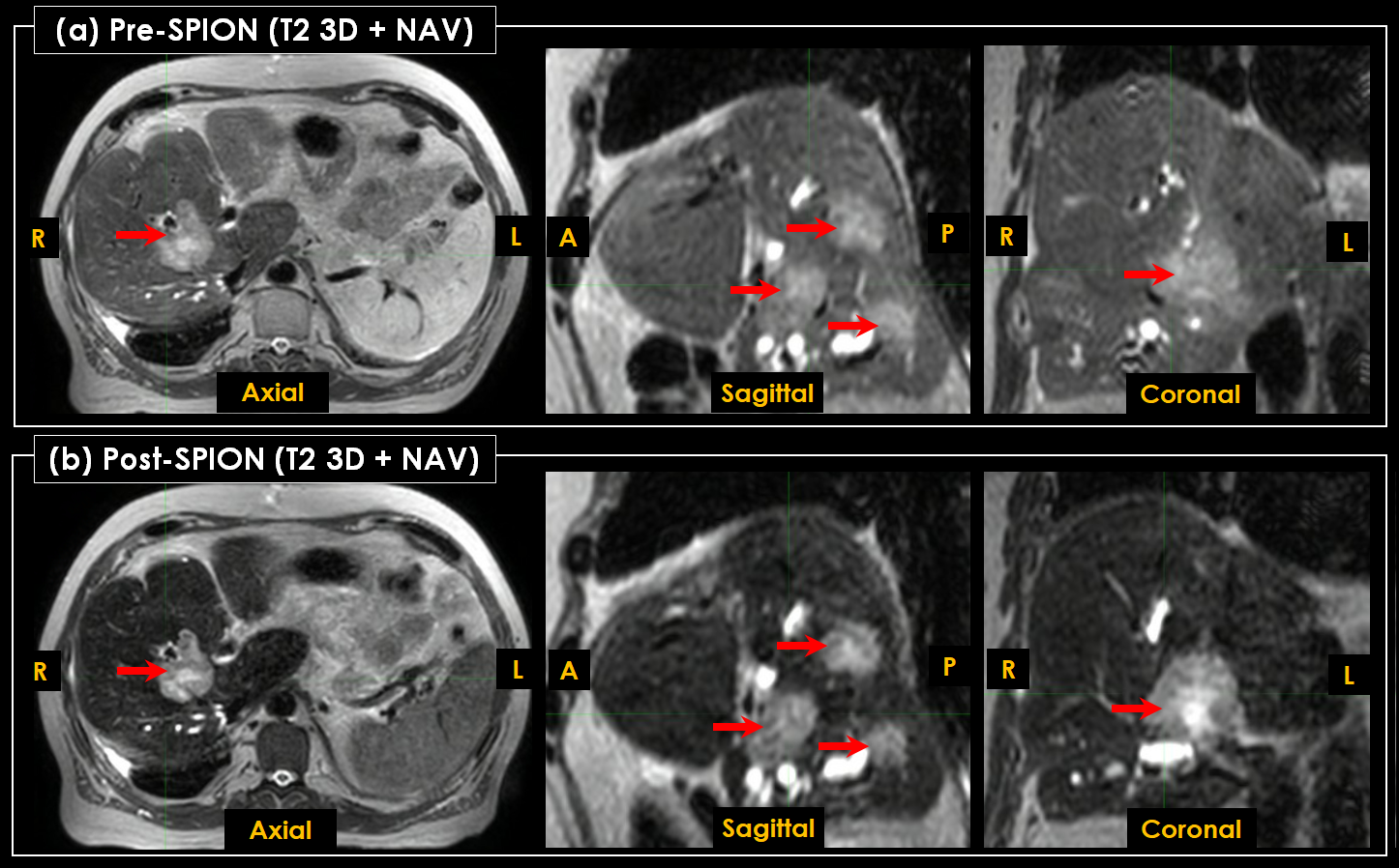
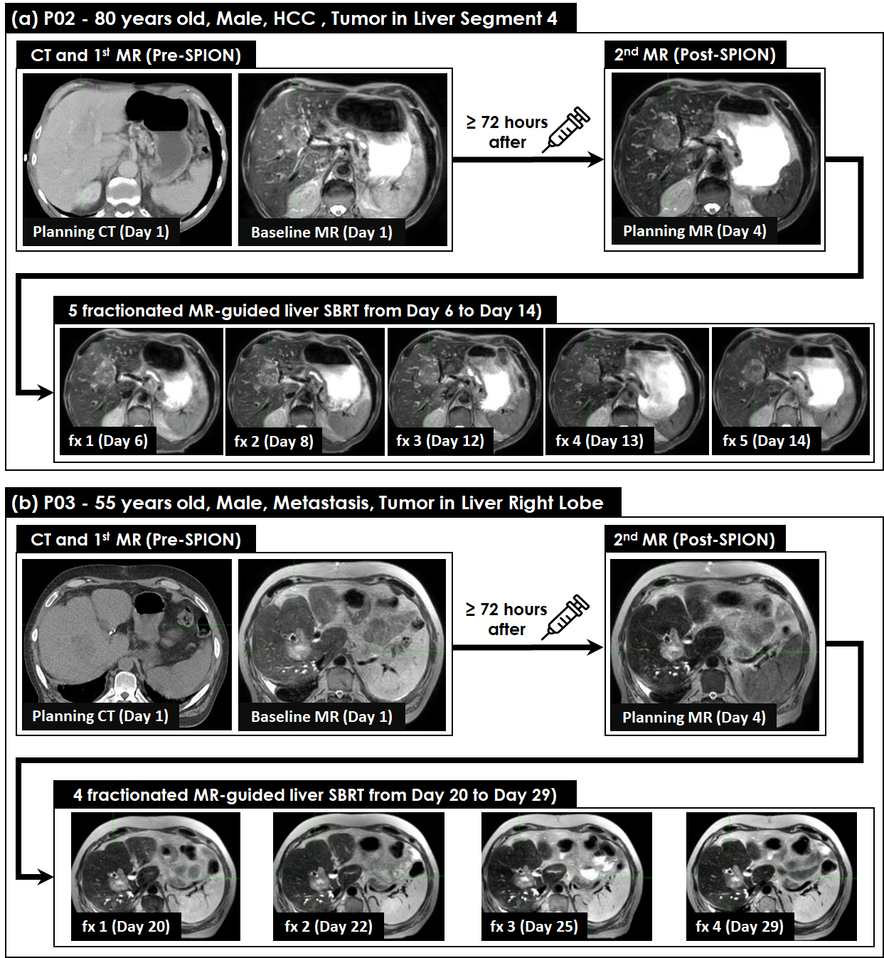
Compared to the pre-SPION image sets (Figure 2(a)), SPION-aided enhancement improved tumor visualization on the post-SPION image sets (Figure 2(b)). Tumor boundary was superiorly clear on post-SPION image sets due to SPION negatively enhanced functional liver parenchyma regions due to that SPION was trapped by resident hepatic macrophages and shortened MR signals. During MR-guided liver SBRT, SPION was retained within the functional liver parenchyma regions and it maintained the consistency of tumor visualization across 3−5 fractions (Figure 3).DISCUSSION:
Online adaptive planning requires a superior tumor-to-liver contrast for rapid and accurate delineation of target tumors, FLPV and OAR. We utilized SPION as an MRI contrast agent and demonstrated a negative enhancement on FLPV due to a shortened T2 relaxation time and thus relatively improved the tumor-to-liver contrast (Figure 2 and 3), which can (1) reducing target volume by eliminating the uncertainty of tumor boundary (Figure 3), (2) be shorten the time of online adaptive planning with rapid tumor delineation, and (3) spare FLPV and OAR with their avoidance during MRI-guided liver SBRT.CONCLUSION:
This is the first study to investigate the impact of SPION on visualization of liver tumors and functional parenchyma using 1.5T MR-Linac. Our results demonstrated that SPION-aided enhancement superiorly improved the tumor-to-liver contrast for clear visualization of tumor boundaries and thus this is a preferable imaging technique for achieving rapid and accurate online adaptive planning for MR-guided liver SBRT.Acknowledgements
We thank the physicians, therapists, nurses, dosimetrists, and staff at Allegheny Health Network and Department of Radiation Oncology for continuous support. We also thank Elekta for the funding.References
- 1. Rosenberg SA, Henke LE, Shaverdian N, et al. A Multi-Institutional Experience of MR-Guided Liver Stereotactic Body Radiation Therapy. Adv Radiat Oncol. 2019;4(1):142-149. 2.
- Kuczmarska-Haas A, Yadav P, Burr A, Witt JS, Blitzer GC, Bassetti MF. MR-Guided Liver Stereotactic Body Radiotherapy (SBRT): To Adapt, or Not to Adapt? International Journal of Radiation Oncology*Biology*Physics. 2020;108(3):S146-S147. 3.
- Rogowski P, von Bestenbostel R, Walter F, et al. Feasibility and Early Clinical Experience of Online Adaptive MR-Guided Radiotherapy of Liver Tumors. Cancers. 2021;13(7):1523. 4.
- Gani C, Boeke S, McNair H, et al. Marker-less online MR-guided stereotactic body radiotherapy of liver metastases at a 1.5 T MR-Linac – Feasibility, workflow data and patient acceptance. Clinical and Translational Radiation Oncology. 2021;26:55-61. 5.
- Poetter-Lang S, Bastati N, Messner A, et al. Quantification of liver function using gadoxetic acid-enhanced MRI. Abdom Radiol (NY). 2020;45(11):3532-3544. 6.
- Maurea S, Mainenti PP, Tambasco A, et al. Diagnostic accuracy of MR imaging to identify and characterize focal liver lesions: comparison between gadolinium and superparamagnetic iron oxide contrast media. Quant Imaging Med Surg. 2014;4(3):181-189. 7.
- Toth GB, Varallyay CG, Horvath A, et al. Current and potential imaging applications of ferumoxytol for magnetic resonance imaging. Kidney International. 2017;92(1):47-66. 8.
- Wojcieszynski AP, Rosenberg SA, Brower JV, et al. Gadoxetate for direct tumor therapy and tracking with real-time MRI-guided stereotactic body radiation therapy of the liver. Radiotherapy and Oncology. 2016;118(2):416-418. 9.
- Hama Y, Tate E. Superparamagnetic iron oxide-enhanced MRI-guided stereotactic ablative radiation therapy for liver metastasis. Rep Pract Oncol Radiother. 2021;26(3):470-474. 10.
- Ahmad F, Treanor L, McGrath TA, Walker D, McInnes MDF, Schieda N. Safety of Off-Label Use of Ferumoxtyol as a Contrast Agent for MRI: A Systematic Review and Meta-Analysis of Adverse Events. J Magn Reson Imaging. 2021;53(3):840-858.
Figures