0443
Quantitative Assessment of Geometric Distortions in MRI-Linac Sequences for Enhanced Radiotherapy Planning1MD Anderson, Houston, TX, United States, 2Elekta, Houston, TX, United States
Synopsis
Keywords: DWI/DTI/DKI, Diffusion/other diffusion imaging techniques, Deformable Image Registration, MR-Linac, Geometric Distortion
Motivation: Geometric distortion in MRI-Linac sequences remains insufficiently characterized, impacting the precision of radiotherapy treatment planning. This study aims to quantify such distortions in EPI, TSE, and SPLICE sequences.
Goal(s): The goal of this study is to quantitatively assess and compare geometric distortion across three common MRI-Linac sequences using deformable image registration and DICE score analysis.
Approach: An in vivo study employed patient T2 and DWI B0 scans. Deformable image registration was conducted using ADMIRE and Elekta-based software, generating deformation vector fields. A Python algorithm calculated RMS values.
Results: The sequences exhibited distinct DSC scores and RMS distortions, revealing registration effectiveness and sequence-dependent variability.
Impact: These findings set the stage for future research on minimizing distortions in MRI-Linac sequences. The developed Python algorithm could be adapted for real-time monitoring, advancing the precision and safety of MRI-guided radiotherapy.
Introduction
Diffusion-weighted imaging (DWI) can be used to predict response to therapy in head and neck cancer (HNC) patients and may be used in the MRI-linear accelerator (MR-Linac) adaptive treatment planning workflows, but further validation is necessary for clinical implementation. Additionally, geometric accuracy is crucial in radiation therapy (RT) to correctly delineate tumors and OARS. The MRI-Linac system enables treatment plan adaptation by redefining the tumor subregions that exhibit higher response to radiation. Traditionally, DWI has been done with Echo Planar Imaging (EPI), but this is known to have some geometric distortions due to EPI being more susceptible to inhomogeneities in the b0. This study aims to quantify and characterize geometric distortion on two Turbo Spin Echo (TSE) based DWI sequences and compare distortions with EPI by conducting an in vivo analysis using patient T2 and corresponding DWI b0 scans.Methods
Five human papillomavirus-positive (HPV+) oropharyngeal cancer patients were imaged in a RT immobilization mask on a 1.5 T MR-linac (Elekta AB, Stockholm, Sweden). All patients provided written informed consent. We acquired EPI, TSE, and SPLICE DWI sequences and a T2-weighted (T2w) sequence as an anatomical reference on the MR-Linac. Deformable image registration (DIR) was performed in ADMIRE, an Elekta-based software, to produce deformation vector fields (DVF). The registration was constrained by the following specific contours: spinal cord, gross tumor volume (GTV), left and right submandibular glands, and left and right parotid glands. The Dice similarity coefficient (DSC), surface Dice similarity coefficient (SDSC), average surface distance (ASD), and Hausdorff distance 95 (H95) were used to evaluate the effectiveness of the registration for each contour and to confirm that the algorithm constrained using the contours. An in-house developed Python tool calculated the root mean square (RMS) to quantify total distortion and provided visualization of the deformation in the DVF. Using the Wilcoxon signed rank statistical analysis test in GraphPad Prism (v10.0.3) to assess statistical significance.Results
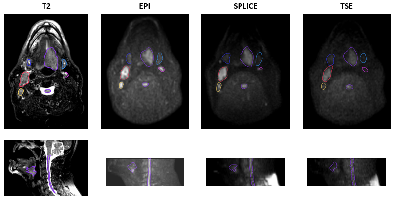
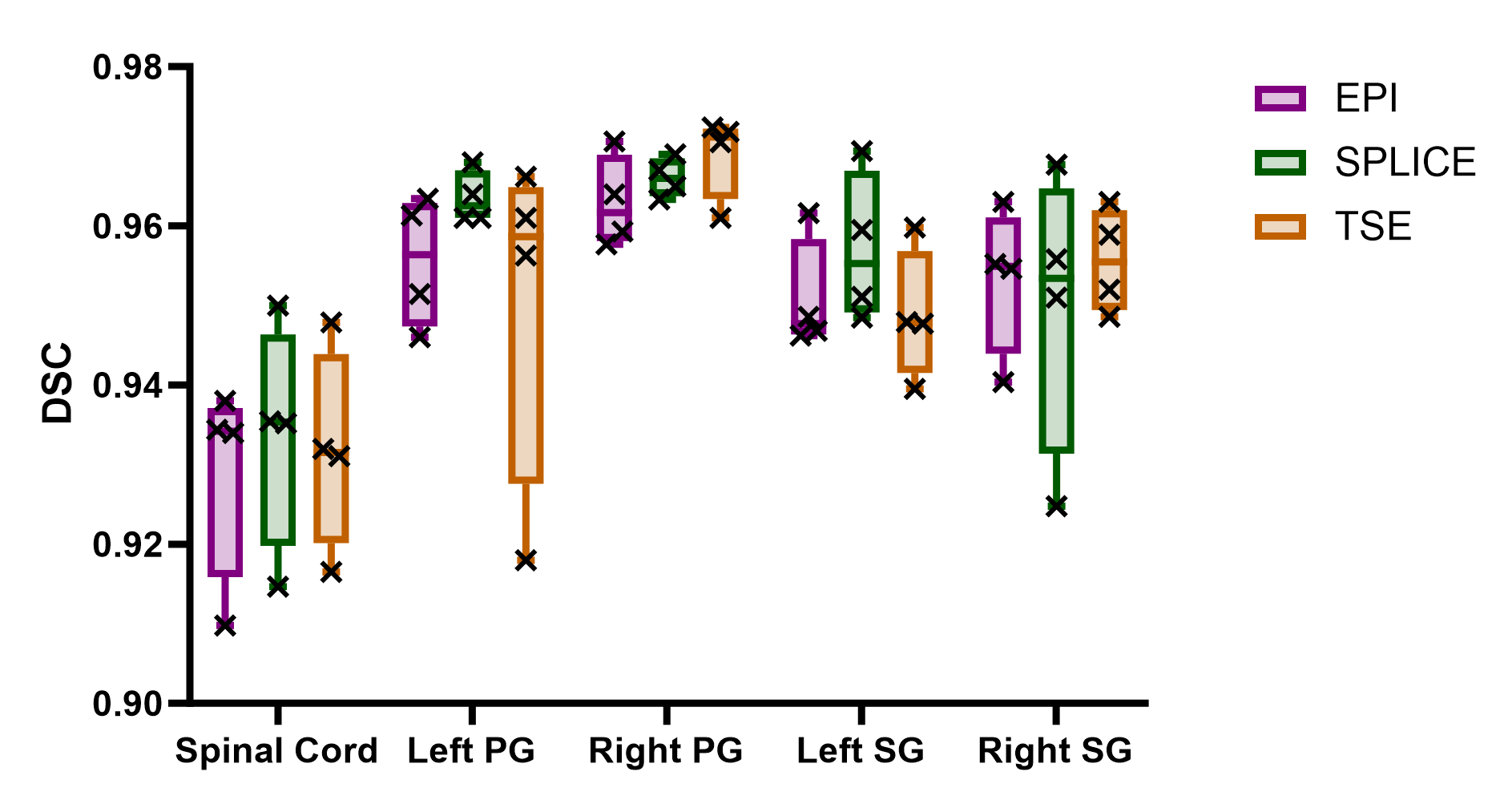
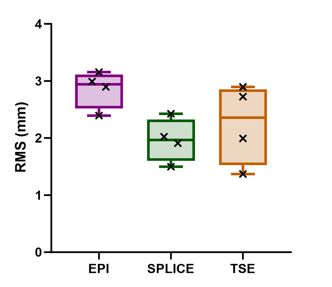
Representative images are shown in figure xxx. Median RMS values for EPI, SPLICE, and TSE were 5.5 +/- 1.09 mm, 2.02 +/- 6.70 mm, and 2.41 +/- 0.55 mm, respectively. The median Dice Similarity Coefficient for the spinal, left submandibular, right submandibular, left parotid and right parotid contours were 0.93 +/-0.01, 0.095 +/- 0.008, 0.95 +/- 0.01, 0.96 +/- 0.013, and 0.967 +/- 0.005 respectively. Results from the Wilcoxon rank test between EPI and SPLICE as well as EPI and TSE showed no statistically significant difference between median RMS values.Discussion
The high DSC values across all contours confirm that the registration effectively worked. Although median RMS values showed no statistically significant difference, results between EPI and SPLICE were trending towards a statistically significant difference with a p value of 0.0571. The implementation of larger voxel sizes in DWI, intended to improve SNR, resulted in a discernible shift close to the magnitude of a voxel in EPI sequences. Such granularity in the analysis provide clinicians with a clearer understanding of the distortion patterns prevalent in these imaging techniques. However, this study acknowledged the inherent uncertainties associated DIR, particularly in the context of contrast variances between T2-weighted and b0 images. Despite these challenges, the constrained registration by anatomical contours demonstrates a reliable approximation of existing geometric distortions, offering a pragmatic approach for clinical application. Recognizing the need for further validation, future research will expand the patient cohort to characterize geometric distortion trends more accurately and will conduct phantom studies to substantiate these preliminary findings. Moreover, leveraging the insights gained from this study, subsequent investigations will aim to develop and refine correction methodologies to enhance the geometric accuracy in DWI, thereby contributing to the precision of adaptive RT planning.Conclusion
In the clinical arena, the implications of this study are multifaced and significant. By advancing our understanding of geometric distortions in DWI sequences, the research directly informs the precision of tumor targeting in adaptive RT. This precision is paramount, as it can substantially influence treatment outcomes and patient morbidity by sparing healthy tissue and more accurately irradiating the tumor. Moreover, the potential for tailored treatment plans based on individual response patterns could refine adaptive therapy approaches, leading to more personalized and effective care. As the study propels developments in distortion correction techniques, it also sets the stage for integrating advanced imaging into routine clinical workflows, thereby enhancing the robustness and adaptability of MR-Linac systems. This study offers crucial insights into geometric distortions in DWI MR-linac sequences. It provides valuable data on DWI geometric distortions within MR-linac sequences, contributing to the optimization of RT planning for HNC patients.Acknowledgements
This project is supported by an academic-industrial partnership R01 grant from the National Institutes of Health (NIH)/National Institute of Dental and Craniofacial Research (NIDCR) (R01DE028290) and NIH/NCI (National Cancer Institute) Image-Guided Cancer Therapy T32 Program (T32CA261856).References
[1] McDonald BA, Salzillo T, Mulder S, Ahmed S, Dresner A, Preston K, He R, Christodouleas J, Mohamed ASR, Philippens M, van Houdt P, Thorwarth D, Wang J, Shukla Dave A, Boss M, Fuller CD. Prospective evaluation of in vivo and phantom repeatability and reproducibility of diffusion-weighted MRI sequences on 1.5 T MRI-linear accelerator (MR-Linac) and MR simulator devices for head and neck cancers. Radiother Oncol. 2023 Aug;185:109717. doi: 10.1016/j.radonc.2023.109717. Epub 2023 May 19. PMID: 37211282; PMCID: PMC10527507.
[2] Naser MA, Wahid KA, Ahmed S, Salama V, Dede C, Edwards BW, Lin R, McDonald B, Salzillo TC, He R, Ding Y, Abdelaal MA, Thill D, O'Connell N, Willcut V, Christodouleas JP, Lai SY, Fuller CD, Mohamed ASR. Quality assurance assessment of intra-acquisition diffusion-weighted and T2-weighted magnetic resonance imaging registration and contour propagation for head and neck cancer radiotherapy. Med Phys. 2023 Apr;50(4):2089-2099. doi: 10.1002/mp.16128. Epub 2022 Dec 29. PMID: 36519973; PMCID: PMC10121748.
[3] Kooreman, Ernst S., et al. "ADC measurements on the Unity MR-linac–A recommendation on behalf of the Elekta Unity MR-linac consortium." Radiotherapy and Oncology 153 (2020): 106-113.
[4] Almansour, Haidara, et al. "Prospective image quality and lesion assessment in the setting of MR-guided radiation therapy of prostate cancer on an MR-linac at 1.5 T: a comparison to a standard 3 T MRI." Cancers 13.7 (2021): 1533.
Figures