0441
Cross-species fMRI reveals transcriptomically and behaviorally-dissociable autism neurosubtypes1IIT, Rovereto, Italy, 2CMI, New York, NY, United States, 3ETH, Zurich, Switzerland
Synopsis
Keywords: Psychiatric Disorders, Brain Connectivity
Motivation: Resting-state fMRI (rsfMRI) studies have revealed atypical patterns of functional connectivity in autism. However, large heterogeneity in the manifestation of these alterations exists across samples and its etiopathological significance remains unclear.
Goal(s): Here, we used cross-species rsfMRI to probe if distinct patterns of functional dysconnectivity observed across 20 genetic models can be identified in rsfMRI scans of individuals with idiopathic autism.
Approach: We mapped whole brain functional connectivity in 20 genetic mouse models and in over 2000 individuals with and without autism.
Results: Our work reveals two autism neurosubtypes characterized by divergent patterns of dysconnectivity, and dissociable transcriptomic and behavioral profiles.
Impact: Connectivity alterations in idiopathic autism encode for etiologically-relevant information.
Introduction
Resting-state fMRI (rsfMRI) studies in individuals with autism have revealed atypical patterns of functional connectivity. However, large heterogeneity in the manifestation of these alterations exists across samples and studies and its etiopathological significance remains unclear. Leveraging recent implementations of rsfMRI mapping in rodents, we showed spatially distinct and divergent dysconnectivity patterns across ASD-relevant genetic models [1]. This suggests that heterogeneous functional connectivity in ASD may encode etiologically-relevant information. Here, we used cross-species rsfMRI to probe if distinct patterns of functional dysconnectivity observed across 20 autism-relevant genetic models can be identified and decoded in rsfMRI scans of individuals with idiopathic autism.Methods
Mouse studies. Mice with 20 autism-related genetic alterations (n=286) and control littermates (n=290) underwent rsfMRI mapping at 7T (n=12 scanned at IIT Rovereto, Italy; n=8 at ETH Zürich, Switzerland). Whole-brain connectivity mapping was carried out [2] and hierarchical clustering was used to group etiologically-relevant rsfMRI patterns. We then used the rodent clustering results to guide the identification of subtypes of idiopathic autism in humans. Human studies. Connectivity mapping was carried out on n=945 low-motion data of individuals with autism and n=1044 controls (6-30 yo) selected from ABIDE-1 [3], ABIDE-2 [4], and an in-house dataset. Inter-site data harmonization was implemented with ComBat [5]. Participants were assigned to discovery and replication datasets. Following preprocessing, we identified individuals with autism showing atypical connectivity patterns in evolutionarily conserved brain regions with dysconnections in the mouse clusters. Then, to further characterize brain systems of the identified subgroups, we used network-based statistics (NBS) upon removal of the dominant hypo- and hyper-connectivity effects. [6] Further, we carried out gene decoding and enrichment to link rsfMRI connectivity maps to genes differentially expressed in autism [7]. Finally, subtypes differences in severity of autistic behaviors was measured by the ADOS-2 scores [8].Results
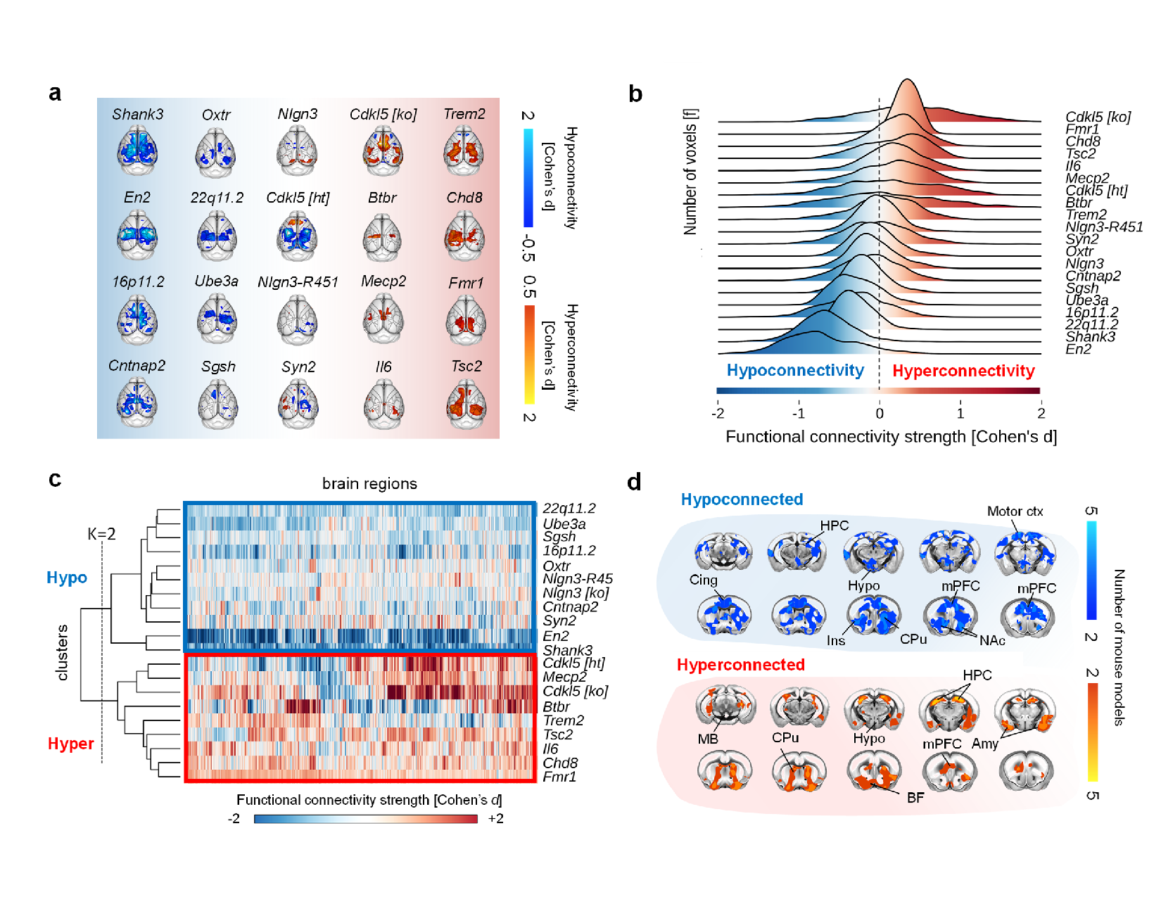
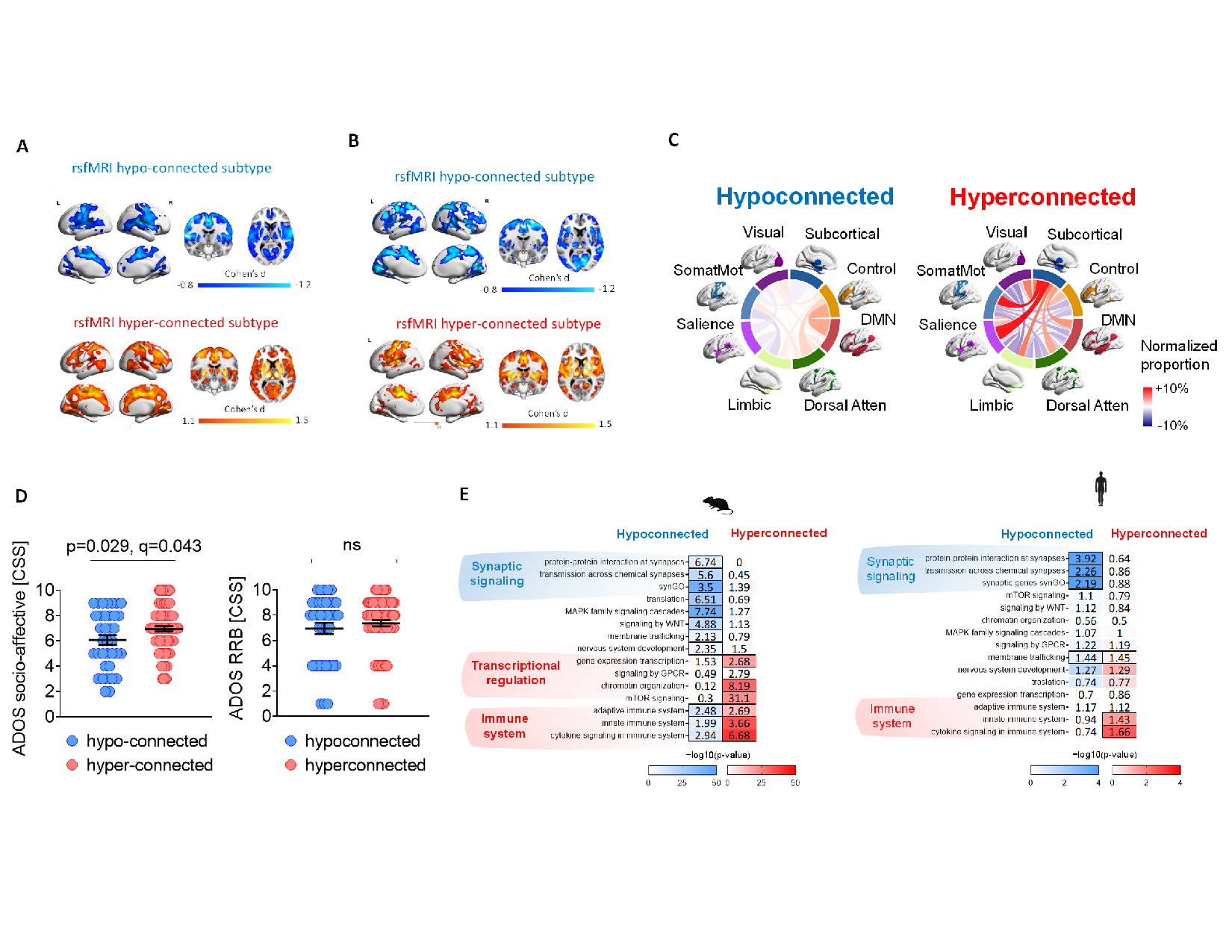
Consistent with our recent work [1], all the 20 autism-relevant genetic models exhibited atypical rsfMRI connectivity (Figure 1A), defining a pseudo-continuous landscape of alterations (Figure 1B). Dominant patterns of hypo or hyper-connectivity and cross-etiological convergences were, however, notable. In line with this observation, cluster analysis revealed two main patterns of dysconnections (Figure 1C); one predominantly characterized by subcortical hyperconnectivity, the other by cortico-striatal hypoconnectivity (Figure 1D). Guided by our mouse findings, a region-specific decoding of rsfMRI connectivity in idiopathic autism revealed two subtypes of participants recapitulating the dominant hypo- and hyper-connectivity identified in the rodent database (8% and 18% of the discovery sample, respectively) (Figure 2A). These patterns of dysconnectivity were highly replicable (Figure 2B). NBS showed rsfMRI hypoconnectivity between cortical networks in the hypo-connected subtype and subcortico-cortical hyper-connectivity in the hyper-connected subtype (Figure 2C). Participants in the hyper-connected subtype showed more severe socio-affective behaviors (p = 0.033, uncorrected) compared to those in the hypo-connected subtype (Figure 2D). In striking resemblance of mouse enrichments, both subtypes showed robust enrichment for molecular pathways associated with autism (Figure 2E).Conclusions
Guided by etiologically-relevant alterations mapped in autism-relevant mouse models, our work reveals two autism neurosubtypes characterized by divergent patterns of dysconnectivity, and by dissociable transcriptomic and behavioral profiles. Our findings suggest that connectivity alterations in idiopathic autism encode for etiologically-relevant information, and support the use of cross species rsfMRI to dissect the connectional, phenotypic and etiological heterogeneity of autism.Acknowledgements
This study was supported by European Union’s Horizon 2020 research and innovation program (Marie Sklodowska‐Curie Global Fellowship – CANSAS, GA845065).References
1. Zerbi, V., et al., Brain mapping across 16 autism mouse models reveals a spectrum of functional connectivity subtypes. Molecular Psychiatry, 2021: p. 1-11.
2. Cole, M.W., S. Pathak, and W. Schneider, Identifying the brain's most globally connected regions. NeuroImage, 2010. 49(4): p. 3132-3148.
3. Di Martino, A., et al., The autism brain imaging data exchange: towards a large-scale evaluation of the intrinsic brain architecture in autism. Mol Psychiatry, 2014. 19: p. 659-667.
4. Di Martino, A., et al., Enhancing studies of the connectome in autism using the autism brain imaging data exchange II. Scientific data, 2017. 4(1): p. 1-15.
5. Johnson, W.E., C. Li, and A. Rabinovic, Adjusting batch effects in microarray expression data using empirical Bayes methods. Biostatistics, 2007. 8(1): p. 118-127.
6. Zalesky, A., A. Fornito, and E.T. Bullmore, Network-based statistic: identifying differences in brain networks. Neuroimage, 2010. 53(4): p. 1197-1207.
7. Gandal, M.J., et al., Broad transcriptomic dysregulation occurs across the cerebral cortex in ASD. Nature, 2022: p. 1-8.
8. Lord, C., et al., The Autism Diagnostic Observation Schedule—Generic: A standard measure of social and communication deficits associated with the spectrum of autism. Journal of autism and developmental disorders, 2000. 30(3): p. 205-223.
Figures