0426
Enhancing Motion Correction in T1-Weighted Abdominal MRI Through Outlier Removal and Overlapping Binning1Radiology, Boston Children's Hospital, Boston, MA, United States, 2Radiology, Boston Children's Hospital and Harvard Medical School, Boston, MA, United States
Synopsis
Keywords: Liver, Motion Correction, T1-weighted imaging, abdomen
Motivation: Respiratory motion creates blurring artifacts in abdominal MRIs for the liver and kidneys, preventing identification of tumors and blood vessels and optimal treatment administration.
Goal(s): We aimed to increase respiratory data density, decrease motion, and maximize data utilization in each motion-state.
Approach: We proposed a motion-correction method to remove respiratory irregularities and sort data into overlapping motion-states. We compared signal density and conspicuity of artifacts and organ structures for images reconstructed from the proposed and traditional methods.
Results: Compared with the traditional method, the proposed method consistently increased data density by over 50% and produced more motion-robust images for inhale-states of irregular-breathing patients.
Impact: Our proposed motion-correction method for T1-weighted abdominal MRIs increases respiratory data density to improve visibility of liver and kidney vessels and boundaries, supporting treatment for irregular-breathing and pediatric patients. This method may enhance dynamic contrast-enhanced MRI with temporal resolution constraints.
Introduction
Respiratory motion often creates artifacts and blurring in abdominal MRI imaging of the liver and kidneys. Such motion may prevent precise identification of blood vessels, tumors, and lesions, inhibiting optimal administration of treatment. Presently, T1-weighted imaging with radial stack-of-stars is employed to mitigate motion [1]. Sinusoidal respiratory data from a navigator can further be sorted into motion-state bins, and regularized image reconstruction (e.g. XD-GRASP [2]) can produce one image per motion-state. Although traditional binning may generate high-quality exhale-state images, inhale-state images still frequently appear blurry with irregular respiratory data, particularly for pediatric patients [3]. Our proposed motion-correction method addresses this challenge. We remove irregularities before binning utilizing data from an FID navigator that has proven superior to traditional k-space center navigators [4][5]. Another challenge arises when more bins are used to accommodate motion, resulting in fewer radial lines per bin and streaking artifacts. We propose an overlapping binning method, allowing bins to share k-space data to increase the number of radial lines in each bin.Methods
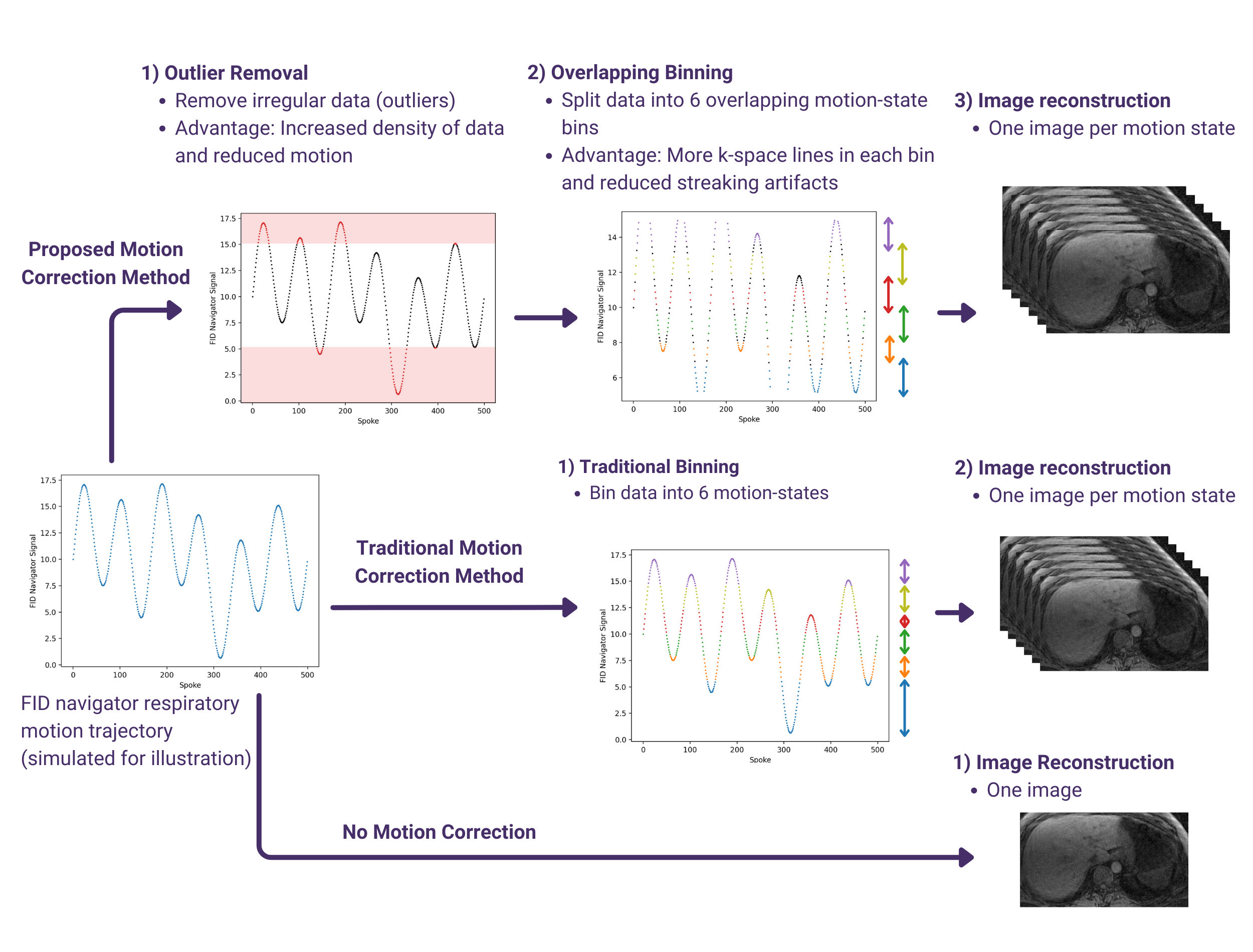
Figure 1 illustrates our proposed method of outlier removal with overlapping binning compared to the traditional binning method.Data acquisition: We performed experiments with informed consent at 3T (Siemens MAGNETOM, Erlangen, Germany), using various imaging parameters for a golden angle-ordered radial stack-of-stars sequence with FIDnavs. We acquired axial and coronal scans without fat saturation (voxel size=1.2x1.2x3mm3) and an axial scan with fat saturation (voxel size=0.9x0.9x5mm3). The scan times were 2.5 and 3.1 minutes for coronal and axial scans. Imaging parameters were TE/TR/FA = 1.49ms/4ms/9˚, 32 coronal slices or 44 axial slices with slice Partial Fourier = 6/8, and 1326 radial spokes. Extracted FIDnav signals underwent principal component analysis and coil clustering for physiological motion signal selection [4]. 800 consecutive spokes from each scan were tested.
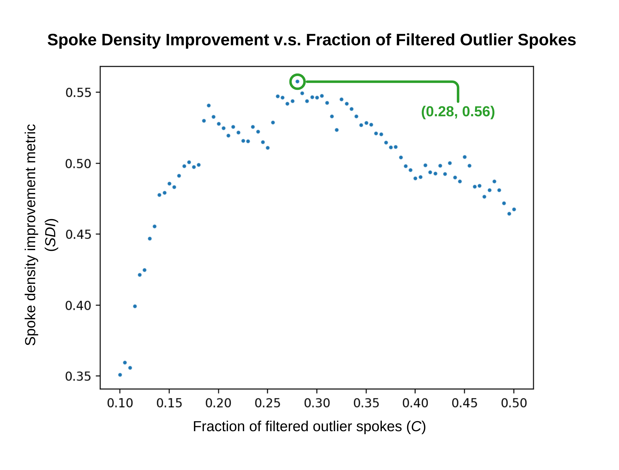
Outlier removal: Our method utilizes Z-scores, a measure of the number of standard deviations a value is from a distribution’s mean, to determine outlier spokes [6]. We filter out a percentage of spokes ($$$C$$$) with the largest absolute Z-scores. We propose a spoke density metric $$$D=\frac{NumberOfSpokes}{NavigatorRange}$$$, where maximum and minimum navigator values determine range. We compute a spoke density improvement metric $$$SDI$$$ for various $$$C$$$ values. We determine the optimal $$$C$$$ as the $$$C$$$ corresponding to the maximum $$$SDI$$$ value (Figure 5).
$$SDI=\frac{D_{OutliersRemoved}-D_{WithOutliers}}{D_{WithOutliers}}$$
Overlapping motion-state binning: We maximize data utilization by selecting particular radial lines to be shared by multiple motion-state bins. First, we divide the navigator signal data into 6 motion-state bins, with each bin containing an equal number of spokes. We construct one gaussian model per bin.
- For each $$$spoke_{i}$$$, we compute a probability density $$$PD_{(i, j)}$$$ that $$$spoke_{i}$$$ belongs to $$$bin_{j}$$$.
- Each $$$PD_{(i, j)}$$$ for $$$spoke_{i}$$$ is normalized to produce a probability $$$P_{(i, j)}=\frac{PD_{(i, j)}}{\sum_{k=1}^6PD_{(i, k)}}$$$, where $$$P_{(i, j)}$$$ is the probability that $$$spoke_{i}$$$ belongs to $$$bin_{j}$$$.
- We identify the two greatest probabilities for $$$spoke_{i}$$$ as $$$P_{(i, a)}$$$ and $$$P_{(i, b)}$$$. If $$$P_{(i, a)}-P_{(i, b)}<T$$$ for an overlap threshold $$$T$$$, $$$spoke_{i}$$$ is assigned to both $$$bin_{a}$$$ and $$$bin_{b}$$$ . Otherwise, $$$spoke_{i}$$$ is solely assigned to $$$bin_{a}$$$.
Reconstruction: Images for each motion state were produced using XD-GRASP reconstruction for the traditional and proposed motion-correction methods.
Results
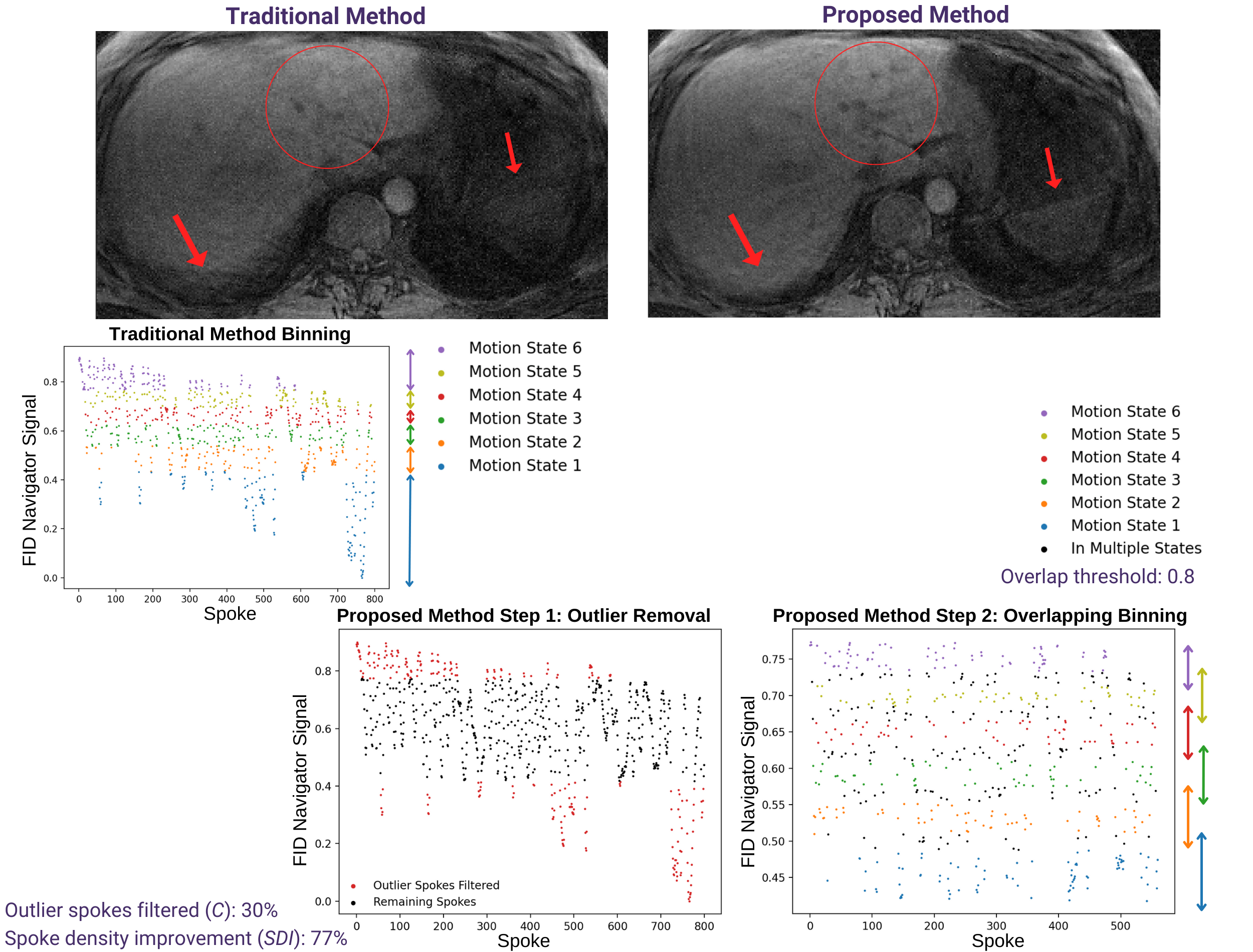
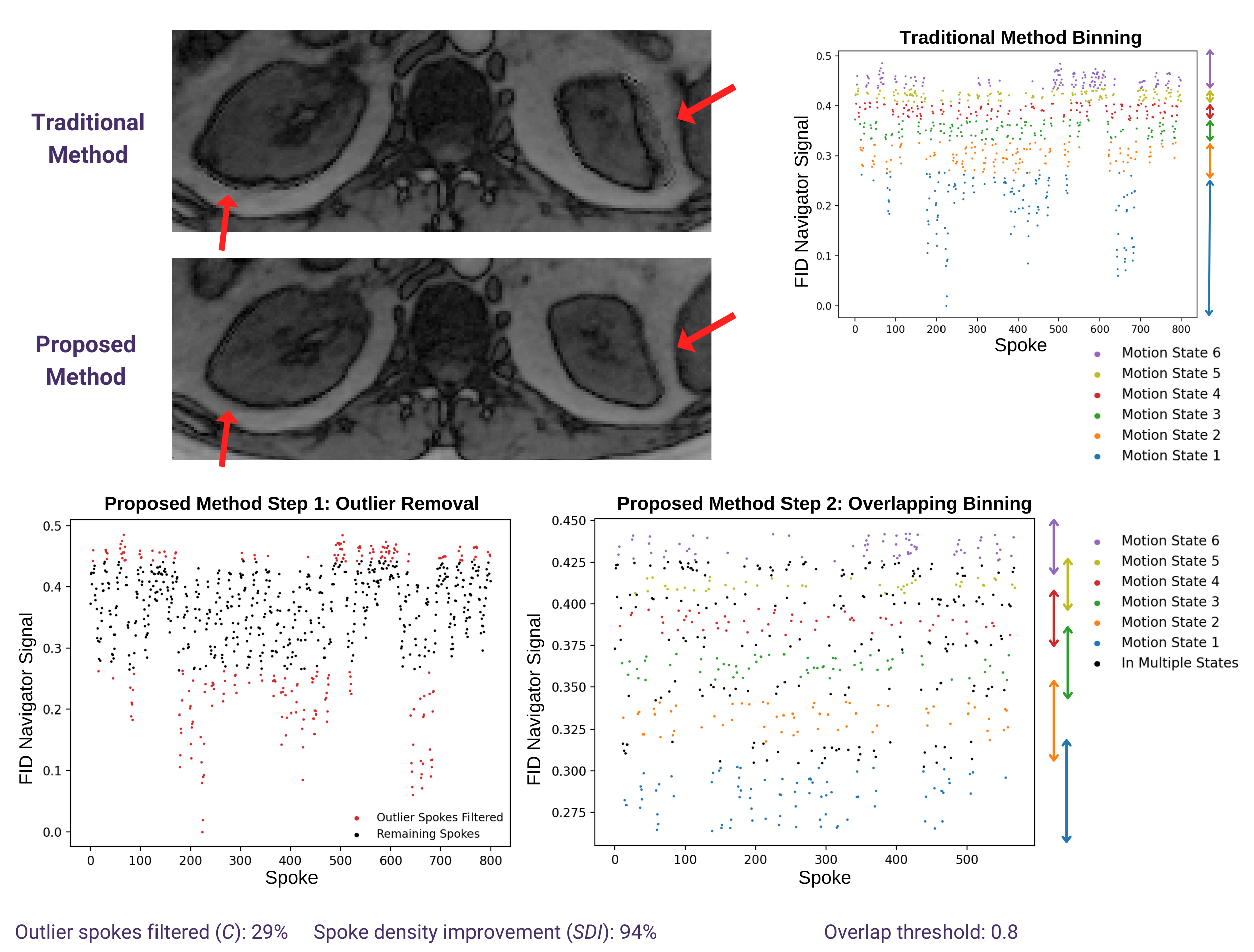
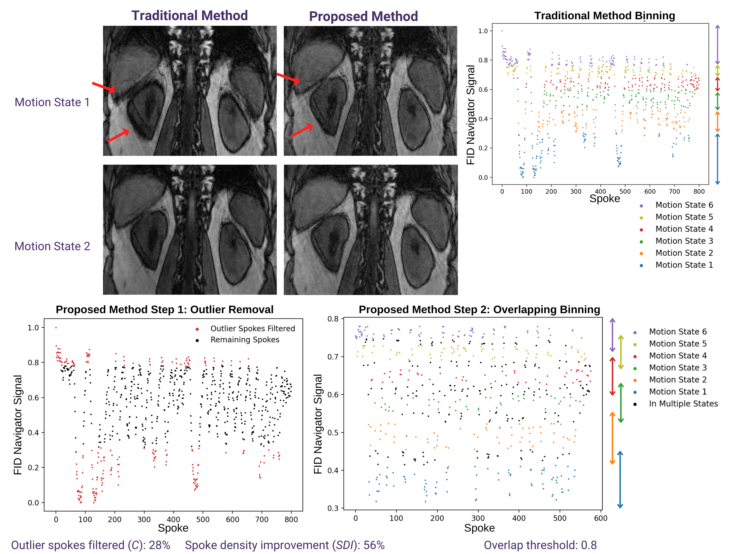
Figures 2-4 depict the original FID navigator signal and the signal obtained after applying our outlier removal and overlapping binning method for three experiments. Our method parameters $$$C$$$ and $$$T$$$ and the resulting $$$SDI$$$ values are also shown. $$$SDI$$$ values consistently exceeded 50%. We compare the conspicuity of motion artifacts as well as organ structures and boundaries in examination of the liver and kidneys for both methods. In inhale-state images, our proposed method shows increased conspicuity of organ structures and boundaries with less motion artifacts and blurring compared to traditional binning. Both methods produce high-quality images of exhale-state bins.Discussion and Conclusion
Our proposed method’s efficacy for inhale-state bins ensures that liver and kidney images are motion-robust across all motion states. Inhale-state images may be considered individually or registered to generate one combined image. Our method is particularly applicable to children and ill or nervous patients exhibiting irregular breathing patterns because we remove abnormalities in the respiratory signal. We share spokes between overlapping bins to maintain the number of radial lines per image. Such overlapping binning can potentially improve dynamic contrast-enhanced MRI imaging, where each bin typically has less radial lines due to temporal resolution constraints. Our balance of removing irregular spokes while sharing select spokes between bins increases precision of the liver and kidney boundaries and visibility for small structures, potentially improving diagnosis confidence and treatment administration.Acknowledgements
This work was supported partially by the National Institute of Diabetic and Digestive and Kidney Diseases (NIDDK), National Institute of Biomedical Imaging and Bioengineering (NIBIB), National Institute of Neurological Disorders and Stroke (NINDS) and National Library of Medicine (NLM) of the National Institutes of Health under award numbers R01DK125561, R21DK123569, R21EB029627, R01NS121657, R01LM013608, S10OD0250111.References
- Block KT, Chandarana H, Milla S, Bruno M, Mulholland T, Fatterpekar G, Hagiwara M, Grimm R, Geppert C, Kiefer B, Sodickson DK. Towards routine clinical use of radial stack-of-stars 3D gradient-echo sequences for reducing motion sensitivity. Journal of the Korean Society of Magnetic Resonance in Medicine. 2014 Jun 1;18(2):87-106.
- Feng L, Axel L, Chandarana H, Block KT, Sodickson DK, Otazo R. XD‐GRASP: golden‐angle radial MRI with reconstruction of extra motion‐state dimensions using compressed sensing. Magnetic resonance in medicine. 2016 Feb;75(2):775-88.
- Chavhan GB, Babyn PS, Vasanawala SS. Abdominal MR imaging in children: motion compensation, sequence optimization, and protocol organization. Radiographics. 2013 May;33(3):703-19.
- Ariyurek C, Wallace TE, Kober T, Kurugol S, Afacan O. Prospective motion correction in kidney MRI using FID navigators. Magnetic Resonance in Medicine. 2023 Jan;89(1):276-85.
- Ariyurek C, Wallace TE, Kober T, Kurugol S, Afacan O. Comparison Of K-Space Center, FID And Pilot Tone Navigators In Abdominal Motion Tracking For Optimal XD-GRASP Reconstruction. Proceedings of Int. Soc. of Magnetic Resonance Imaging. 2023
- Patel VR, Mehta RG. Impact of outlier removal and normalization approach in modified k-means clustering algorithm. International Journal of Computer Science Issues (IJCSI). 2011 Sep 1;8(5):331.
Figures