0423
MRI4All: A Week-Long Hackathon for the Development of an Open-Source Ultra-Low-Field MRI System1Biomedical Engineering, Case Western Reserve University, Cleveland, OH, United States, 2Bernard and Irene Schwartz Center for Biomedical Imaging, Department of Radiology, New York University Grossman School of Medicine, New York, NY, United States, 3Center for Advanced Imaging Innovation and Research (CAI2R), Department of Radiology, New York University Grossman School of Medicine, New York City, NY, United States, 4Icahn School of Medicine at Mount Sinai, New York City, NY, United States, 5Bernard and Irene Schwartz Center for Biomedical Imaging, Department of Radiology, New York University Grossman School of Medicine, New York City, NY, United States, 6Radiology, Stanford., Stanford, CA, United States, 7Department of Electrical Engineering and Computer Sciences, University of California, Berkeley, CA, United States, 8LaGuardia Studio, New York City, NY, United States, 9A. A. Martinos Center for Biomedical Imaging, Boston, MA, United States, 10Department of Neurosurgery, Baylor Scott & White Medical Center, Temple, TX, United States, 11Accessible MR Laboratory, Biomedical Engineering, and Imaging Institute, Dept. of Diagnostic, Molecular and Interventional Radiology, Mount Sinai Hospital, New York City, NY, United States, 12BARNLabs, Muenzkirchen, Austria, 13Biomedical Engineering, University of Michiga, Ann Arbor, MI, United States, 14Department of Biomedical Engineering, Columbia University in the City of New York, New York City, NY, United States, 15Bayer, Cambridge, MA, United States, 16NIST: National Institudes of standards and Techonology, Boulder, CO, United States, 17Division of Vascular & Interventional Radiology, Department of Radiology, Duke University Medical Center, Durnham, NC, United States, 18Department of Chemistry, New York University, New York City, NY, United States, 19Department of Biomedical Engineering, Yale University, New Haven, CT, United States, 20Vanderbilt University, Nashville, TN, United States, 21New York University, New York City, NY, United States, 22Columbia University, New York City, NY, United States, 23Electrical & Computer Engineering, University of Texas at Austin, Austin, TX, United States, 24Department of Computer Science, Rutgers University, Pitscataway, NJ, United States, 25Harvard University, Cambridge, MA, United States, 26Massachusetts Institute of Technology, Boston, MA, United States
Synopsis
Keywords: Low-Field MRI, Low-Field MRI, open-source
Motivation: To break the accessibility barrier of high-field MRI, we demonstrate that hardware and software systems necessary for an affordable MRI system, can be designed and constructed within a week using open-source tools and conventional 3D printing approaches.
Goal(s): To illustrate the realization of an ultra low field MRI system fully operational using open source tools
Approach: Over the course of a week, researchers across the USA have assembled to build the main magnet, field homogenization, gradient, radio-frequency (RF), and software systems needed for the creation of MRI.
Results: We present, for the first time, a community-driven open-source MRI system built within a week.
Impact: to introduce a community-driven open-source MRI low-field systems have the capability to widely democratize MRI throughout the community and the world.
Introduction
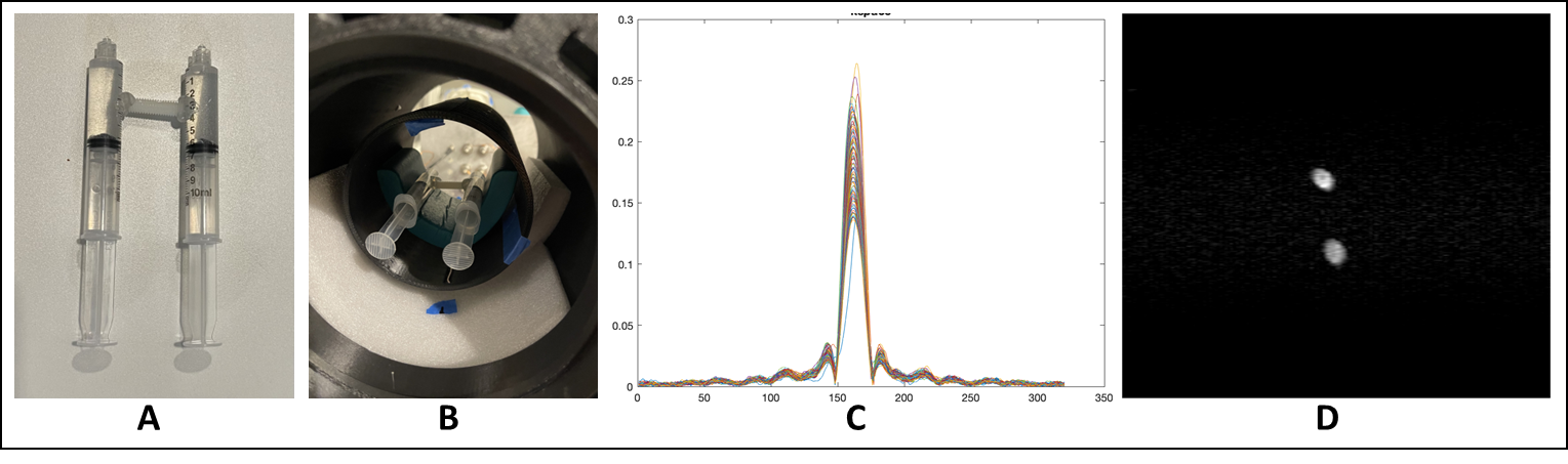
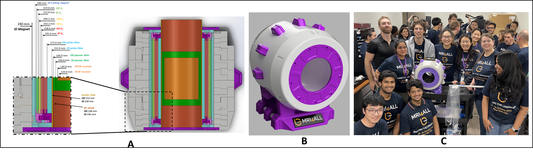
The MRI4ALL Hackathon represents a collaborative endeavor to gather students and experts from the NMR/MRI community to encourage teamwork, problem solving and collaboration. The goal of the MRI4ALL Hackathon 2023 was to build an open-source, low-field MRI system from scratch and use it to get the image of a phantom. All 50+ participants were divided into teams based on their preference and skills as such: Magnet, Gradient, RF Coils & Software teams. Each team worked on separate locations and communicated to each other in the meantime with a final assembly of all individual components 6 days after the beginning of the hackathon. A community vote named the MRI as Zeugmatron Z1, which comes from the Greek meaning “Joining electrical instrument,” and Z1 represented the first version of this scanner. All used materials, components, 3D designs, and software were made open-source on the Github github.com/mri4allMain Magnet
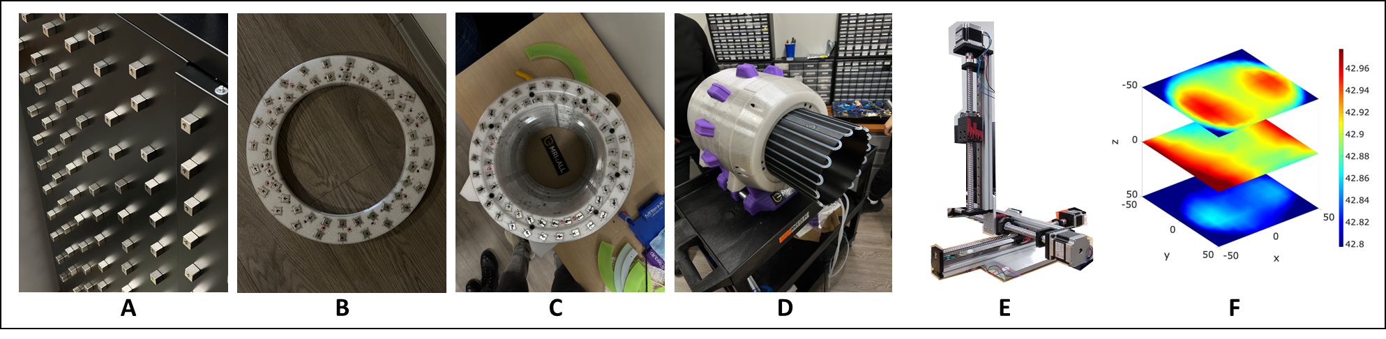
The 44mT Halbach array magnet was constructed by 990 N40UH small half-inch magnets which were simulated and modeled in an array using a combination of Magpylib[1] python packages in order to optimize the resulting main magnetic field and its homogeneity. The magnet design and construction was done by marking the polarity of the half-inch magnets and placing them inside individual 3D-printed rings which were later stacked together. A 3D field mapping device was built using a magnetometer mounted on a system with 3 robotic arms for x,y, and z components. The shimming team could use the information of the field measurement to shim the field through active/passive shimming. A water cooling system with a silicone tubing was built to minimize the temperature change of the gradient coils.Field Homogenization
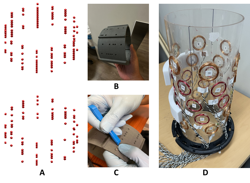
Active and passive shim inserts were designed to homogenize the generated main magnetic field. Since major inhomogeneity was produced along the length of the final magnet, dual shim sleeves located at the ends of the magnet were used to correct the field inhomogeneity. The passive and active shim optimizations were conducted using magpylib on the measured magnetic field produced by the main magnet using the self-built automated field-mapping robot. The active shimming was also accomplished by running a genetic algorithm that was used to compute the optimal locations for the coils. Upon convergence, thirty 35mm diameter hand-wound coils with 15 turns were used, each, fixed onto a 152 mm diameter acrylic outer tube. The simulated field inhomogeneity improved from 2635 Hz to 55 Hz over a DSV of 10cm.Gradient and RF system
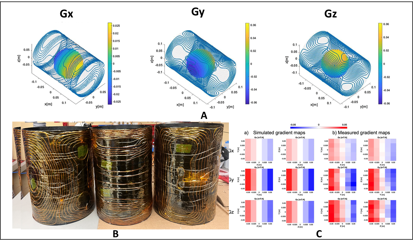
The work done by the gradient team included, the simulation, construction of three gradients for each main axis, building gradient filters, configuring gradient power amplifiers (GPAs) that were compatible with MaRCoS[2] and setting up and testing the individual components. The coil was designed to configure an open source target field using the CoilGen open-source gradient coil generation software[3]. For the RF system, a 20-turn, single-channel RF coil made out of a Litz wire with an outer radius of 66.5 mm was tuned to 1.825 MHz and placed between the passive shim sleeves at 40 mm and 235 mm from the front of the magnet. A 1kW RF power amplifier (RFPA) was used to drive the transmitter and the receiver used a preamplifier with a 0.5 dB noise figure. To allow for transmitting and receiving with the same coil, a custom made passive TR-switch was built.Software platform
The goal of the software team was to develop an open-source software platform for imaging that resembles conventional MRI scanner systems. RF transmit and receive ports in Red Pitaya were used to generate and receive pulses. Pulse’s .seq files were executed through MaRCoS and pypulseq[4]. The Red Pitaya was responsible for driving the scanner components and recording the raw data. A UI service was developed to do the patient registration, sequence configuration, interactive adjustment steps, data visualization and outgoing DICOM connection. All software was made open-source on: https://github.com/mri4allConclusion
The costs for MRI system, especially with higher field strength, can be enormous. There is a high demand in public healthcare systems to expand the accessibility of MRI exams to a greater public, and therefore democratize MR technology. The MR research community shapes the future of MRI and therefore is able to improve the impact to this demand and advance the access to diagnostic imaging throughout the world. With this first-of-its-kind low-field MRI system, that is made completely open-source and will be maintained for further improvement, the democratization of MRI can take the next leap. Visit https://github.com/mri4all and expand the open-source goal for easier accessible MRI to everybody.Acknowledgements
The authors would like to thank the ISMRM community with special thanks to the organizers and sponsors. The Hackathon was supported by the NYU School of Medicine. Data and code availability: all tools and design for this project can be found at https://github.com/mri4all.References
[1] Ortner, Michael; Bandeira, Lucas Gabriel Coliado "Magpylib: A free Python package for magnetic field computation" 11 (2020) 100466
[2] Negnevitsky Vlad, et al. “MaRCoS, an open-source electronic control system for low-field MRI”Journal of Magnetic Resonance (2023) 350.107424
[3] Amrein, Philipp, et al. "CoilGen: Open‐source MR coil layout generator." Magnetic Resonance in Medicine 88.3 (2022): 1465-1479
[4] Keerthi Sravan Ravi, et al. “Pulseq-Graphical Programming Interface: Open source visual environment for prototyping pulse sequences and integrated magnetic resonance imaging algorithm development”. Magnetic Resonance Imaging (2018) 52:9-15
Figures