0422
Portable Ultra-Low Field MRI is Sensitive to Distinct Profiles in Brain Development of Malnutrition and Nutritional Intervention1Institute for Social Sciences, University of Stavanger, Stavanger, Norway, 2Department of Statistics, University of California Davis, Davis, CA, United States, 3Centre for Neuroimaging Sciences, King's College London, London, United Kingdom, 4Department of Forensic and Neurodevelopmental Sciences, King's College London, London, United Kingdom, 5Department of Perinatal Imaging and Health, King's College London, London, United Kingdom, 6Department of Computer Science, UCL, London, United Kingdom, 7Scientific Data Division and Biological Systems & Engineering Division, Lawrence Berkeley National Laboratory, University of California Berkeley, Berkeley, CA, United States, 8Helen Wills Neuroscience Institute and Redwood Center for Theoretical Neuroscience, University of California Berkeley, Berkeley, CA, United States, 9Department of Pediatrics, Division of Developmental Medicine, University of California San Francisco, San Francisco, CA, United States, 10Maternal, Newborn, and Health Discovery Tolls, Bill and Melinda Gates Foundation, Seattle, WA, United States, 11School of Public Health, Makerere University, Kampala, Uganda
Synopsis
Keywords: Neuro, Pediatric, Low-Field MRI
Motivation: The first years are essential for a child’s development and adverse factors, including malnutrition, can affect neurodevelopment and survival rates. Access to high-field MRI scanners in Sub-Saharan Africa is highly limited.
Goal(s): To detect distinct profiles in brain development of malnutrition and nutritional intervention using ultra-low field MRI.
Approach: We used ultra-low field MRI in a pediatric cohort in Uganda of 71 infants (<1.5 years) with and without history of malnourishment, imaged before and after receiving an intervention.
Results: Using PACE brain-for-age growth percentiles, we demonstrate that ultra-low field MRI is sensitive to distinct profiles in brain development of malnutrition and nutritional intervention.
Impact: The distinct profiles of early malnourishment on neurodevelopment and their changes after nutritional intervention derived by ultra-low field MRI could allow for more appropriate neurodevelopmental burden estimates in LMIC pediatric populations and support early intervention evaluation.
Introduction
The first 1000 days in a child’s life display a critical time window for rapid brain growth1 and skill development2. However, this developmental period is highly vulnerable and can be affected by a variety of child health inequalities, such as malnutrition and infectious diseases, which in turn result in underweight and malnourishment3. The prevalence of undernutrition exceeds 800 million people worldwide4. Most of these children live in low to middle income countries (LMICs), where 3.5 million childhood deaths below the age of 5 are attributable to it5. Under- and malnutrition has been linked with lower cognitive performance6, which is notoriously difficult to assess in those under three years of age. In addition, most studies have been conducted in developed countries and very few have been taken place in LMICs7, due to high MRI scanner cost and low availability. To address this, a new generation of portable ultra-low field MRI scanners has emerged, allowing for safe and mobile scanning at a much lower cost point8. However, the sensitivity of these new scanners to LMIC focused problems, such as malnutrition, and their relation to early brain development has yet to be established.Here, we use ultra-low field MRI to investigate a longitudinal two-armed LMIC pediatric (<1.5 years) cohort from Uganda: one with malnourished children before and after nutritional intervention, and one without malnourishment scanned and weighted at the same time points. We applied PACE brain-for-age growth percentiles to investigate whether ultra-low field MRI can detect variations in brain development of never malnourished children and previously malnourished children before and after receiving an intervention.
Methods
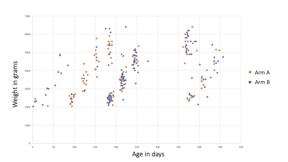
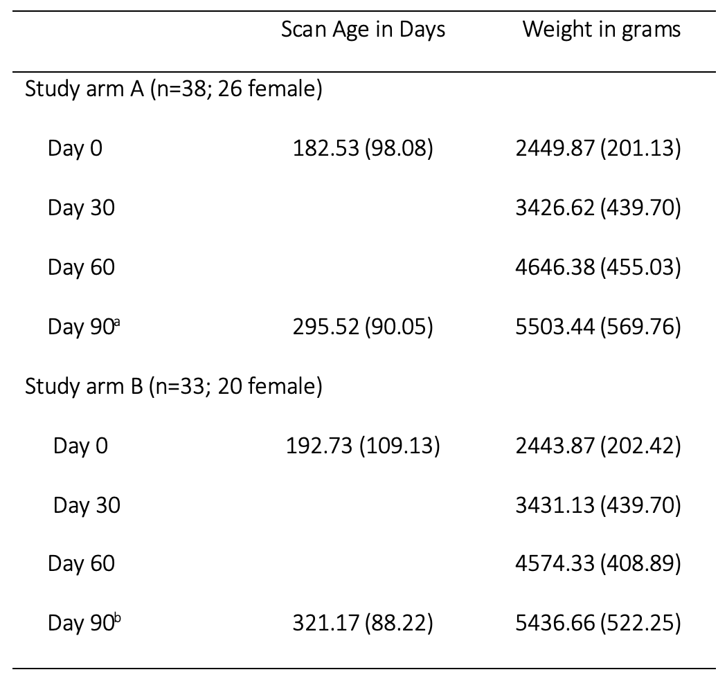
Longitudinal whole-brain T1- and T2-weighted MRI data were collected in 71 children (38 female) on a 64mT Hyperfine Swoop imaging system, following previously documented scanning parameters9. Participant demographics and weight at 30-day increments can be found for both groups in Table 1, Figure 1. Following data acquisition and image reconstruction, images were non-linearly aligned to age-corresponding anatomical templates in MNI space, and an atlas-based segmentation approach was used to delineate whole-brain white matter (WM), grey matter (GM), cerebral spinal fluid (CSF), as described elsewhere9. PACE brain-for-age growth percentiles were created for proportional and absolute volumes of CSF, WM, GM, and regression slope functions with 95% pointwise confidence intervals to identify critical windows of brain development10.Results
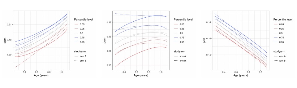
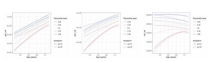
Following successful nutritional intervention, weight increased with time in both groups (Figure 1). Age-based dynamic proportional (p) and absolute volumes of GM, WM and CSF percentiles differed between both arms, but aligned with time and intervention (Figures 2, 3).Discussion
In this study, we were able to demonstrate that ultra-low field MRI is sensitive to detect distinct patterns in brain development of never malnourished and previously malnourished children that aligned after receiving a nutritional intervention. While strong improvements can be seen specifically for the vulnerable lowest 25th percentile for pGM volume development with weight gain over time, pWM showed the largest initial differences across percentiles between both groups (Figure 2). These differences are even more pronounced when looking at absolute volumes (Figure 3).Healthy brain development has been identified as a key predictor of current and future cognitive development11 and under- and malnutrition has been linked with lower cognitive performance6. Differences in WM integration and cognitive functions have been linked to children who suffered early neglect, potentially indicating high sensitivity of this structure to early adverse factors influencing early brain development12,13. While cognitive performance is only emerging in the first two years of life and with assessments often limited by language dependence at that age, the sensitivity of our method to effects of malnourishment on brain development could be a promising tool for more appropriate neurodevelopmental burden estimates in LMIC pediatric populations and support early intervention evaluation.
Our study focused on early childhood (<1.5 years old), a period during which the brain undergoes fast and important changes10, establishing the ground for higher cognition and language development. Using percentiles helps to place individual brain development trajectories, informing about “where on the curve” a child is relative to the population. The importance of well-timed interventions and monitoring of neurocognitive health underlines the importance of early non-invasive assessment of neurodevelopment. We plan to investigate the relationship of early neurodevelopment in LMIC settings using ultra-low field MRI and link them to cognitive development.
Acknowledgements
We would like to thank the Bill and Melinda Gates Foundation (INV005774 and INV-047885) for their continuous support of this work, and all of the participants of the PRIMES study.References
1. Fox SE, Levitt P, Nelson CA 3rd. (2010). How the timing and quality of early experiences influence the development of brain architecture. Child Dev, 81(1),28-40. 2. Bruchhage, MMK, Ngo, GC, Schneider, N, D’Sa, V, Deoni, SCL (2020). Functional connectivity correlates of infant and early childhood cognitive development. Brain structure and Function. Doi:10.1007//s00429-020-02027-4 3. Walker, S. P., Wachs, T. D., Grantham-McGregor, S., Black, M. M., Nelson, C. A., Huffman, S. L., et al. (2011). Inequality in early childhood: risk and protective factors for early child development. The Lancet, 378:1325–38. 4. The State of Food Insecurity in the World (2015). Meeting the 2015 international hunger targets: taking stock of uneven progress. FAO; Rome, Italy. 5. Black, R. E., Allen, L. H., Bhutta, Z. A., Caulfield, L. E., de Onis, M., Ezzati, M., Mathers, C., Rivera, J. (2008). Maternal and child undernutrition: global and regional exposures and health consequences. Lancet, 371, 243–260. 6. Repositioning Nutrition as Central to Development A Strategy for Large-Scale Action. The World Bank; Washington, DC, USA: 2006. 7. Ogbole GI, Adeyomoye AO, Badu-Peprah A, Mensah Y, Nzeh DA. (2018). Survey of magnetic resonance imaging availability in West Africa. Pan Afr Med J., 30, 240. 8. Marques, JP, Simonis, FFJ, Webb, AG (2019), Low-field MRI: An MR physics perspective. J. Magn. Reson. Imaging, 49, 1528-1542. 9. Deoni, SCL, Bruchhage, MMK, Beauchemin, J, Volpe, A, D'Sa, V, Huentelman, M, Williams, SCR. (2021). Accessible pediatric neuroimaging using a low field strength MRI scanner. NeuroImage, 238, 118273. 10. Chen, Y, Dubey, P, Müller, H-G, Bruchhage, MMK, Wang, J-l, Deoni, SCL (2021). Modeling sparse longitudinal data in early neurodevelopment. NeuroImage, 237:118079. 11. Gilmore, J. H., Knickmeyer, R. C., & Gao, W. (2018). Imaging structural and functional brain development in early childhood. Nature Reviews Neuroscience, 19(3), 123–137. 12. Hanson, J. L., Adluru, N., Chung, M. K., Alexander, A. L., Davidson, R. J., & Pollak, S. D. (2013). Early neglect is associated with alterations in white matter integrity and cognitive functioning. Child Development, 84(5), 1566–1578. 13. Zhu, C., Chen, Y., Müller, H.-G., Wang, J.-L., O'Muircheartaigh, J., Bruchhage, M., & Deoni, S. (2023). Trajectories of brain volumes in young children are associated with maternal education. Human Brain Mapping, 44(8), 3168–3179.Figures