0414
NORMAL APPEARING BREAST TISSUE ON BREAST MRI HAS ALTERED CHEMISTRY CONSISTENT WITH “SWITCHED-ON” STATES IN WOMEN WITH INVASIVE CARCINOMA1Griffith University, Southport, Australia, 2Datchem Pty Ltd, Brisbane, Australia, 3Princess Alexandra Hospital, Woolloongabba, Australia, 4Radiology, Princess Alexandra Hospital, Woolloongabba, Australia, 5The Breast Centre, Gateshead, Australia, 6Jones Radiology, Adelaide, Australia, 7Datchem, Brisbane, Australia
Synopsis
Keywords: Breast, Spectroscopy, lipids, cholesterol
Motivation: Adenoma-carcinoma cell models examined using 2D COSY recorded altered triglyceride, cholesterol and metabolites prior to malignant transformation.
Goal(s): Investigate if such chemical profiles are recorded in vivo in MRI apparently normal tissue in women with invasive cancer.
Approach: Nineteen women with invasive breast cancer were compared with healthy low risk controls.
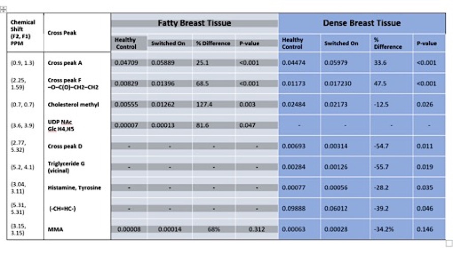
Results: Compared to controls, MRI normal tissue in cancer patients with low-density breasts recorded increases in cross peak F (68%), cholesterol (127%), and tumor promotor UDP-GlcNAc (81%). For dense breasts, increases recorded in cross peak F (47%), decreases in cholesterol (12%), triglyceride (56%) and double bonds (40%) in "switched-on" tissue.
Impact: Altered chemistry states, consistent with mechanisms leading to development of invasive carcinoma, are recorded in vivo from breast tissue distant to the cancer compared to controls. These states are referred to as "switched-on" tissue and differ according to breast density.
INTRODUCTION
The concept of a sequence associated with tumor development was introduced in 1990 (1). Magnetic resonance spectroscopy (MRS) of colon and ovarian cell models showed that biological and genetic characteristics directly correlated with the chemical analysis of whole cells and membranes (2-4). In particular, the biomarkers of adenoma-carcinoma sequence were molecules active on the MR timescale and included lipids, cholesterol and metabolites (4). The two-dimensional COrrelated SpectroscoPY (2D COSY) sequence showed specific chemical changes occurring prior to and at malignant transformation (3).Our hypothesis is that breast tissue deemed healthy on conventional imaging shows early change(s) associated with cancer development, a "switched-on" state, in women diagnosed with invasive breast cancer elsewhere and when compared to healthy women at low risk.
The goal is to investigate whether chemical profiles consistent with a "switched-on" state can be recorded in vivo using 2D COSY MRS in the normal appearing breast tissue in women bearing invasive cancer.
METHODS
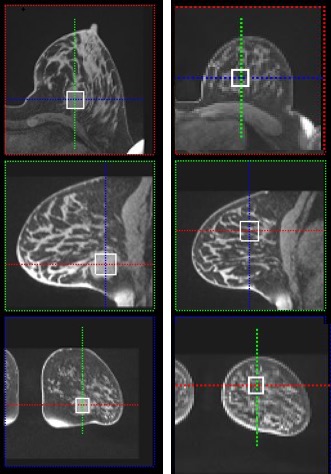
Institutional review board approval and written informed consent obtained. A cross-sectional study with prospective data collection was carried out at Princess Alexandra Hospital, Brisbane, Australia.Nineteen women (mean age, 51; min-max, 31-71) with histologically proven invasive breast cancer were recruited. Breast MRI and 2D COSY (5-7) was performed before treatment. For MRS acquisition, one voxel was placed over the cancer and another one over normal appearing breast tissue away from the lesion (Figure 1). Wherever the cancer was extensive or the breast was small, the second voxel was placed in the contralateral breast. This cohort was compared to healthy women at low risk (n=66; mean age, 43). The healthy cohort was recruited based on NICE guidelines (5). Some data from the healthy cohort has been reported previously (8,9).
The 2D COSY sequence parameters were TR 2000 ms, TE initial of 30 ms, 96 t1 increments at 0.8 ms, 6 averages per increment, f2 bandwidth 2000 Hz, vector size of 1024 points and RF offset frequency set on 3.2 ppm. ‘WET’ water suppression (8) applied. Processing undertaken as reported (9).
For statistical analysis, correlation of measured intensity of these peaks to age, breast density, and menopausal status used Kendall’s tau or Pearson’s method. For mean comparison between low risk, and switched on tissue using one-way ANOVA was used. The mean of switched on tissue of cancer patients and their tumor was compared using paired t-test.
RESULTS
When comparing tissue chemistry between healthy cohort, apparently healthy tissue in cancer bearing patient (switched-on tissue) and the tumor (Figure 2), breast density showed to impact on tissue chemistry (Table 1).Low-density breasts: Several significant increases were recorded between low risk cohort and the switched-on tissue: a) Cross peak A, the methyl to methine coupling of the fatty acyl chain increases by 25%, b) Cross peak F (-O-C(O)-CH2-CH2-) increases by 68%, c) the methyl on the cholesterol increases by 127%, d) methylmalonic acid (MMA) increases by 68%. Additionally, new in vivo assignment is made for Uridine-diphosphate N-acetylglucosamine (UDP-GlcNAc, H4,H5) at (F2: 3.6, F1: 3.9ppm) which increased by 81% in the switched-on low-density breast tissue. Comparison between the "switched-on" tissue and the tumor showed large increases in these same cross peaks with a further 8 species increasing in the tumor tissue, that did not get to significance level.
Dense breasts: Significant increases were also recorded in Cross peak A (34%) and Cross peak F (47%) , and a decrease in cholesterol methyl (12%), cross peak D (55%), triglyceride (56%), histamine/tyrosine (28%), the double bond index (39%) and MMA (34%). Compared to the tumor chemistry, large non-significant changes are recorded with a further 9 species.
DISCUSSION
We coin the term “switched-on” to describe chemical state(s) measured in vivo in breast tissue that is different from healthy tissue and is found in women with invasive breast cancer elsewhere. The chemical profiles have been reported previously in cell models of tumor development (4). These changes are seen here in vivo and differ according to breast density.The UDP-GlcNAc is is a known marker of tumorigenicity (10,11) and another difference between the healthy and the “switched-on” tissue in low-density breasts. The tumor promotor MMA is recorded in dense breast tissue in the "switched-on" tissue.
SUMMARY
Chemically altered states referred to as “switched-on” tissue, consistent with the mechanisms leading to development of breast cancer, are recorded from normal appearing tissue distant from tumor and differ according to breast density.Acknowledgements
This study was supported by the Queensland Government under the Advance Queensland initiative.References
1. Fearon ER, Vogelstein B. A genetic model for colorectal tumorigenesis. Cell 1990; 61: 759-767.
2. Mackinnon WB, Dyne M, Hancock R, Grant AJ, Russell P, Mountford CE. Malignancy-related characteristics of wild type and drug-resistant Chinese hamster ovary cells. Pathology 1993; 25: 268-276.
3. Mackinnon WB, Huschtscha L, Dent K, Hancock R, Paraskeva C, Mountford CE. Correlation of cellular differentiation in human colorectal carcinoma and adenoma cell lines with metabolite profiles determined by 1H magnetic resonance spectroscopy. Int J Cancer 1994; 59: 248-261.
4. Mackinnon WB, May GL, Mountford CE. Esterified cholesterol and triglyceride are present in plasma membranes of Chinese hamster ovary cells. Eur J Biochem 1992; 205: 827-939.
5. Ramadan S, Arm J, Silcock, J, et al. Lipid and Metabolite Deregulation in the Breast Tissue of Women Carrying BRCA1 and BRCA2 Genetic Mutations. Radiology, 2015; 275: 675-682.
6. Thomas MA, Binesh N, Yue K, DeBruhl N. Volume-localized two-dimensional correlated magnetic resonance spectroscopy of human breast cancer. J Magn Reson Imaging 2001;14: 181-186.
7. Thomas MA, Lipnick S, Velan SS, eta al. Investigation of breast cancer using two-dimensional MRS. NMR in Biomed 2009; 22:77-91.
8. Santamaría G, Naude N, Bennett I, et al. In vivo assignment of methylmalonic acid in breast tissue using 2D MRS and relationship with breast density, menopausal status and cancer risk. NMR in Biomed 2023; 36: e4851.
9. Santamaría G, Naude N, Watson, J, et al. Breast tissue chemistry measured in vivo in healthy women correlate with breast density and breast cancer risk. J Magn Reson Imaging 2022; 56:1355-1369.
10. Gomes AP, Age-induced accumulation of methylmalonic acid promotes tumour progression. Nature 2020; 585:283-287.
11. Ruegenberg S, Horn M, Pichlo C, Allmeroth K, Baumann U, Denzel MS.Loss of GFAT-1 feedback regulation activates the hexosamine pathway that modulates protein homeostasis. Nat Commun 2020; 11: 687.
Figures
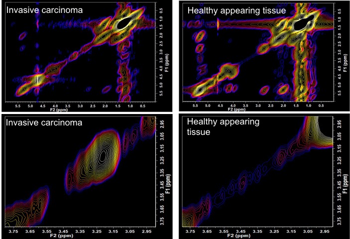
Processed 2D-COSY spectrum (FelixNMR) of both invasive carcinoma (left) and healthy tissue (right). Displayed field of view: (0.0-6.0/0.0-6.0)ppm at the top and (2.8-3.8/2.8-3.8)ppm (F2/F1) at the bottom.
Note the increased spectral volumes in the metabolite region in the carcinoma (2.8-3.8/2.8-3.8)ppm compared to the healthy tissue, and the markedly increased metabolic activity in the carcinoma around the choline resonance (at 3.22-3.22ppm).