0402
Predicting IDH Mutation and MGMT Methylation Status in Glioma Patients at the Voxel Level using CEST-Based Deep Learning1Department of Electronic Science, Fujian Provincial Key Laboratory of Plasma and Magnetic Resonance, School of Electronic Science and Engineering, National Model Microelectronics College, Xiamen University, Xiamen, China, 2Department of Radiology, Union Hospital, Tongji Medical College, Huazhong University of Science and Technology, Wuhan, China
Synopsis
Keywords: CEST / APT / NOE, Tumor
Motivation: Predicting glioma subtypes based on molecular profiles is crucial for treatment decisions and predicting survival rates.
Goal(s): We proposed a CEST-based deep learning method to predict IDH mutation and MGMT methylation status in glioma patients at the voxel level.
Approach: 86 patients were recruited for CEST experiments on 3T MRI scanner. A CEST-based deep learning method, composed of a 1D convolutional neural network, was proposed for different types of status prediction at the voxel level. The confusion matrix and ROC were conducted to evaluate the performance of the proposed method.
Results: Our method achieves higher accuracy compared to existing CEST-based prediction methods.
Impact: The proposed method may facilitate the application of CEST MRI in the diagnosis of glioma.
Introduction
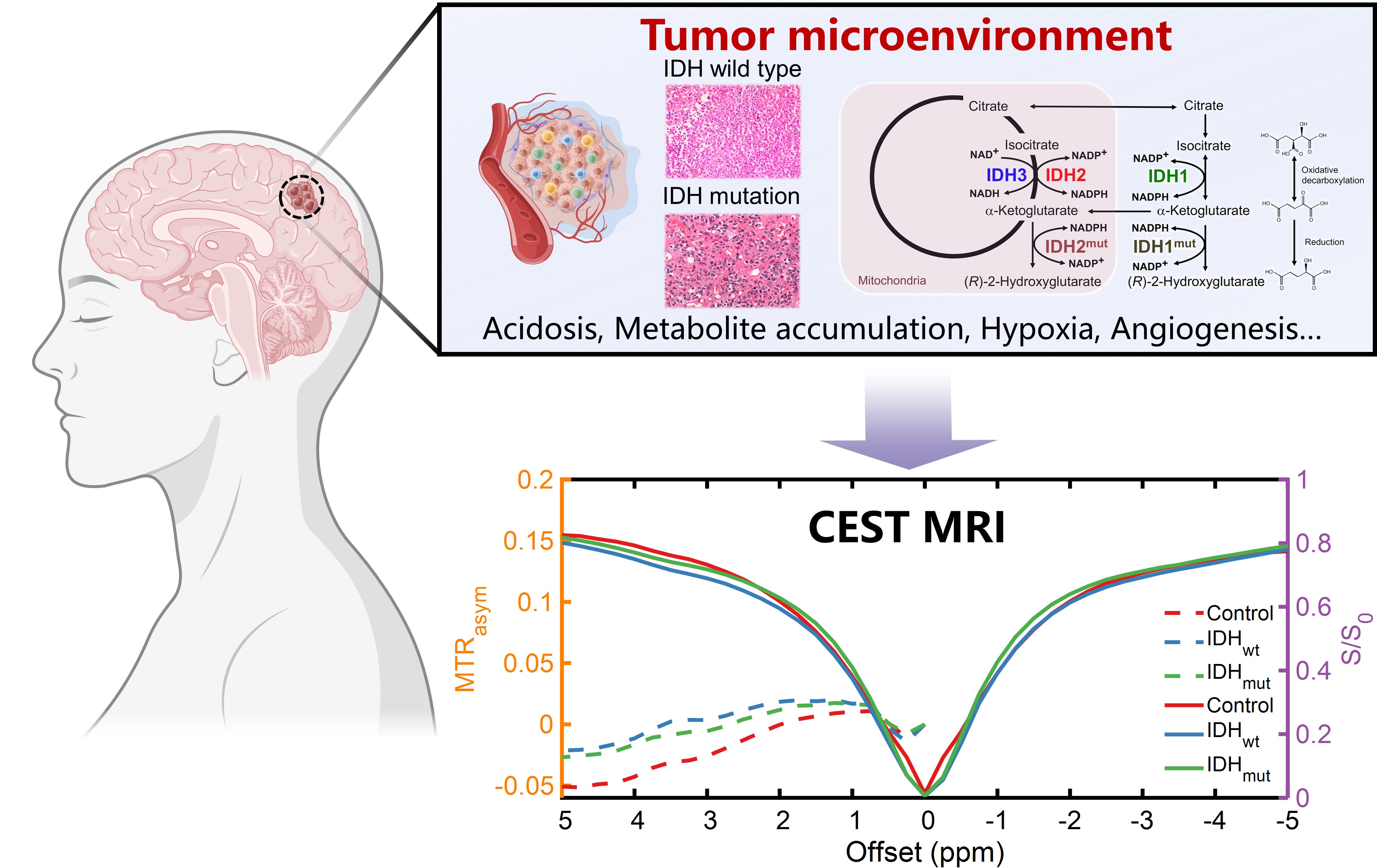
The fifth edition of the 2021 World Health Organization (WHO) Classification of Tumors of the Central Nervous System (WHO CNS5) emphasizes the importance of classifying glioma subtypes based on molecular profiles, which is crucial for treatment decisions and predicting survival rates1. Previous studies reveal that isocitrate dehydrogenase (IDH) mutation and O6-methylguanine-DNA methyltransferase (MGMT) promoter methylation lead to distinct tumor microenvironments, including acidosis, and metabolite accumulation, which can be exploited to predict glioma subtypes2,3. Chemical exchange saturation transfer (CEST) enables the non-invasive detection of endogenous metabolites and the cellular microenvironment, which can be used for differentiating glioma subtypes, as illustrated in Fig. 14. Nevertheless, its effectiveness is influenced by the choice of quantification method, and various quantification methods may produce conflicting results. Furthermore, most current deep learning tumor classification methods are image-based, necessitating a significant amount of training data for neural network training, which can be challenging to obtain in a clinical setting.Here, a CEST-based deep learning method, consisting of 1D convolutional neural network (CNN), was proposed for glioma subtype prediction at the voxel level, aiming to improve prediction accuracy, reduce the requirement for substantial training data, and enhance prediction speed.
Methods
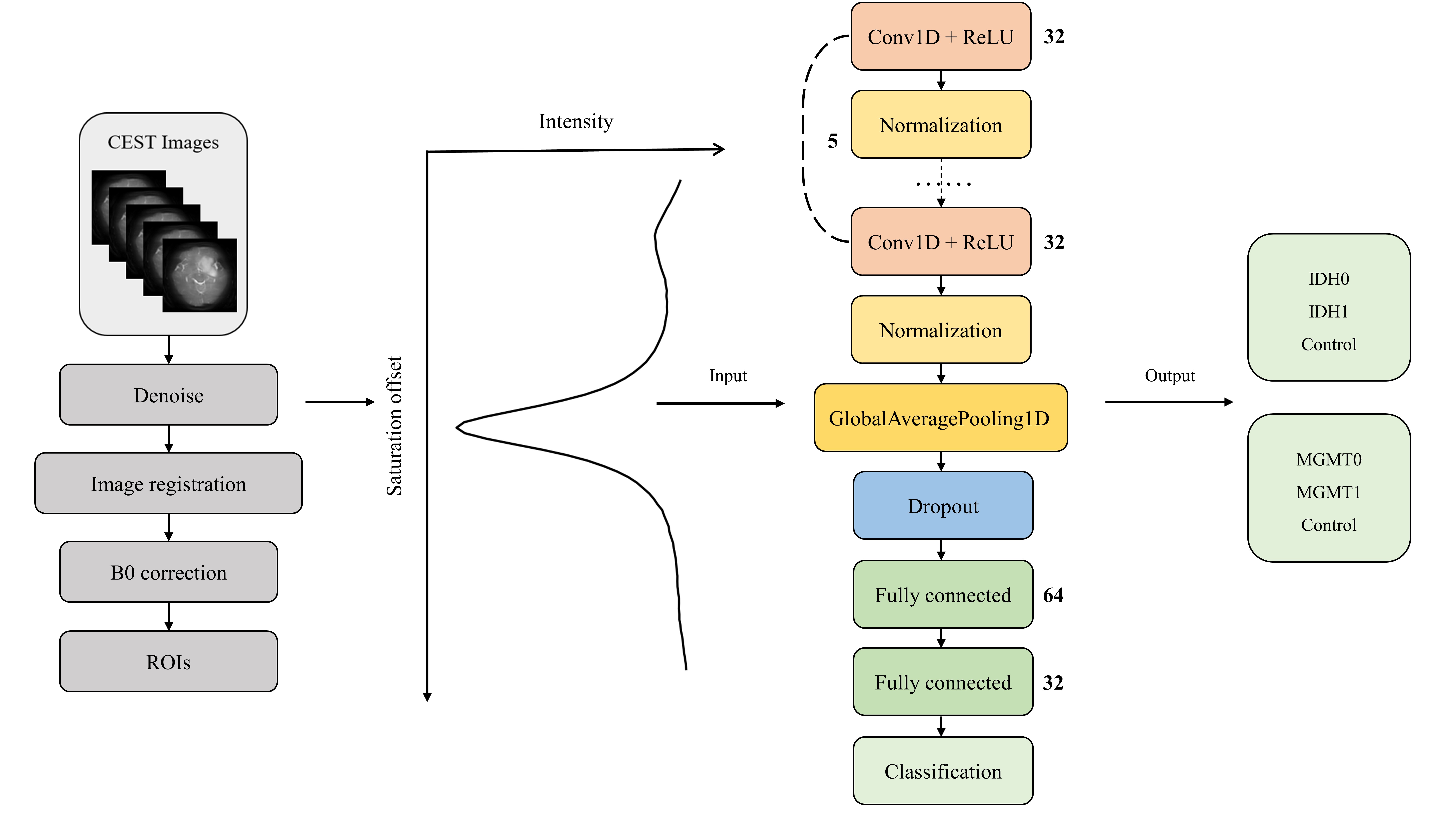
A total of 86 patients, including 31 IDH wild type (IDH0), 32 IDH mutation (IDH1), 11 MGMT unmethylation (MGMT0), and 12 MGMT methylation (MGMT1), were recruited for the study with their written consent. All patients were confirmed by histopathological examination after surgical resection according to the World Health Organization 2021 criteria1. The CEST experiments were conducted on a 3 T scanner with saturation offsets ranging from -30 to +30 ppm. Preprocessing, including CEST denoising and image registration, was performed. The regions of the tumor and control were delineated by experienced neuro-radiologists. Two 1D CNN-based classification models, with voxel-based Z-spectrum as input, were proposed for IDH and MGMT classification, respectively. The flowchart of the proposed method is shown in Fig. 2. Eight patient data were randomly selected for testing, while the remaining data were used for neural network training. The performance of the proposed method was evaluated using a confusion matrix and ROC analysis.Results and Discussion
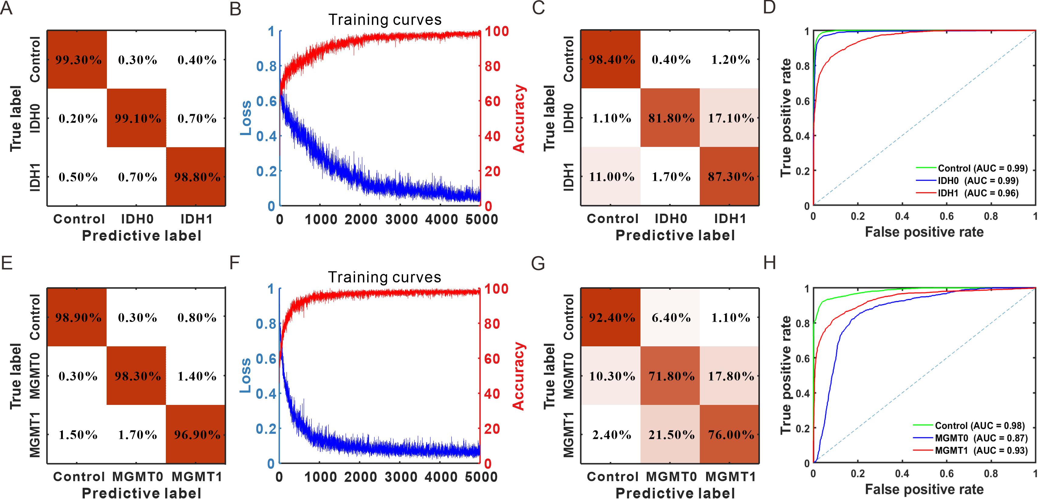
From the training confusion matrix and curves (Fig. 3A, B, E&F), it is evident that the neural networks for both IDH and MGMT prediction have converged after 5000 iterations. For the testing data, the confusion matrix in Fig. 3C shows true positive (TP) rates of 98.4%, 81.8%, and 87.3% for contralateral normal tissue, IDH wild type, and IDH mutant, respectively. The confusion matrix in Fig. 3G displays TP rates of 92.4%, 71.8%, and 76.0% for contralateral normal tissue, MGMT unmethylation, and MGMT methylation, respectively. In Figure 3D, the AUC values for contralateral normal tissue, IDH wild type, and IDH mutant in the IDH status classification model were 99%, 99%, and 96%, respectively, which outperform the work from Jiang et al., who reported an AUC of 89% using APTw MRI at 3T5.The previous study by Jiang et al. suggests that APTw MRI with asymmetric analysis can differentiate MGMT status6. However, Paech et al7. report that the CEST contrast obtained using Lorentzian fitting and AREX is unable to predict MGMT status. Given that the tumor microenvironment, including pH and metabolite accumulation, can affect both CEST peaks and the entire background region, quantification methods based on specific assumptions (e.g. asymmetric analysis or Lorentzian fitting) might overlook this influence, resulting in varying prediction performance. In this study, the complete Z-spectrum, containing all available information, was inputted into the neural network. The training results indicate that the neural network effectively leverages the distinctions between Z-spectra from various MGMT subtypes and applies the acquired knowledge for prediction.
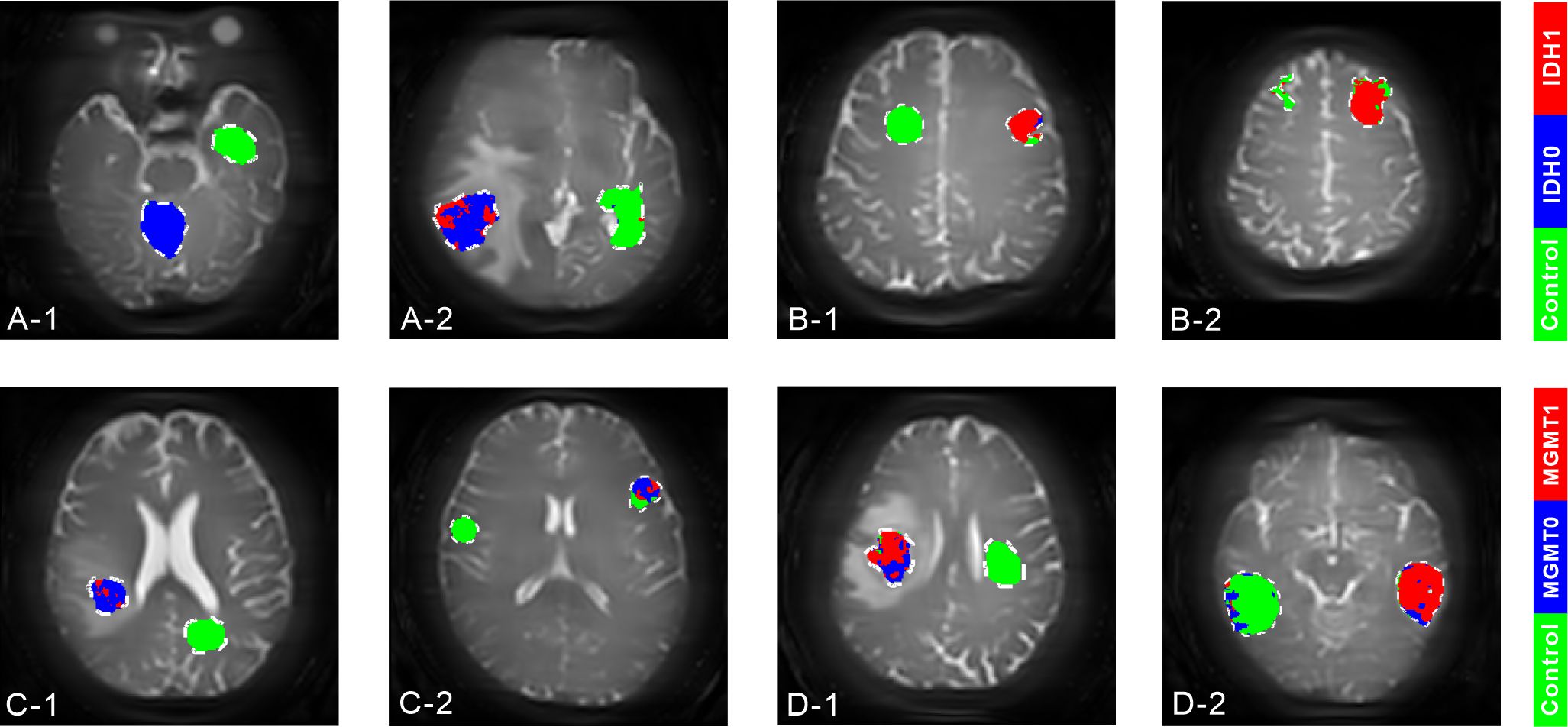
The prediction maps for 8 testing patient data are displayed in Fig. 4. The results indicate that the proposed method provides overall satisfactory prediction performance. However, the edge region of the ROI shows degraded accuracy due to partial volume effects and tumor inhomogeneity.
The use of a 1D CNN in the proposed method significantly reduces training time in comparison to working with 2D or higher-dimensional data. Moreover, when compared to image-based methods, voxel-based methods are much more accessible for gathering ample data for neural network training. Finally, the absence of the need for CEST quantification in the proposed method leads to a rapid prediction process, which is completed in just a few seconds on a personal computer.
Conclusion
In this study, we developed a CEST-based deep learning method for predicting glioma subtypes, which offers rapid predictions and enhanced accuracy compared to existing approaches, potentially aiding in treatment decisions and the prediction of survival rates.Acknowledgements
This work is supported by the National Natural Science Foundation of China, Grants Number:82302151, 82371945; Shenzhen Science and Technology Program, Grant/Award Number: JCYJ20220818101213029; Fujian Province Science and Technology Project, Grant/Award Number: 2022J05013; Xiamen University Nanqiang Outstanding Talents Program.References
1.Louis, D.N., et al., The 2021 WHO Classification of Tumors of the Central Nervous System: a summary. NEURO-ONCOLOGY, 2021. 23(8): p. 1231-1251.
2.Siegal, T., Clinical Relevance of Prognostic and Predictive Molecular Markers in Gliomas. Advances and technical standards in neurosurgery, 2016(43): p. 91-108.
3.Martínez-Reyes, I. and N.S. Chandel, Cancer metabolism: looking forward. NATURE REVIEWS CANCER, 2021. 21(10): p. 669-680.
4.Zhou, J.Y., et al., Review and consensus recommendations on clinical APT-weighted imaging approaches at 3T: Application to brain tumors. MAGNETIC RESONANCE IN MEDICINE, 2022. 88(2): p. 546-574.
5.Jiang, S.S., et al., Predicting IDH mutation status in grade II gliomas using amide proton transfer-weighted (APTw) MRI. MAGNETIC RESONANCE IN MEDICINE, 2017. 78(3): p. 1100- 1109.
6.Jiang, S.S., et al., Discriminating MGMT promoter methylation status in patients with glioblastoma employing amide proton transfer-weighted MRI metrics. EUROPEAN RADIOLOGY, 2018. 28(5): p. 2115-2123.
7.Paech, D., et al., Assessing the predictability of IDH mutation and MGMT methylation status in glioma patients using relaxation-compensated multipool CEST MRI at 7.0 T. NEURO- ONCOLOGY, 2018. 20(12): p. 1661-1671.
Figures