0400
A targeted Fe-based MR contrast agent for glioma imaging1Innovation Academy for Precision Measurement Science and Technology, Chinese Academy of Sciences, WuHan, China, 2Huazhong University of Science and Technology, WuHan, China
Synopsis
Keywords: Contrast Agents, Brain, glioma
Motivation: Neurological adverse effects caused by the deposition of gadolinium-based contrast agents in the brain remain uncertain, and it is necessary to develop a biocompatible alternative molecule for evaluation of glioma malignancy and site occupancy.
Goal(s): To demonstrate that Fe-based contrast agents have superior safety, stability, and longevity in glioma imaging compared to gadolinium-based contrast agents.
Approach: The T7-Fe-PyC3A was synthesized, and its stability was validated through in vitro kinetic thermodynamic experiments. Furthermore, A in situ glioma model mice was choosen for investigating the imaging ability.
Results: A targeted Fe-based MR contrast agent provides clear glioma contours with a 15-60 minutes post-dose imaging window.
Impact: Although gadolinium-based contrast agents are widely used in the clinic, iron-based contrast agents targeting gliomas show the enhanced safety and stability profile. The extended imaging window allows them as preferable alternative for gliomas imaging.
Introduction
Gliomas, particularly glioblastoma multiforme (GBM), are aggressive cancers of the brain and spinal cord with poor prognosis [1-2]. Neuroimaging routine examination mainly includes CT and MRI, of which, MRI is better than CT at providing detailed images. Routine MRI scans include T1-weighted images, T2-weighted images, and FLAIR sequence imaging [3]. In particular, the dynamic contrast-enhanced MRI (DCE-MRI) can accurately determine the extent of the lesion, as well as any edema or tumor vascular flow signals. The most frequently utilized contrast agents for gliomas in clinical settings are Gd-based, such as Omniscan and Magnevist. While current studies have not established the relationship between intracerebral gadolinium deposition and relevant neurological adverse effects, there is still significant interest in discovering safer contrast agents with lengthier imaging windows to supplant gadolinium-based contrast agents.Methods
The contrast agent T7-Fe-PyC3A and Fe-PyC3A were synthesized. In addition, the relaxivity profile of T7-Fe-PyC3A was assessed in pH 7.4 Tris buffer at 0.5 and 9.4 T. The stability of the T7-Fe-PyC3A for 72 hours was monitored by HPLC and potentiometric titration. An in-situ glioma model in nude mice was established by injecting U87-MG cells into the nude mouse brain via a mouse brain stereotaxic apparatus. T1-weighted MR images were acquired by monitoring the brain of U87-MG tumor-bearing mice following the administration of T7-Fe-PyC3A and Gd-DTPA through the tail vein at 9.4 T imaging system.Results
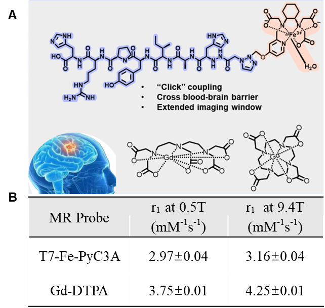
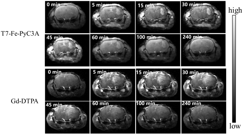
T7-Fe-PyC3A (Figure 1A) and its control molecule without the T7 peptide (Fe-PyC3A) were synthesized. The longditudinal relaxivity of T7-Fe-PyC3A at 9.4 T was 3.16 ± 0.04 mM−1 s−1 which is comparable to clinical used Gd-DTPA. To demonstrate its higher stability, the complex has been incubated under various condition at 37 ℃ for 72 hours. Notably, the solution's color, longitudinal relaxation rate, and the retention time in the HPLC profile remained largely unchanged. Following the tail vein injection of this complex into a glioma model in mice, the T1-weighted signal intensity within the tumor tissue increased rapidly, reaching its peak at the 45-minute time point with a 1.6-fold enhancement compared to the pre-injection state. In comparison, the control group of Gd-DTPA shows the peak around 30 minutes in the brain and metabolized rapidly in about 2 hours.Discussion
The obtained contrast agent of T7-Fe-PyC3A possesses the longitudinal relaxivity of 3.16 mM-1s-1 at 310K, similar to that of Gd-DTPA, indicating its potential as a MR contrast agent. Moreover, the stability results and the inherent “iron-based” contrast agent greatly reduced toxicity issue. In vivo imaging experiments have demonstrated that the contrast agent can cross the blood-brain barrier and image in situ gliomas in mice, with a clear outline of the glioma and a window of imaging between 15 and 60 minutes after injection by i.v. The results suggest that the contrast agent can outline gliomas with occupying properties, but the information on blood flow and neovascularization around the tumors is still unclear and needs to be further investigated.Conclusion
A targeted Fe-based MR contrast agent for glioma imaging was developed, and it shows the ability to draw the outline of in situ gliomas in vivo and image vascular conditions in the tumor area. Preliminary results have confirmed that this contrast agent can cross the blood-brain barrier and be enriched in situ gliomas, which can prolong the imaging time and show the specific occupancy of tumor. That will provide an imaging reference for preoperative diagnosis. We expect that this contrast agent will be used as an alternative to gadolinium-based contrast agents with safety, stability, and longevity in glioma imaging, avoiding the concern of gadolinium-based contrast agents for brain deposition.Acknowledgements
This work is supported by National Key R&D Program of China (2018YFA0704000), and National Natural Science Foundation of China (U21A20392, 82127802, 21921004, and 81901737).References
[1] Capper D, Reifenberger G. Classification of gliomas. Current progress and perspectives. Nervenarzt. 2015; 86(6): 672-683.
[2] Chen R, Smith-Cohn M, Cohen AL, et al. Glioma Subclassifications and Their Clinical Significance. Neurotherapeutics. 2017; 14(2): 284-297.
[3] Upadhyay N, Waldman AD. Conventional MRI evaluation of gliomas. Br J Radiol. 2011; 84(2): 107-111.
Figures