0398
Deep Learning Algorithm for Prediction of Molecular Subtypes and Grades in Adult-type Diffuse Gliomas: According to the 2021 WHO Updates1School of Electrical and Electronic Engineering, Yonsei University, Seoul, Korea, Republic of, 2Radiology, Yonsei University College of Medicine, Seoul, Korea, Republic of, 3Center for Healthcare Robotics, Korea Institute of Science and Technology, Seoul, Korea, Republic of, 4Department of Oral and Maxillofacial Radiology, Yonsei University College of Dentistry, Seoul, Korea, Republic of
Synopsis
Keywords: Tumors (Pre-Treatment), Cancer
Motivation: Noninvasive prediction of molecular subtype and grade in adult-type diffuse gliomas based on 2021 WHO classification can aid in clinical practice.
Goal(s): To establish a robust and interpretable deep learning model for molecular subtyping and grading in adult-type diffuse gliomas.
Approach: Institutional multiparametric MRI data (n=1,053) were used to train deep learning models, including 2D CNN and Vision Transformer. Our models were externally validated on the TCGA dataset (n=200). Explainable AI methods were used to interpret the predictions of our models.
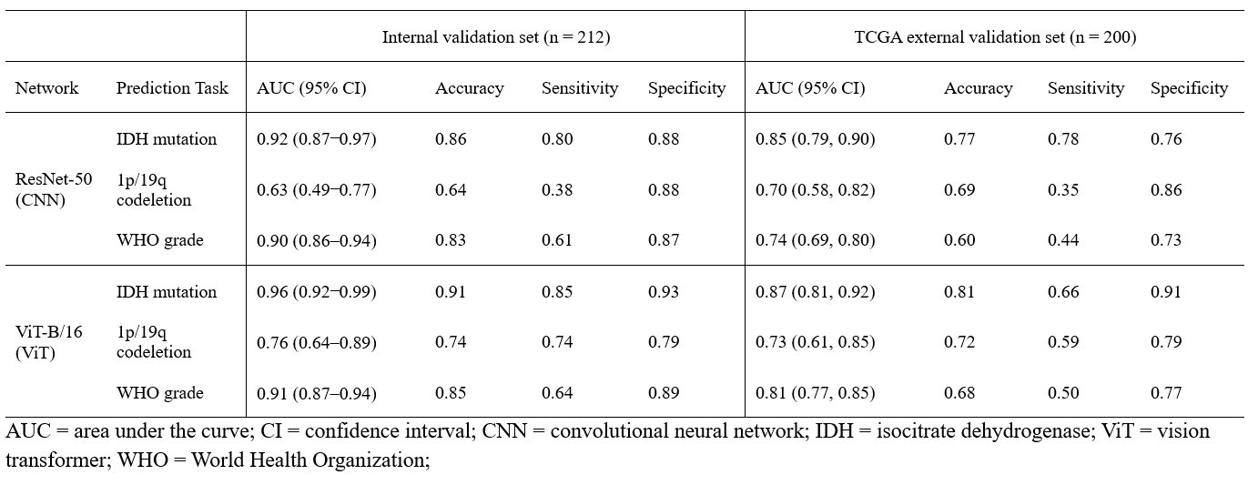
Results: ViT outperformed CNN with AUCs of 0.87, 0.73, and 0.81 for prediction of IDH mutation, 1p/19q codeletion, and grading, respectively.
Impact: Our study demonstrates that Vision Transformer provides reliable and interpretable prediction of molecular subtype and grades in adult-type diffuse gliomas based on the 2021 WHO classification using multiparametric MRI data.
Introduction
Predicting the molecular subtype and grade of adult-type diffuse gliomas noninvasively holds significant potential for improving treatment planning and prognosis. The World Health Organization (WHO) 2021 classification1 for central nervous system tumors provides a more simplified categorization, differentiating it from the 2016 WHO classification2 that encompassed multiple entities. The updated standard categorizes the adult-type diffuse gliomas into three different types based on the Isocitrate Dehydrogenase (IDH) mutation and 1p/19q codeletion status: oligodendroglioma, IDH-mutant diffuse astrocytoma, and IDH-wildtype glioblastoma. Recent deep learning (DL) studies for predicting the molecular subtype and grade of gliomas have predominantly used convolutional neural networks (CNNs) and adhered to the WHO 2016 standard3-5. However, with advancements in computer vision, Vision Transformers (ViTs)6 have shown enhanced capabilities, especially in capturing long-range spatial dependencies compared to CNNs. Therefore, the objective of this study is to explore the potential efficacy of ViTs and develop a robust and interpretable model for molecular subtyping and grading of adult-type diffuse gliomas according to the WHO 2021 classification.Methods
Data and PreprocessingMultiparametric MRI (T1, T1C, T2, FLAIR) data of 1,053 patients with adult-type diffuse gliomas (144 oligodendrogliomas, 157 IDH-mutant astrocytomas, and 752 IDH-wildtype glioblastomas) according to the 2021 WHO classification were included in the institutional development set. We splited the institutional development set into train and validation sets at an 8:2 ratio. A total of 200 adult-type diffuse patients (29 oligodendrogliomas, 57 IDH-mutant astrocytomas, and 114 IDH-wildtype glioblastomas) were included as an external validation from The Cancer Genome Atlas (TCGA). Figure 1 shows the patient flowchart for the institutional development and TCGA external validation sets. In the image preprocessing stage, we applied isovoxel resampling to 1 mm³, performed N4 bias field correction, and coregistered T1, T2, and FLAIR images to T1C images using Advanced Normalization Tools (ANTs). We also performed skull stripping using HD-BET7 and normalized signal intensities to z-scores.
Model Architecture and Training
The framework designed for the prediction of molecular subtyping and grading of adult-type diffuse gliomas is illustrated in Figure 2. We used ResNet-508 for CNN-based model, and ViT-B/166 for ViT-based model. Both models were initialized with weights from networks pretrained on ImageNet9 dataset and trained respectively to predict the IDH mutation status, 1p/19q codeletion status, and grade of the tumor.
Performance Evaluation and Interpretability
To evaluate the performance of molecular subtyping and grading of adult-type diffuse gliomas, we used the area under the curve (AUC), accuracy, sensitivity, and specificity. For the interpretation and analysis of the DL features contributing to prediction of IDH mutation status of adult-type diffuse gliomas, we used GradCAM10 for CNN and attention visualization method11 for ViT.
Results
Classification PerformanceThe performance of DL models is summarized in Table 1. On internal validation, ViT showed superior performance with AUC scores of 0.96, 0.76, and 0.91 for the prediction of IDH mutation, 1p/19q codeletion, and tumor grading, respectively. The CNN, however, achieved lower AUC scores of 0.92 for IDH mutation, 0.63 for 1p/19q codeletion, and 0.90 for tumor grading. On external validation, ViT outperformed the CNN, with AUC scores of 0.87 for IDH mutation, 0.73 for 1p/19q codeletion, and 0.81 for grading, compared to the CNN's scores of 0.85 for IDH mutation, 0.70 for 1p/19q codeletion, and 0.74 for grading.
Interpretation of DL models
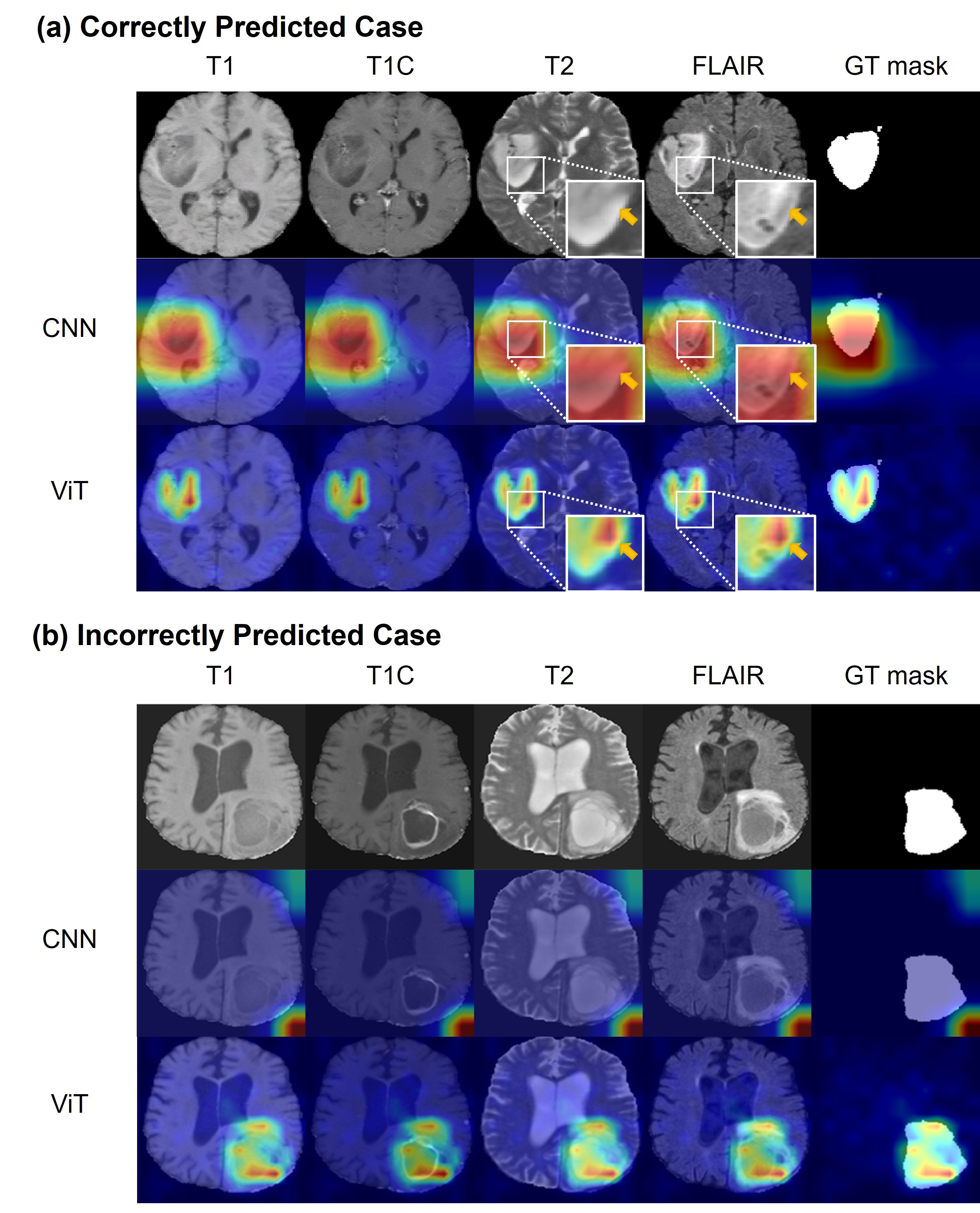
The interpretation of DL models is illustrated in Figure 3. Figure 3a shows the correctly predicted IDH-mutant astrocytoma, Central Nervous System (CNS) WHO grade 2. The attention map from ViT shows better activation in the tumor region compared to the CNN. Notably, the attention map from ViT highlights T2-FLAIR mismatch12 area of the tumor, which is a highly specific imaging signature for IDH-mutant astrocytomas. Figure 3b shows the incorrectly predicted case of IDH-mutant astrocytoma, CNS WHO grade 4. Despite the incorrect prediction, the attention map from ViT still reliably activates the tumor region, unlike the CNN’s map. However, ring enhancement and necrosis, representative characteristics of IDH-wildtype glioblastoma, can also be present in IDH-mutant astrocytomas of WHO CNS grade 4. Therefore, as shown in Figure 3b, classifying IDH mutation status using conventional imaging can be challenging, even though ViT highlights the tumor regions accurately.
Conclusion
This study shows that ViTs have outperformed CNNs in predicting the subtypes and grade of adult-type diffuse glioma based on the latest 2021 WHO classification. Consequently, future research should be conducted using advanced models and adhere to the newest standards for practical clinical application.Acknowledgements
This research was supported by Basic Science Research Program through the National Research Foundation of Korea funded by the Ministry of Science and ICT (2021R1A4A1031437, 2022R1A2C2008983), Artificial Intelligence Graduate School Program at Yonsei University [No. 2020-0-01361], the KIST Institutional Program (Project No.2E32271-23-078), and partially supported by the Yonsei Signature Research Cluster Program of 2023 (2023-22-0008).References
1. Louis DN, Perry A, Wesseling P, et al. The 2021 WHO Classification of Tumors of the Central Nervous System: a summary. Neuro Oncol. 2021; 23(8):1231-1251.
2. Louis DN, Perry A, Reifenberger G, et al. The 2016 World Health Organization Classification of Tumors of the Central Nervous System: a summary. Acta Neuropathol. 2016; 131(6):803-820.
3. Choi, Yoon Seong, et al. "Fully automated hybrid approach to predict the IDH mutation status of gliomas via deep learning and radiomics." Neuro-oncology 23.2 (2021): 304-313.
4. Cluceru, Julia, et al. "Improving the noninvasive classification of glioma genetic subtype with deep learning and diffusion-weighted imaging." Neuro-oncology 24.4 (2022): 639-652.
5. van der Voort, Sebastian R., et al. "Combined molecular subtyping, grading, and segmentation of glioma using multi-task deep learning." Neuro-oncology 25.2 (2023): 279-289.
6. Dosovitskiy, Alexey, et al. "An image is worth 16x16 words: Transformers for image recognition at scale." arXiv preprint arXiv:2010.11929 (2020).
7. Isensee F, Schell M, Pflueger I, et al. Automated brain extraction of multisequence MRI using artificial neural networks. Hum Brain Mapp. 2019; 40(17):4952-4964.
8. He, Kaiming, et al. "Deep residual learning for image recognition." Proceedings of the IEEE conference on computer vision and pattern recognition. 2016.
9. Deng, Jia, et al. "Imagenet: A large-scale hierarchical image database." 2009 IEEE conference on computer vision and pattern recognition. Ieee, 2009.
10. Selvaraju, Ramprasaath R., et al. "Grad-cam: Visual explanations from deep networks via gradient-based localization." Proceedings of the IEEE international conference on computer vision. 2017.
11. Chefer, Hila, Shir Gur, and Lior Wolf. "Transformer interpretability beyond attention visualization." Proceedings of the IEEE/CVF conference on computer vision and pattern recognition. 2021.
12. Patel, Sohil H., et al. "T2–FLAIR mismatch, an imaging biomarker for IDH and 1p/19q status in lower-grade gliomas: a TCGA/TCIA project." Clinical cancer research 23.20 (2017): 6078-6085.
Figures
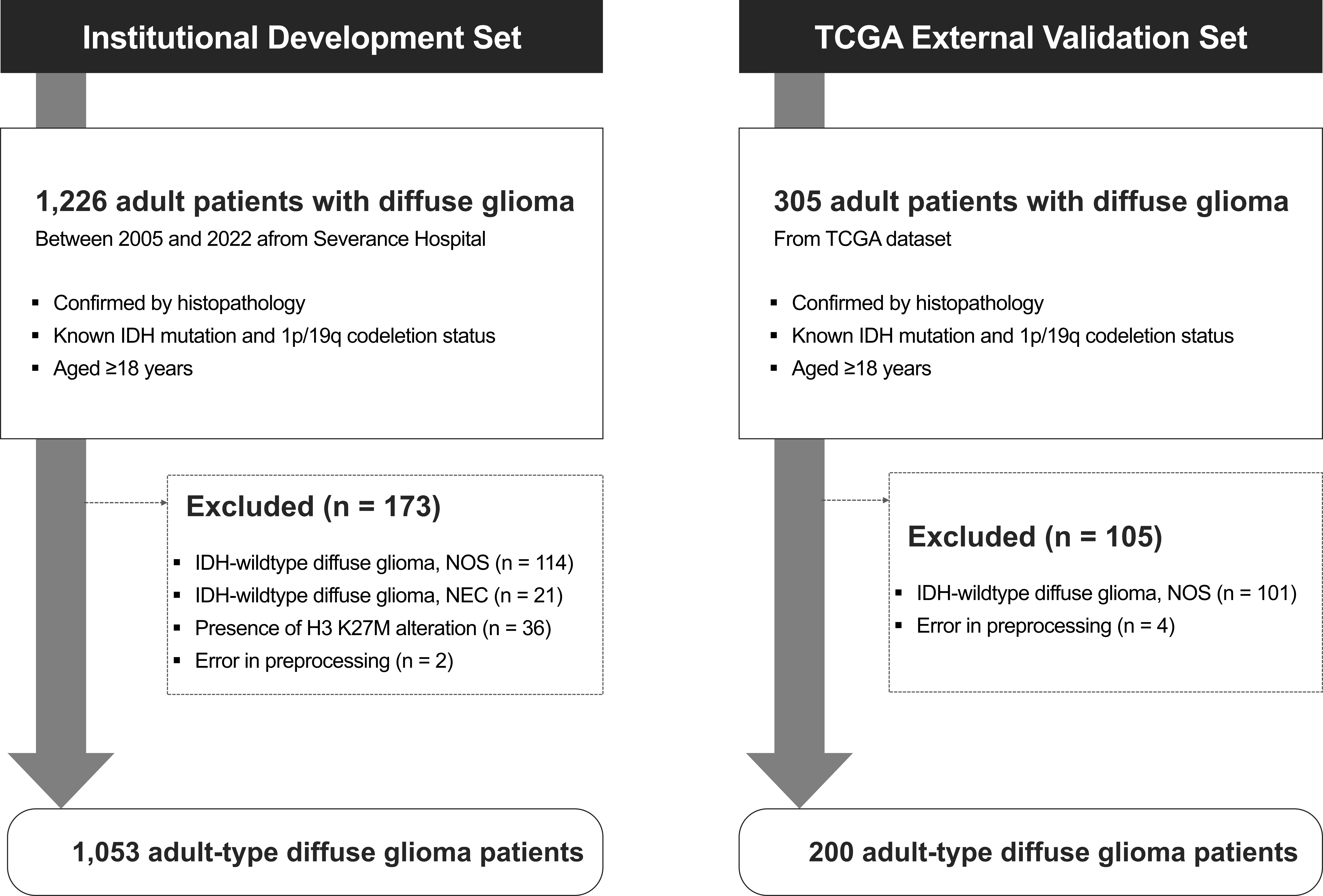
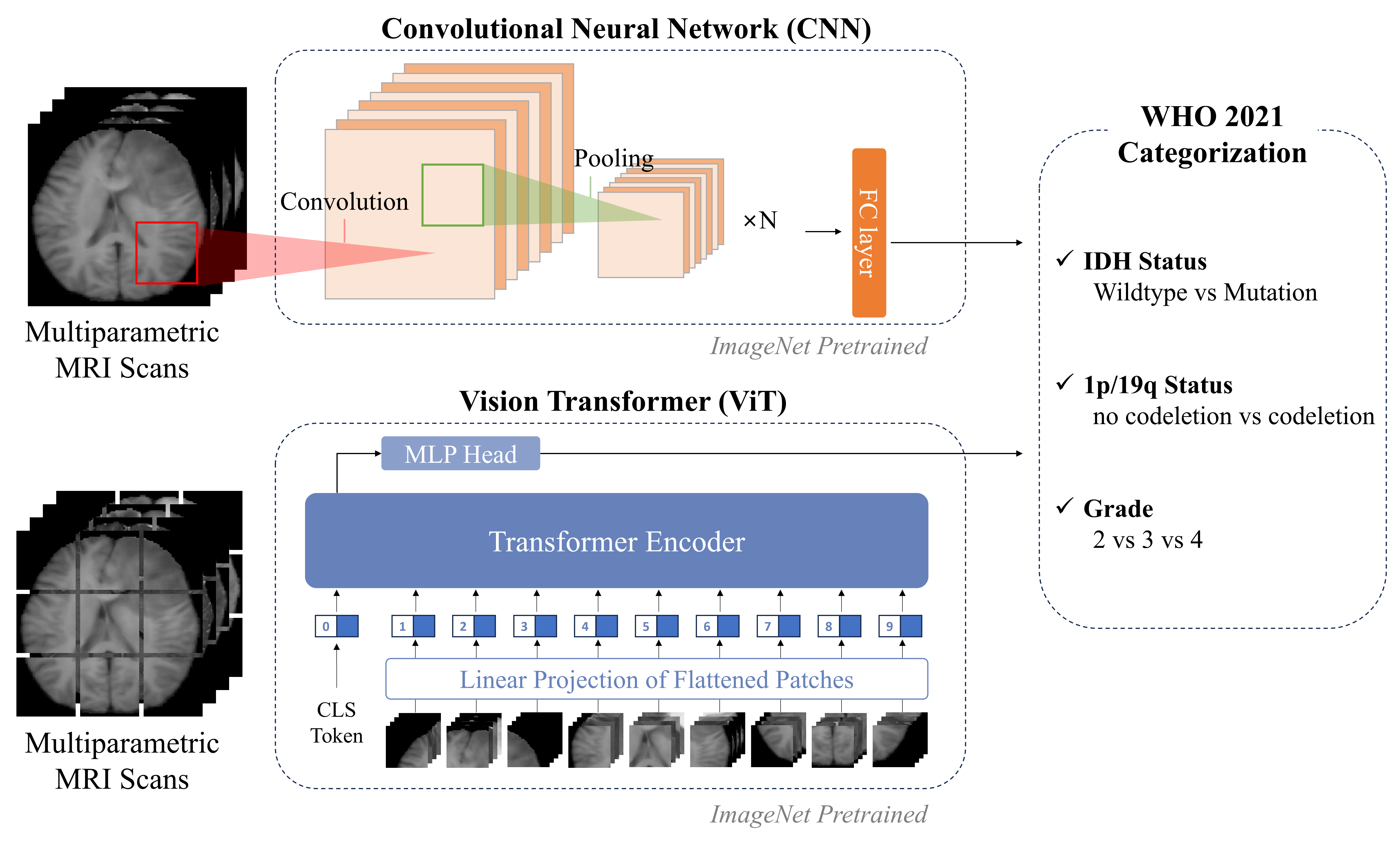
Figure 2. Overview of deep learning framework for molecular subtyping and grade classification of adult-type diffuse glioma using multiparametric MRI scans according to WHO 2021 categorization. The framework utilizes a Convolutional Neural Network (CNN), which processes images through convolution and pooling operations, and a Vision Transformer (ViT) that divides images into patches and employs a self-attention mechanism. Each network is trained to perform several classification tasks, including the prediction of IDH status, 1p/19q status, and WHO grade.