0394
Contrast-Optimized Basis Functions for Self-Navigated Motion Correction in 3D quantitative MRI1Bernard and Irene Schwartz Center for Biomedical Imaging, Department of Radiology, New York University Grossman School of Medicine, New York, NY, United States, 2Center for Advanced Imaging Innovation and Research (CAI2R), Department of Radiology, New York University Grossman School of Medicine, New York, NY, United States, 3Vilcek Institute of Graduate Biomedical Sciences, New York University Grossman School of Medicine, New York, NY, United States
Synopsis
Keywords: Motion Correction, Motion Correction, MR Fingerprinting, qMT, low rank
Motivation: Motion-induced artifacts are a significant barrier to achieving clinically acceptable image quality for multi-compartment quantitative MRI techniques, e.g. a 2-pool magnetization transfer model.
Goal(s): To develop a self-navigating approach to estimating motion parameters in an MR-fingerprinting-like acquisition.
Approach: We optimize a subspace that maximizes the contrast-to-noise ratio between brain parenchyma and cerebrospinal fluid for a low-resolution, time-segmented low-rank reconstruction used to estimate motion.
Results: Compared to the typical SVD basis, the contrast-optimized basis improves the smoothness of the motion estimates and the apparent resolution of the reconstructed coefficient images and quantitative maps.
Impact: The proposed retrospective, self-navigating motion correction technique does not require any sequence modifications and/or additional scan time. It can therefore be applied to many quantitative MRI techniques where the signal's variation over time can be well-described in a low-rank subspace.
Introduction
Motion-induced artifacts pose severe challenges to clinical MRI. This problem is amplified by long scan times, as are typical for quantitative techniques. Retrospective motion correction1,2,3,4,5 uses either navigator images or images derived from a subset of the actual data to estimate motion parameters. The latter approach, commonly referred to as self-navigation, has been proposed for MR-Fingerprinting flavored quantitative MRI2. In MR-fingerprinting, similar to many other quantitative MRI approaches like inversion-recovery or multi-echo spin echo, the signal varies over time, and Kurzawski et al.2 reconstructed images from these dynamics in an SVD-based subspace and used the first coefficient image for motion estimation. While the SVD promotes high signal intensity in the first coefficient, it does not maximize contrast between different tissue types, which can impede the motion estimation. We build on Kurzawski's approach and propose to utilize a generalized eigendecomposition to explicitly promote a high contrast-to-noise ratio between different tissue types.Methods
Inspired by the formalism introduced for region optimized virtual coils6, we utilize a generalized eigendecomposition to find a basis that maximizes the signal of one set of fingerprints while minimizing the signal from another set. Here, we simulated the signal typical for brain parenchyma, i.e., white and gray matter, for the first set and cerebrospinal fluid (CSF)-typical signal for the other.The proposed approach entails a two-step procedure for generating the desired basis. First, we pooled all simulated fingerprints and performed a truncated SVD to the first 3 basis functions, thus deriving a sub-space that covers most of the signal intensity and minimizes artifacts in the reconstructed images that would otherwise arise from unmodeled signal. Second, we rotated this sub-space to maximize the contrast-to-noise ratio between brain parenchyma and CSF in the first coefficient image.
To this end, we calculated the generalized eigenvalue decomposition of the mean auto-correlation matrices corresponding to each set of simulated signals. The rotated sub-space maximizes the parenchyma and minimizes the CSF signal in the first coefficient, while the last coefficient has the opposite properties. Last, we enforced orthogonality of the three basis functions with the Gram-Schmidt process while leaving the first basis function unchanged.
We tested our motion correction approach with a 3D hybrid-state sequence7, which was optimized for quantitative magnetization transfer imaging8. The sequence utilizes a 3D radial koosh-ball readout with golden-angle increments9,10, has a nominal resolution of 1mm isotropic, and a total scan time of 12 mins. It entails a 4-second long pattern of variable flip angles that are repeatedly acquired with different k-space spokes. We aggregate all spokes from one 4-second cycle to reconstruct a low-resolution image ($$$4\times 4\times 4\,\mathrm{mm}$$$) directly in the space of the contrast-optimized basis or, for comparison, the original SVD11,12. To reduce undersampling artifacts, we penalized the total variation (TV) along time.
In vivo experiments were performed with a 3T Siemens Prisma scanner after obtaining informed consent in accordance with our IRB. The volunteer was not instructed to move, but large unintentional movements occurred in the second half of the scan.
Results
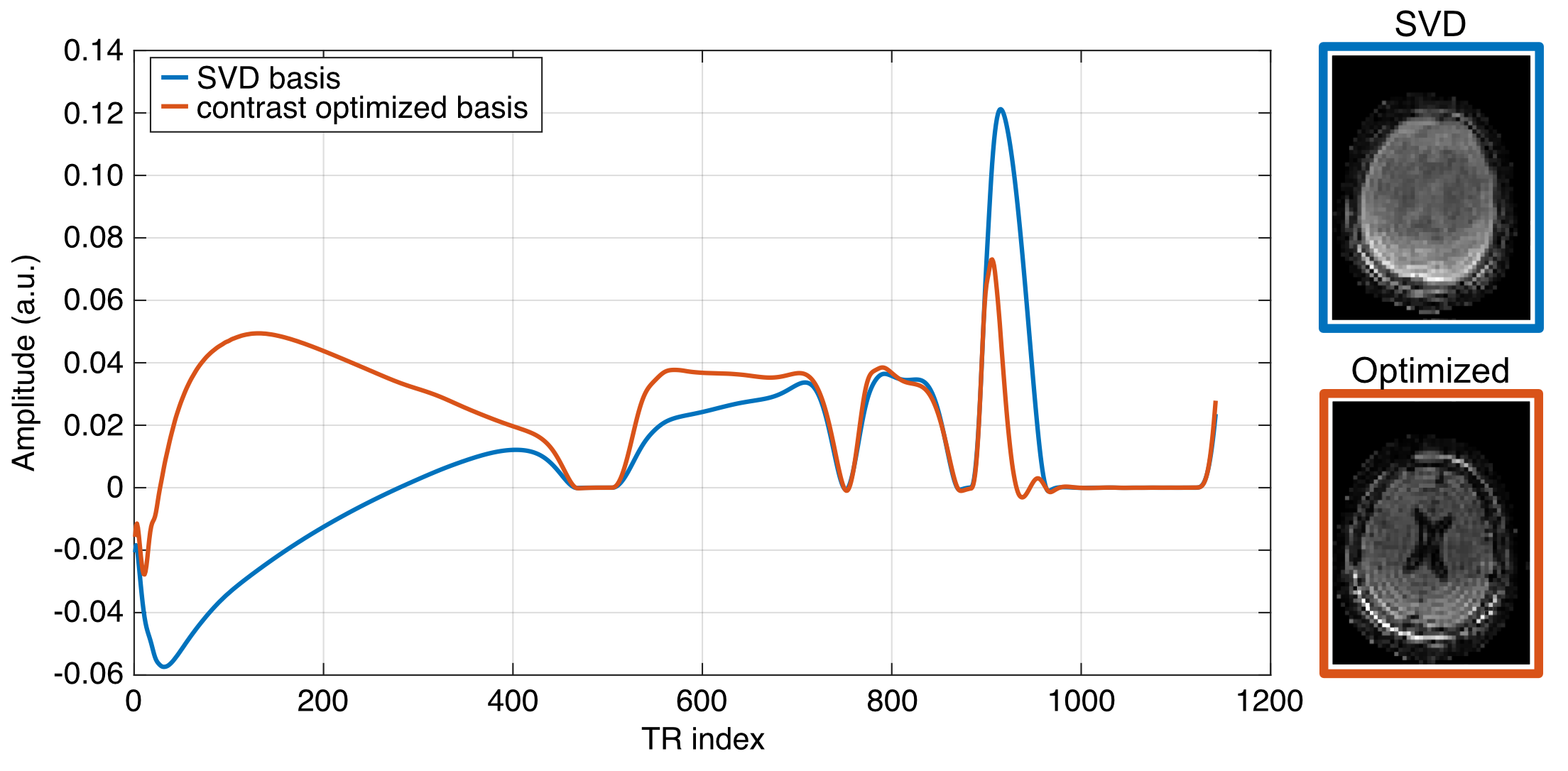
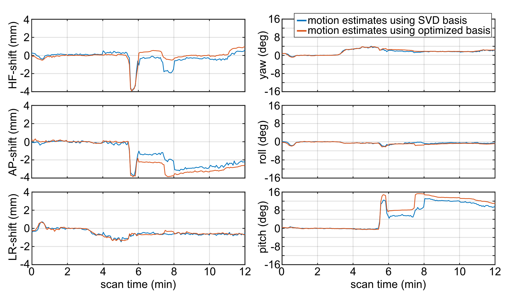
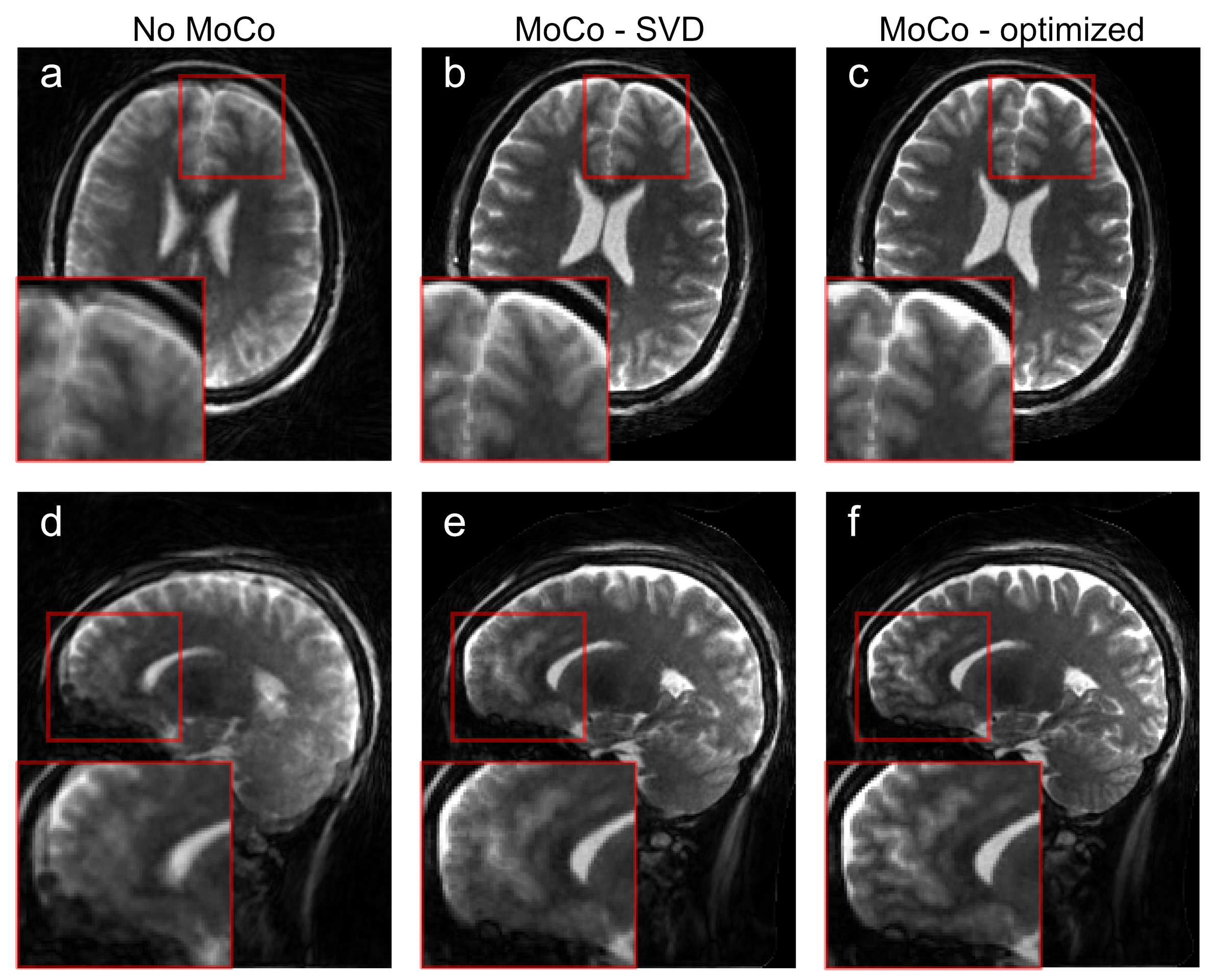
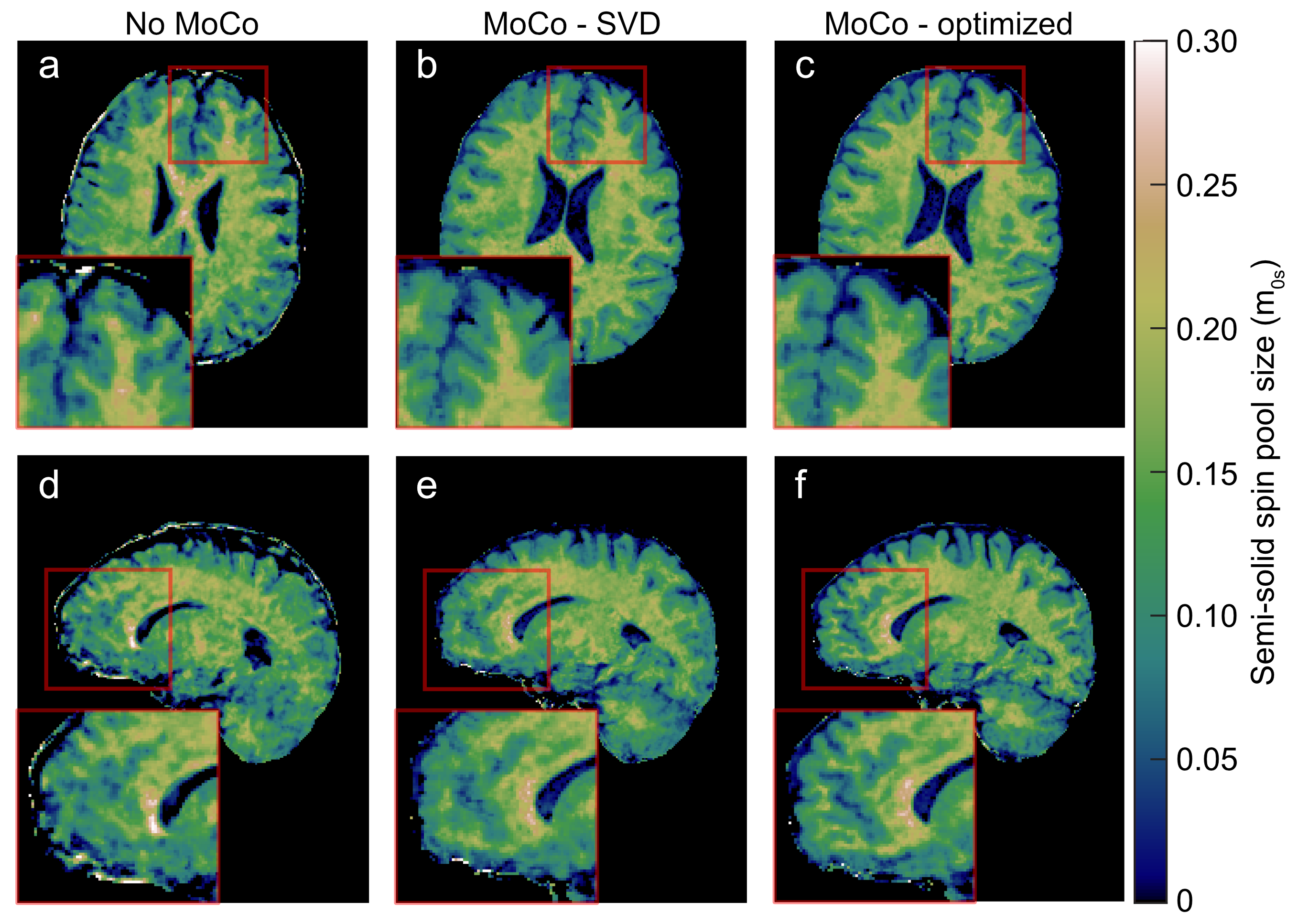
Fig. 1 shows the first basis function of the original SVD and the proposed contrast-optimized basis alongside the corresponding low-resolution coefficient images used for motion estimation. The contrast-optimized basis greatly improves the parenchyma-CSF contrast. Fig. 2 illustrates that this increased contrast results in smoother motion estimates and Figs. 3, 4 demonstrate that this translates to improved image quality in the motion-corrected reconstruction and, ultimately, in the quantitative maps. Reduced artifacts and improved apparent resolution is observed in both the coefficient images (Fig. 3) and quantitative maps (Fig. 4).Discussion
The proposed approach to design bases for self-navigated motion correction shows great efficacy in enhancing the precision of the motion estimates. The approach is tailored to transient-state quantitative MRI approaches, e.g., inversion-recovery, multi-echo spin echo, or MR-Fingerprinting17, in which the same dynamics are acquired repeatedly while filling k-space. Key ingredients for successful implementation are 3D imaging to avoid through-slice motion and a k-space trajectory with sufficient coverage in each cycle of the spin dynamics for a time-segmented reconstruction. Here, we used a koosh-ball trajectory with golden-angle increments---which repeatedly samples the center of k-space---combined with a TV-regularized low-resolution reconstruction. We tested the proposed method in three scans with varying degrees of motion and showed only the most extreme case, though the motion-corrected image quality was comparable between all three scans. Future work will involve the evaluation of the approach in a larger cohort.Acknowledgements
This work was performed under the rubric of the Center for Advanced Imaging Innovation and Research (CAI2R, www.cai2r.net), an NIBIB National Center for Biomedical Imaging and Bioengineering (NIH P41 EB017183). A.M. receives support from NIH grants F30 AG077794 and T32 GM136573.References
[1] Gastão Cruz, Olivier Jaubert, Torben Schneider, Rene M. Botnar, and Claudia Prieto. Rigid motion-corrected magnetic resonance fingerprinting. Magnetic Resonance in Medicine, 2019.
[2] Jan W. Kurzawski, Matteo Cencini, Luca Peretti, Pedro A. Gómez, Rolf F. Schulte, Graziella Donatelli, Mirco Cosottini, Paolo Cecchi, Mauro Costagli, Alessandra Retico, Michela Tosetti, and Guido Buonincontri. Retrospective rigid motion correction of three-dimensional magnetic resonance fingerprinting of the human brain. Magnetic Resonance in Medicine, 2020.
[3] Zhongbiao Xu, Huihui Ye, Mengye Lyu, Hongjian He, Jianhui Zhong, Yingjie Mei, Z. Chen, Ed. X. Wu, Wufan Chen, Qianjin Feng, and Yanqiu Feng. Rigid motion correction for magnetic resonance fingerprinting with sliding-window reconstruction and image registration. Magnetic resonance imaging, 2019.
[4] Zidan Yu, Tiejun Zhao, Jakob Assländer, Riccardo Lattanzi, Daniel K.Sodickson, and Martijn A. Cloos. Exploring the sensitivity of magnetic resonance fingerprinting to motion. Magnetic Resonance Imaging, 2018.
[5] Siyuan Hu, Yong Chen, Xiaopeng Zong, Weili Lin, Mark Griswold, and Dan Ma. Improving motion robustness of 3d mr fingerprinting with a fat navigator. Magnetic Resonance in Medicine, 2023.
[6] Daeun Kim, Stephen F. Cauley, Krishna S. Nayak, Richard M. Leahy, and Justin P. Haldar. Region-optimized virtual (rovir) coils: Localization and/or suppression of spatial regions using sensor-domain beamforming. Magnetic Resonance in Medicine, 2021.
[7] Jakob Assländer, Dmitry S. Novikov, Riccardo Lattanzi, Daniel K.Sodickson, and Martijn A. Cloos. Hybrid-state free precession in nuclear magnetic resonance. Communications Physics, 2019.
[8] Jakob Assländer, Andrew Mao, Erin S Beck, Francesco La Rosa, Robert W Charlson, Timothy M Shepherd, and Sebastian Flassbeck. On multi-path longitudinal spin relaxation in brain tissue, 2023.
[9] Stefanie Winkelmann, Tobias Schaeffter, Thomas Koehler, Holger Eggers,and Olaf Doessel. An Optimal Radial Profile Order Based on the Golden Ratio for Time-Resolved MRI. IEEE Trans. Med. Imaging, 2007.
[10] Philipp Ehses, Nicole Seiberlich, Dan Ma, Felix A. Breuer, Peter M. Jakob, Mark A. Griswold, and Vikas Gulani. IR TrueFISP with a golden-ratio-based radial readout: Fast quantification of T1, T2, and proton density. Magn. Reson. Med., 2013.
[11] Jonathan I. Tamir, Martin Uecker, Weitian Chen, Peng Lai, Marcus T.Alley, Shreyas S. Vasanawala, and Michael Lustig. T 2 shuffling: Sharp, multicontrast, volumetric fast spin-echo imaging. Magnetic Resonance in Medicine, 2017.
[12] Jakob Assländer, Martijn A Cloos, Florian Knoll, Daniel K Sodickson,Jürgen Hennig, and Riccardo Lattanzi. Low rank alternating direction method of multipliers reconstruction for MR fingerprinting. Magnetic Resonance in Medicine, 2018.
[13] UCL Queen Square Institute of Neurology Functional Imaging Laboratory. Spm 12 - https://www.fil.ion.ucl.ac.uk/spm/software/spm12/, 2020.
[14] Andrew Hoopes, Jocelyn S. Mora, Adrian V. Dalca, Bruce Fischl, and Malte Hoffmann. Synthstrip: skull-stripping for any brain image. NeuroImage, 2022.
[15] Andrew Mao, Sebastian Flassbeck, Cem Gultekin, and Jakob Assländer.Cramér-Rao bound optimized temporal subspace reconstruction in quantitative mri, 2023.
[16] Xiaoxia Zhang, Quentin Duchemin, Kangning Liu, Cem Gultekin,Sebastian Flassbeck, Carlos Fernandez-Granda, and Jakob Assländer.Cramér–Rao bound-informed training of neural networks for quantitative MRI. Magnetic Resonance in Medicine, 2022.
[17] Dan Ma, Vikas Gulani, Nicole Seiberlich, Kecheng Liu, Jeffrey L. Sunshine, Jeffrey L. Duerk & Mark A. Griswold, Magnetic resonance fingerprinting, Nature, 2013
Figures