0393
Real-time motion correction and multicoil shim array B0 update for whole-brain MR spectroscopic imaging1Radiology, Martinos Center for Biomedical Imaging, Harvard Medical School, Massachusetts General Hospital, Boston, MA, United States, 2Electrical Engineering and Computer Science, Massachusetts Institute of Technology, Cambridge, MA, United States, 3Siemens Medical Solutions, Boston, MA, United States
Synopsis
Keywords: Motion Correction, Motion Correction, Real-time shimming, Multicoil shim array, Metabolic Imaging
Motivation: Very high quality of MR spectroscopic imaging (MRSI) data is needed for robust and reproducible metabolite quantification. This critically depends on the B0 shimming and scan stability. Integrated RF-receive/B0-shim arrays significantly improve spectral quality.
Goal(s): Real-time motion correction and multicoil shimming update with an integrated RF-receive/B0-shim array for robust whole-brain MRSI.
Approach: We developed a rapid navigator for head tracking and B0 fieldmapping in combination with rapid processing for real-time update of multicoil shim currents and MRSI localization.
Results: Real-time motion correction and multicoil shimming provides significantly narrower linewidth, higher signal-to-noise, reduced quantification errors and reproducible metabolic imaging.
Impact: Whole-brain MRSI is a unique method for non-invasive mapping of brain neurochemistry, and in combination with real-time motion correction and multicoil shim array update provides robust and reproducible quantitative metabolic imaging for clinical use.
INTRODUCTION
Magnetic resonance spectroscopic imaging (MRSI) allows non-invasive mapping of in-vivo metabolism [1]. High resolution whole-brain MRSI has great value to investigate healthy brain and disease pathology [2] but its performance is severely limited when spectral resolution and signal-to-noise is not adequate to resolve overlapping metabolite peaks. Low MRSI data quality due to motion artifacts and suboptimal B0 shimming hamper metabolite quantification [3]. Integrated RF-receive/B0-shim arrays (AC/DC) provide ultra-fast switchable high-order shimming to improve B0 field homogeneity [4,5]. However, real-time update of AC/DC multicoil shim currents is needed for preserving B0 field homogeneity in the presence of head motion to maintain consistently high MRSI quality, which is the goal and newly demonstrated in this work.METHODS
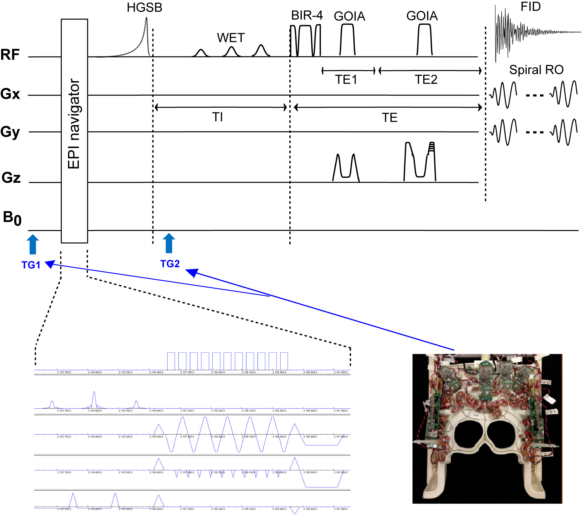
Pulse sequence:Whole-brain 3D MRSI pulse sequence using adiabatic spin echo and spiral spatial-spectral encoding was interleaved with an accelerated dual-echo EPI volume navigator (vNav) for real-t×ime motion correction and shimming [5]. The measurements were performed on a 3T whole-body MRI system (MAGNETOM Prisma, Siemens Healthcare, Erlangen, Germany) with a 32-channels AC/DC coil. 3D MRSI was acquired in 4:08 min with TR = 1.8 s, TE = 97 ms, FOV = 220×220×73 mm3, matrix 30×30×10, isotropic voxel 7.3 mm3, spectral window 1350 Hz, and 5ms×20kHz GOIA pulses for slab selection. The dual-echo EPI volume navigator was accelerated 8x with GRAPPA parallel imaging and acquired in 378 ms, using TR = 12 ms, TE1/TE2 = 3.9/6.3 ms, FOV = 240×240×150 mm3, matrix 48×48×30, isotropic voxel 5 mm3, flip angle 2°. Details of the pulse sequence are presented in Figure 1.
Real-time motion correction and multicoil shimming update:
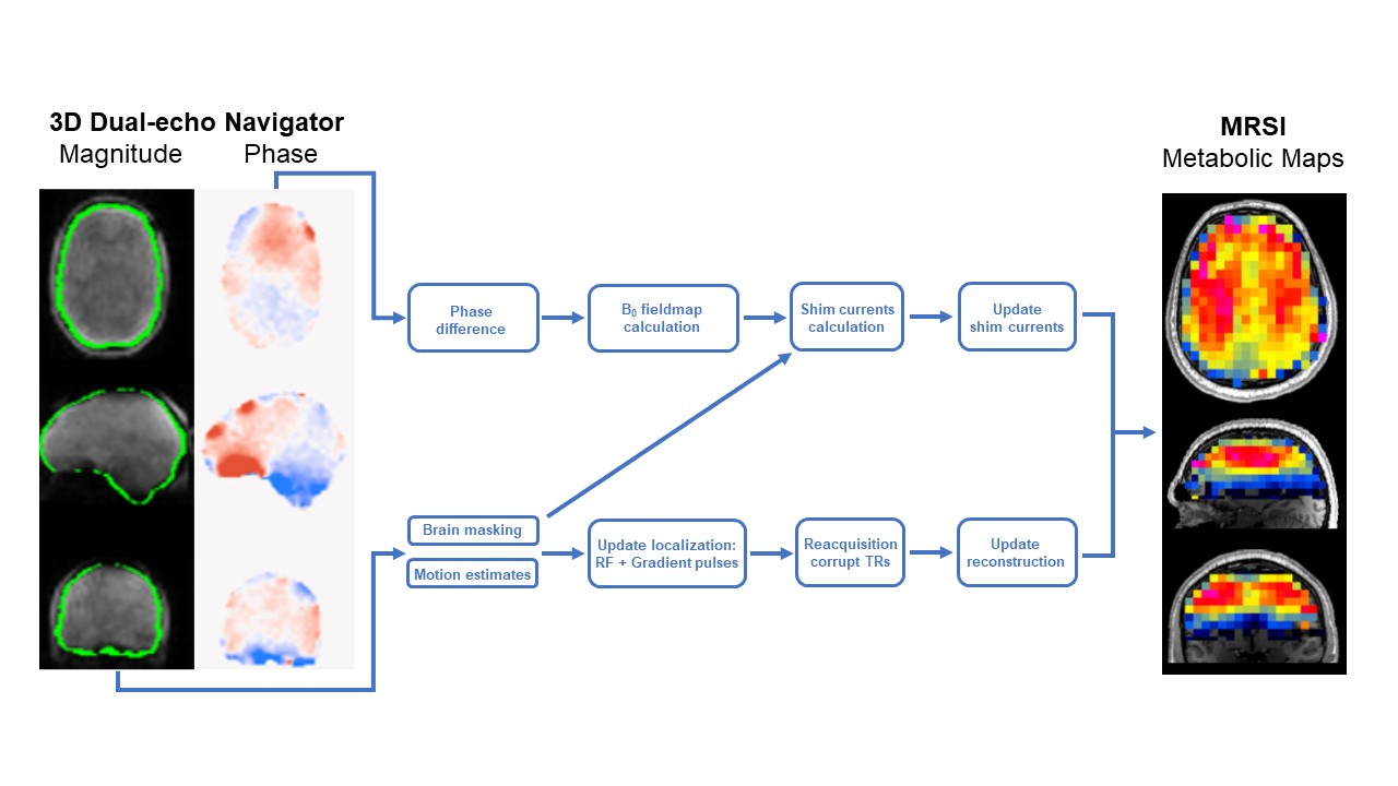
For each TR, pose changes are computed online by co-registering the magnitude image of the latest vNav to the initial vNav, using PACE (Prospective Acquisition Correction [6]), and this was used to update all imaging gradients and RF pulses, and the brain mask [7] used for shimming. B0 field maps were obtained from difference phase images by PRELUDE (Phase Region Expanding Labeller for Unwrapping Discrete Estimates [8]). The shim currents for ACDC were calculated [9,10] over the brain mask by linearly constrained quadratic optimization problem, solved with OSQP [11] which took less than 100 ms each TR. The total processing time for all the steps needed for localization and shim update was 600 ms which was inserted between the end of vNav and the start of MRSI. Details of the pipeline are presented in Figure 2.
Human experiments:
Four healthy volunteers were scanned with informed consent. Each volunteer had five MRSI measurements: 1) resting with second order spherical harmonics shimming (Rest 2SH) using scanner hardware, 2) resting with AC/DC shimming (Rest ACDC), 3) head motion with real-time motion correction and AC/DC shim update (Motion RT-ACDC), 4) head motion with real-time motion correction but no AC/DC shim update (Motion ST-ACDC), 5) head motion with no correction (Motion NoCo). For head-motion experiments subjects were instructed to consistently reproduce in each measurement the right-left and nodding movements that are typical in clinically scans.
RESULTS
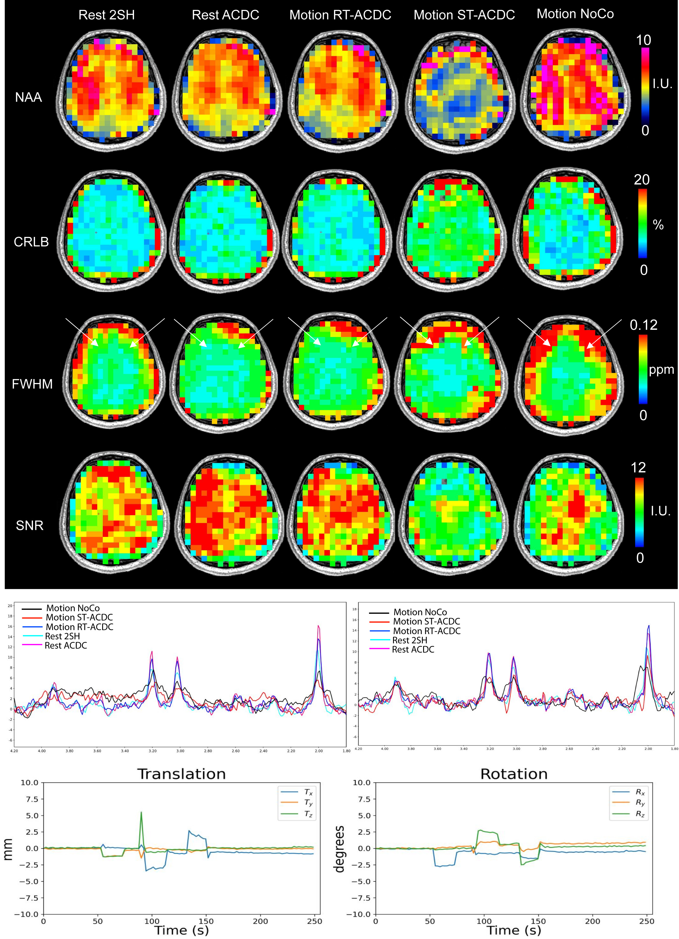
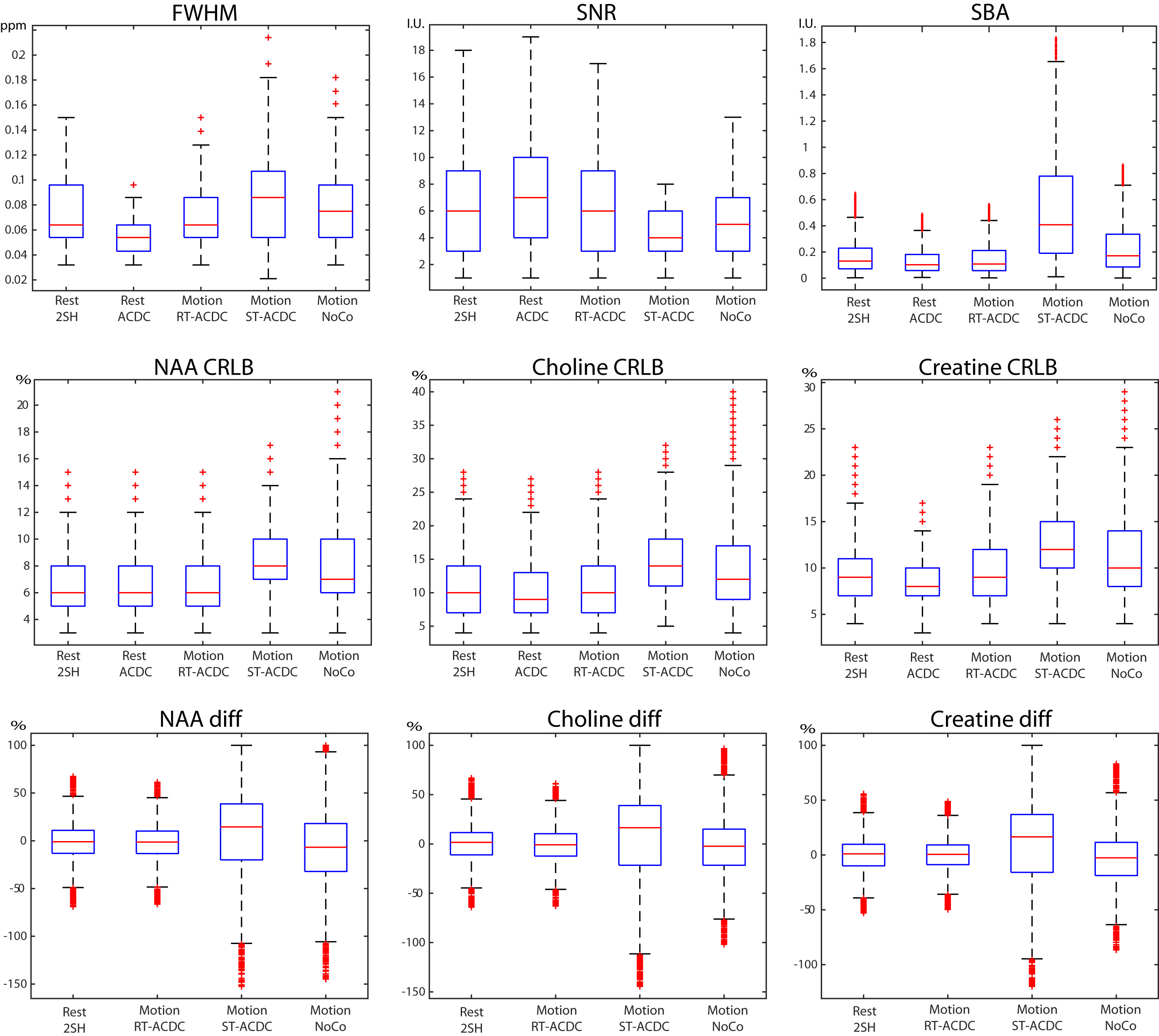
Figure 3 compares MRSI results of the five measurements obtained from a healthy volunteer. Maps of metabolic concentration, spectral linewidth, signal-to-noise and fitting error (CRLB) show that data obtained during motion with full corrections (Motion RT-ACDC) is comparable with data obtained under resting (Rest 2SH and Rest ACDC) conditions. By contrast, data obtained during motion with no correction (Motion ST-ACDC and Motion NoCo) are significantly worse than resting scans. Examples of the spectra for all scans show that spectral quality under motion is maintained for Motion RT-ACDC, but is heavily degraded for Motion ST-ACDC and Motion NoCo.Figure 4 quantitatively compares the spectral quality and reproducibility of metabolite quantification in all four subjects across the five measurements. The spectral quality and reproducibility of Motion RT-ACDC is similar to the resting scans and substantially improves over Motion ST-ACDC and Motion NoCo. In particular, the reproducibility of metabolite quantification of the last two scans has a large ±100% variability.
CONCLUSIONS
We present a rapid navigator framework which is compatible with real-time motion correction and multicoil AC/DC shim array update to improve the quality of MRSI and the reproducibility of quantitative metabolic imaging. It is expected that this methodology will improve the robustness of MRSI in clinical routine and augment its clinical utility.Acknowledgements
Dr. Aaron Hess and Dr. Dylan Tisdall for developing parts of the navigator software for real-time motion and shim correction. Dr Wolfgang Bogner, Dr Bernhard Strasser and Dr Borjan Gagoski for developing parts of the MRSI software for real-time motion and shim correction. Funding from the NIH grants R01CA255479, 2R01CA211080-06A1, R21EB029641, R01AG079422, R01HD110152.References
1. Maudsley, A. A. et al (2020). "Advanced magnetic resonance spectroscopic neuroimaging: Experts' consensus recommendations." NMR Biomed Special Issue: Advanced methodology for in vivo magnetic resonance spectroscopy, 34(5):e4309.
2. Oz, G. et al (2014). "Clinical proton MR spectroscopy in central nervous system disorders." Radiology. 270(3): 658-679.
3. Hess, A. T., et al (2012). "Real-time Motion and B0 correction for LASER MRSI using EPI volumetric navigators." NMR Biomedicine 25(2): 347-358.
4. Stockmann, J.P. et al. (2016) A 32-channel combined rf and b0 shim array for 3t brain imaging. Magnetic resonance in medicine, 75(1):441–451.
5. Strasser, B. et al (2022). "Improving D-2-hydroxyglutarate MR spectroscopic imaging in mutant isocitrate dehydrogenase glioma patients with multiplexed RF-receive/B0-shim array coils at 3 T.” NMR in Biomedicine, 35(1):e4621.
6. Thesen, S. et al (2000). “Prospective acquisition correction for head motion with image-based tracking for real-time fMRI.” Magn Reson Med 44, 457-465.
7. Hoffmann M., et al (2021). “Rapid head-pose detection for automated slice prescription of fetal-brain MRI.“ International Journal of Imaging Systems and Technology. 2021 Sep;31(3):1136-1154.
8. Jenkinson, M. (2003). “Fast, automated, N-dimensional phase-unwrapping algorithm. Magn Reson Med 49, 193-197.”
9. Arango, N.S. et al (2022), “Real-time Motion Compensated ∆B0 Shimming with an AC/DC Shim Coil and Dual-Echo vNavs“, ISMRM Meeting, London, 7-12 May 2022, #856.
10. Arango, N.S. et al (2023), “Motion-Compensated Slice-by-Slice ∆B0 Shimming with an AC/DC Shim Coil and Dual-Echo vNavs“, ISMRM Meeting, Toronto, 3-8 June 2023, #1012.
11. Stellato B., et al. (2020). “OSQP: an operator splitting solver for quadratic programs.” Mathematical Programming Computation, 12(4):637–672.
Figures