0391
PHIMO: Physics-Informed Motion Correction of GRE MRI for T2* Quantification1Institute of Machine Learning in Biomedical Imaging, Helmholtz Munich, Munich, Germany, 2School of Computation, Information and Technology, Technical University of Munich, Munich, Germany, 3School of Medicine and Health, Department of Diagnostic and Interventional Neuroradiology, Technical University of Munich, Munich, Germany, 4School of Medicine and Health, TUM-Neuroimaging Center, Technical University of Munich, Munich, Germany, 5Philips GmbH Market DACH, Hamburg, Germany, 6School of Medicine and Health, Clinic for Neurology, Technical University of Munich, Munich, Germany, 7Biomedical Engineering Department, School of Biomedical Imaging and Imaging Sciences, King’s College London, London, United Kingdom
Synopsis
Keywords: Motion Correction, Quantitative Imaging, Motion Correction, Deep Learning, Brain
Motivation: T2* quantification from GRE-MRI is particularly impacted by subject motion due to its sensitivity to magnetic field inhomogeneities. The current multi-parametric quantitative BOLD motion correction method depends on additional k-space acquisition, extending overall acquisition times.
Goal(s): To develop a learning-based motion correction method tailored to T2* quantification that avoids redundant data acquisition.
Approach: PHIMO leverages multi-echo T2* decay information to identify motion-corrupted k-space lines and excludes them from a data-consistent deep learning reconstruction.
Results: We are able to correct motion artifacts in subjects with stronger motion, approaching the performance of the current motion correction method, while substantially reducing the acquisition time.
Impact: PHIMO reduces strong motion artifacts in T2* maps by utilizing T2* decay information in an unrolled DL reconstruction. PHIMO avoids redundant data acquisition compared to a current correction method and reduces the acquisition time by over 40%, facilitating clinical applicability.
Introduction
Patient motion remains a major challenge for MRI. T2* quantification from gradient echo (GRE) MRI is particularly sensitive to motion due to the influence of magnetic field inhomogeneities, especially for larger echo times.1 When acquired as part of the oxygenation-sensitive multi-parametric quantitative BOLD (mqBOLD) protocol,2 motion-related errors propagate into derived parameters, such as the susceptibility-related R2’ relaxation rate.3 The current mqBOLD motion correction (MoCo) method4 relies on a redundant k-space acquisition, significantly increasing acquisition time. Deep learning-based MoCo approaches for brain MRI have been proposed,5 but are mostly developed for high resolution (e.g. 3D-MPRAGE) acquisitions6,7 or - in the context of T2* quantification - do not incorporate data consistency.8 In this work, we propose PHIMO, a PHysics-Informed Motion cOrrection method, which utilizes information from the T2* decay to exclude motion-corrupted k-space lines from a data-consistent reconstruction.Methods
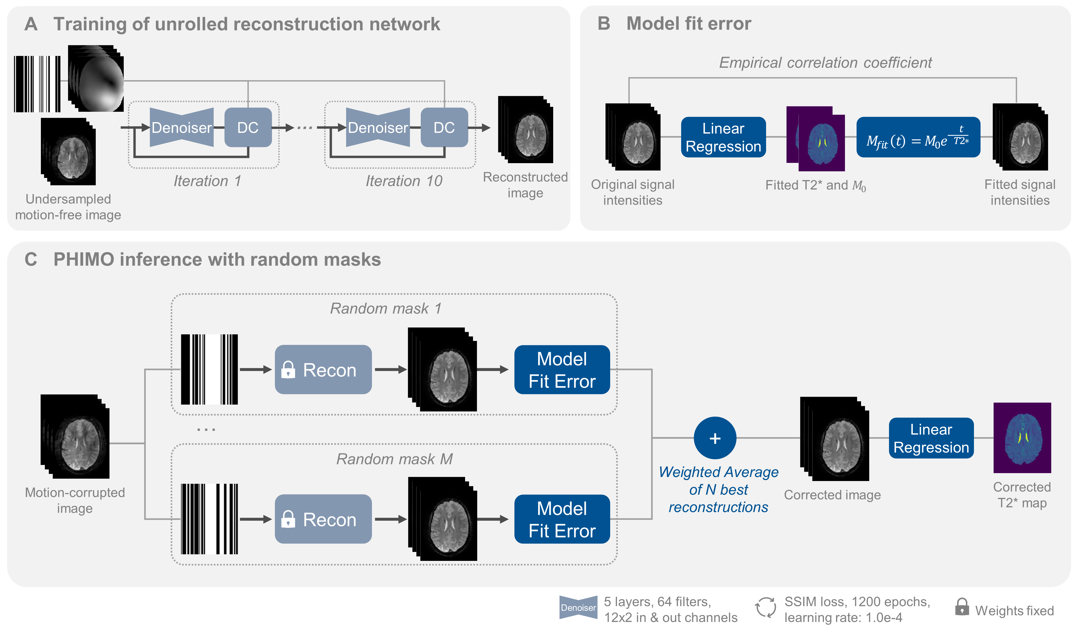
PHIMO is inspired by MoCo through bootstrap aggregation9 and consists of two steps (Figure 1). First, we train an unrolled reconstruction network10,11 on variable-density undersampled motion-free data. The reconstruction alternates 10 times between a CNN-based denoiser and a gradient descent layer, without weight sharing. Second, we apply M randomly generated masks to the motion-corrupted image, feed it through the fixed reconstruction network and combine the reconstructions - in contrast to Oh et al.9 not by simple averaging, but based on the model fit error. The latter is calculated as empirical correlation coefficient (ECC) between original signal intensities and “fitted” intensities after inserting the fitted parameters into the T2* decay model (Figure 1B). The result of PHIMO is a weighted average of the N reconstructions with the highest ECC. Below, we show the results for M=1000/N=20.We acquire multi-coil k-space data from 14 volunteers (26.7 ± 2.9y, 5 females) on a 3T Philips Elition X MR scanner (Philips Healthcare, Best, The Netherlands), using a multi-slice 2D GRE sequence (12 echoes, TE1=ΔTE=5 ms, TR=2300 ms, voxel size: 2×2×3mm3, 32-channel head coil). We perform repeated scans without and with voluntary head motion, where the subject was instructed to randomly move and, e.g. imitate sneezing or coughing. Additionally, we acquire half- and quarter-resolution data in both conditions to compare to the current mqBOLD MoCo4 (“HR/QR-MoCo”). The datasets are divided subject-wise into train, validation, and test sets (2/6/2 subjects), including only slices with more than 10% brain voxels, resulting in 193/61/189 train/validation/test slices.
For the evaluation, we manually exclude inferior slices to disentangle motion from severe susceptibility artifacts, which can be addressed separately by background field correction12 or high-resolution 3D acquisition.13 We register all images to the motion-free acquisition (in-plane). HR/QR-MoCo and segmentation of anatomical scans are performed in MATLAB (R2022b) and SPM12 with custom programs14. All other computations are performed in Python 3.8.12, using Pytorch and MERLIN15. For statistical testing we use Wilcoxon signed rank tests and False-Discovery Rate correction for multiple comparisons. The code is available at https://github.com/HannahEichhorn/PHIMO.
Results
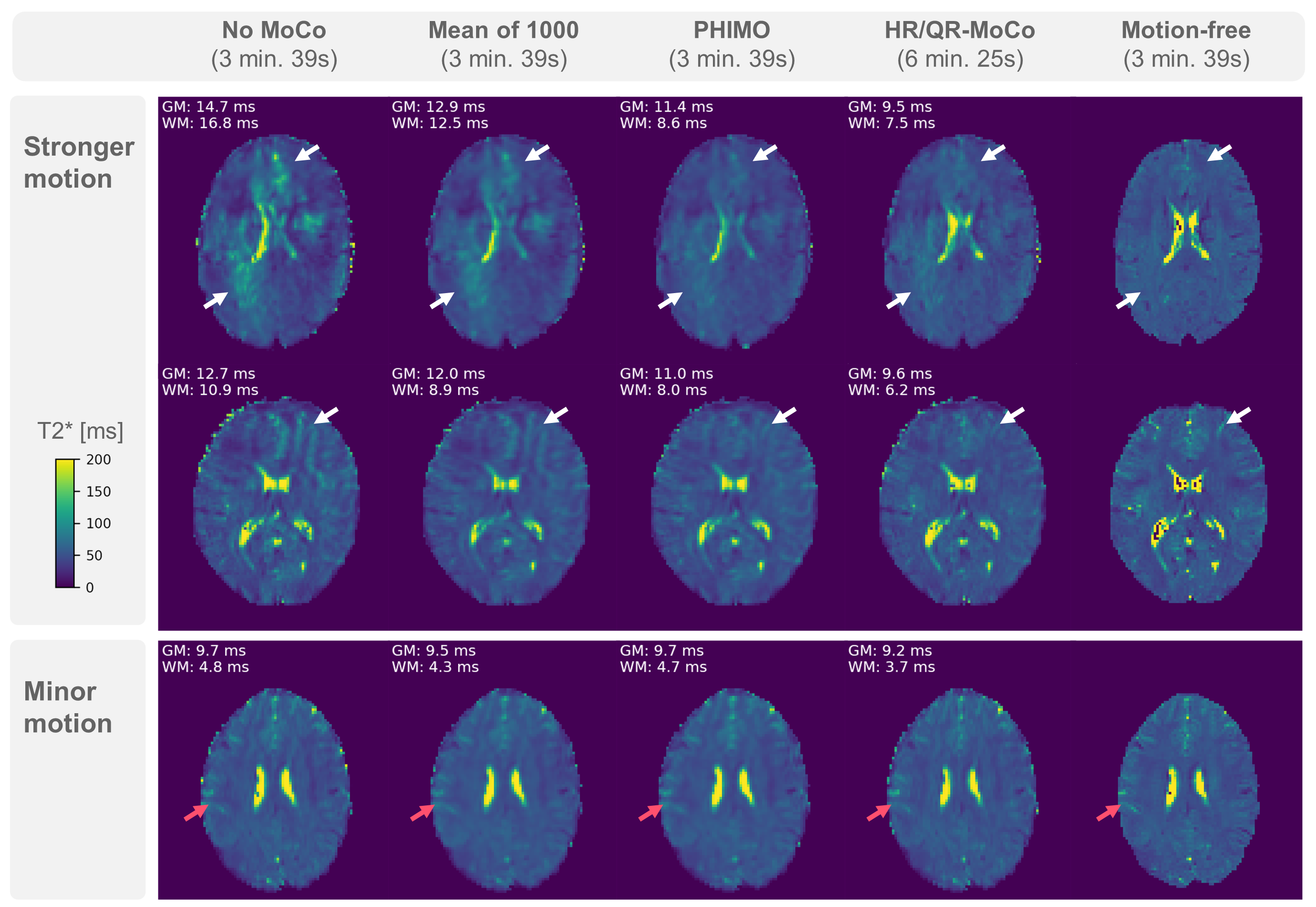
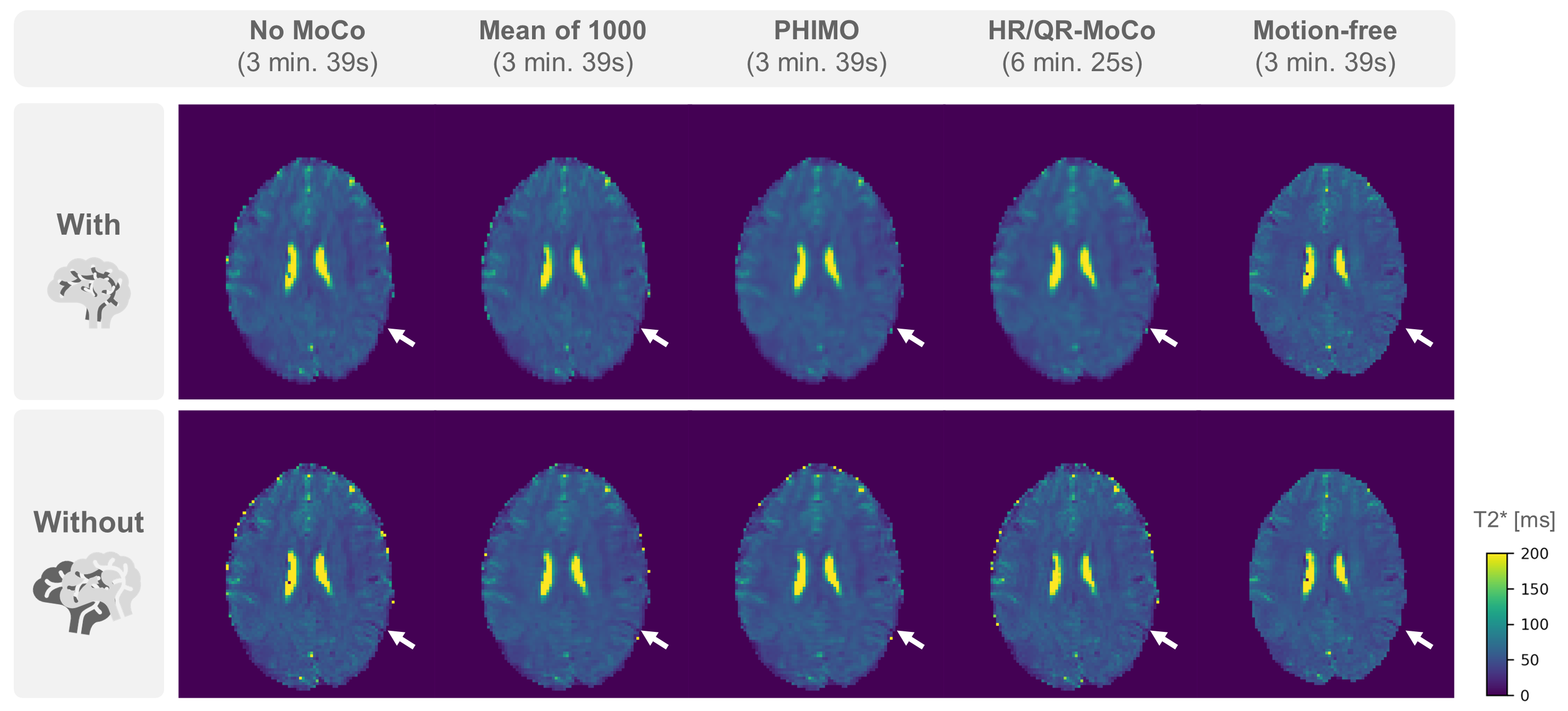
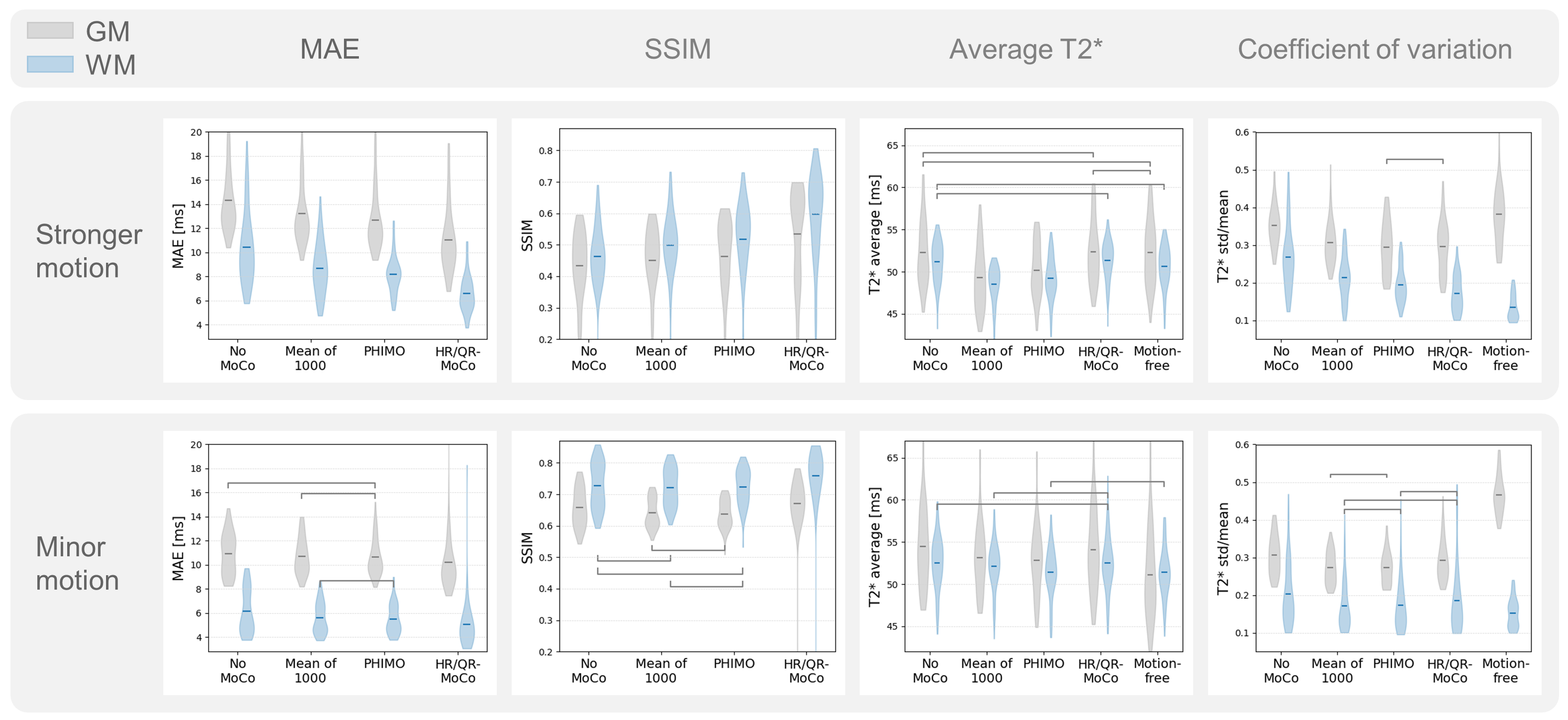
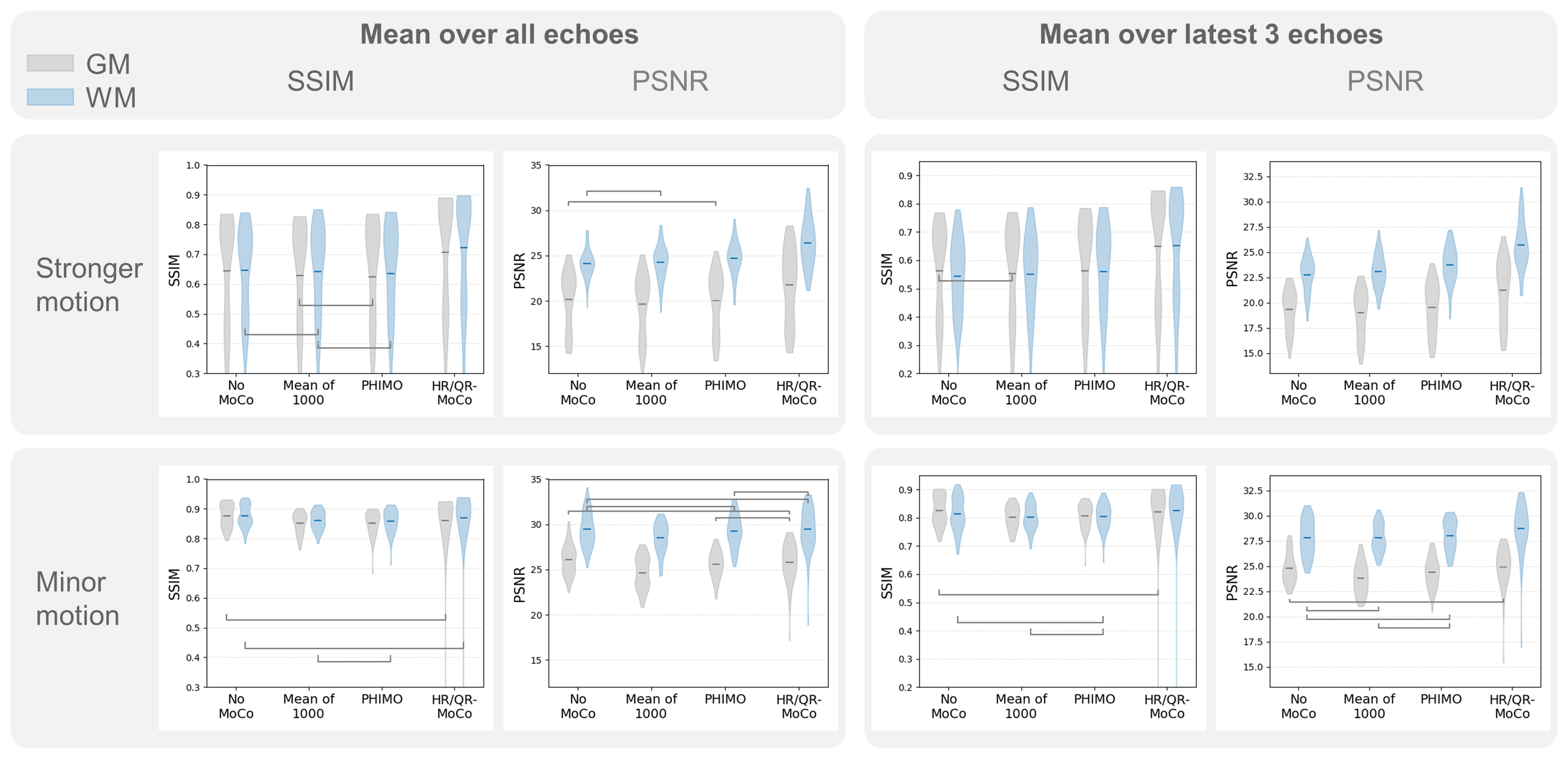
Example T2* maps in Figure 2 demonstrate that (compared to the mean-aggregation) incorporating model fit information into the bootstrap aggregation leads to a clear reduction of strong motion artifacts, close to the performance of HR/QR-MoCo. However, PHIMO reconstructions show residual blurring, particularly in subjects with minor motion. Note that some blurring is introduced by interpolation during registration (Figure 3). The qualitative observations are supported by mean absolute error (MAE) and structural similarity index (SSIM) between T2* maps, as well as variation in gray/white matter (Figure 4). SSIM and peak-signal-to-noise ratio (PSNR) on T2*-weighted images show only small improvements by all investigated methods (Figure 5).Discussion and Conclusion
PHIMO allows us to reduce strong motion artifacts in T2* maps, close to the performance of HR/QR-MoCo4, while avoiding multi-resolution data acquisition and significantly reducing the acquisition time by over 40% (6min 52s vs. 3min 39s). In contrast to mean-aggregation,9 PHIMO incorporates T2* decay information and performs a data-consistent reconstruction. We observe residual blurring, which influences quantitative evaluations in particular for minor motion. While some blurring is introduced by interpolation-based co-registration, the remaining blurring might originate from averaging the N best samples and could be avoided by further optimizing for the best mask, subject to future work. Note that both PHIMO and HR/QR-MoCo4 build on the assumption of randomized motion events during the acquisition, and their performance depends on the exact motion pattern. Similar to MoCo by mean-aggregation9, PHIMO does not require motion-corrupted data for training.In conclusion, we demonstrated that PHIMO effectively incorporates information from T2* model fitting and reduces strong motion artifacts. Additionally, PHIMO significantly decreases the overall acquisition time, enhancing the clinical applicability of GRE-based T2* quantification.
Acknowledgements
H.E. and V.S. were supported in part by the Helmholtz Association under the joint research school ”Munich School for Data Science - MUDS”.References
[1] Magerkurth, J., Volz, S., Wagner, M. et al. “Quantitative T * 2 -Mapping Based on Multi-Slice Multiple Gradient Echo Flash Imaging: Retrospective Correction for Subject Motion Effects: Movement Correction in T*2 Mapping.” Magnetic Resonance in Medicine 66, no. 4 (2011): 989–97.
[2] Hirsch, N. M., Toth, V., Förschler, A. et al. “Technical Considerations on the Validity of Blood Oxygenation Level-Dependent-Based MR Assessment of Vascular Deoxygenation: BOLD-based assessment of vascular deoxygenation.” NMR in Biomedicine 27, no. 7 (2014): 853–62.
[3] Eichhorn, H., Hammernik, K., Epp S. M. et al. “Investigating the Impact of Motion and Associated B0 Changes on Oxygenation Sensitive MRI through Realistic Simulations”. Proceedings of the 2023 ISMRM & ISMRT Annual Meeting & Exhibition (2023).
[4] Nöth, U., Volz, S., Hattingen, E. et al. “An improved method for retrospective motion correction in quantitative T2* mapping”. NeuroImage 92 (2014): 106–119.
[5] Spieker, V., Eichhorn, H., Hammernik, K. et al. "Deep Learning for Retrospective Motion Correction in MRI: A Comprehensive Review". IEEE Transactions on Medical Imaging [Early Access].
[6] Haskell, M. W., Cauley, S. F., Bilgic, V. et al. “Network Accelerated Motion Estimation and Reduction (NAMER): Convolutional neural network guided retrospective motion correction using a separable motion model”. Magnetic Resonance in Medicine 82, no. 4 (2019): 1452–1461.
[7] Hossbach J., Splitthoff, D. N., Cauley, S. et al. “Deep learning-based motion quantification from k-space for fast model-based magnetic resonance imaging motion correction”. Medical Physics 50, no. 4 (2023): 2148-2161.
[8] Xu, X., Kothopalli, S. V. V. N., Liu, J. et al. “Learning-based motion artifact removal networks for quantitative R2∗ mapping”. Magnetic Resonance in Medicine 88, no. 1 (2022): 106–119.
[9] Oh, G., Lee, J. E. and Ye, J. C. “Unpaired MR Motion Artifact Deep Learning Using Outlier-Rejecting Bootstrap Aggregation.” IEEE Transactions on Medical Imaging 40, no. 11(2021): 3125–3139.
[10] Schlemper, J., Caballero, J., Hajnal, J. H. et al. “A Deep Cascade of Convolutional Neural Networks for Dynamic MR Image Reconstruction”. IEEE Transactions on Medical Imaging 37, no. 2 (2018): 491–503.
[11] Hammernik, K., Klatzer, T., Kobler, E. et al. “Learning a variational network for reconstruction of accelerated MRI data”. Magnetic Resonance in Medicine 79, no. 6 (2018): 3055–3071.
[12] Hirsch, N.M., and Preibisch, C.. “T2* Mapping with Background Gradient Correction Using Different Excitation Pulse Shapes.” American Journal of Neuroradiology 34, no. 6 (2013): E65–68.
[13] Saks, E., Hoffmann, G., Eichhorn, H. et al. “Accelerated High-Resolution 3D Gradient Echo with DL-Based Reconstruction Improves T2* Mapping for Oxygenation-Sensitive MRI”. Submitted to Proceedings of the International Society for Magnetic Resonance in Medicine (2023).
[14] Kaczmarz, S., Hyder, F., and Preibisch, C.. “Oxygen Extraction Fraction Mapping with Multi-Parametric Quantitative BOLD MRI: Reduced Transverse Relaxation Bias Using 3D-GraSE Imaging.” NeuroImage 220 (2020): 117095.
[15] Hammernik, K., and Küstner, T. "Machine Enhanced Reconstruction Learning and Interpretation Networks (MERLIN)". Proceedings of the International Society for Magnetic Resonance in Medicine (2022).
Figures