0389
Integrating scout and guidance line-based retrospective motion correction into a 3D deep learning reconstruction for fast and robust brain MRI1Siemens Healthineers, Erlangen, Germany, 2Siemens Medical Solutions, Boston, MA, United States, 3Siemens Shenzhen Magnetic Resonance Ltd., Shenzhen, China, 4Massachusetts General Hospital, Boston, MA, United States, 5A. A. Martinos Center for Biomedical Imaging, Boston, MA, United States
Synopsis
Keywords: Alzheimer's Disease, MR Value
Motivation: Rising medical imaging utilization and increasing use of automated support systems demand high-quality, fast, and reproducible/robust MRI techniques. Despite rapid scanning afforded by deep learning, motion remains a common source of artifacts.
Goal(s): Integrate retrospective motion correction into a deep learning reconstruction to facilitate high-quality, fast, and motion-robust brain imaging.
Approach: Scout and guidance line-based motion correction was implemented into MPRAGE, SPACE and SWI to enable rapid motion trajectory estimation. A data-consistency driven neural network reconstruction was adapted to perform network regularized motion correction.
Results: Improved SNR and reduced motion artifacts are demonstrated in vivo using 4-6-fold accelerated scans with instructed subject motion.
Impact: Retrospective motion correction was integrated into a deep learning reconstruction to facilitate fast and motion-robust 3D brain imaging across T1, T2, T2 FLAIR and T2*/SWI. This should add clinical value to routine brain exams and emerging neuro-degenerative screening protocols (ARIA).
Background
In an era of rising medical imaging utilization (e.g., regular MR screenings for Alzheimer’s drug treatment) and increasing use of quantitative disease biomarkers & clinical support systems (e.g., brain morphometry, and hemorrhage, edema, tumor identification/segmentation), there is demand for high-quality, fast, and reproducible/robust MRI techniques. Motion during MRI examinations remains one of the largest sources of image quality degradation, especially in patients with neuro-degenerative diseases. This can negatively affect the radiologist’s image interpretation/diagnosis but also the quality of automated post-processing algorithms1. Deep learning image reconstruction has enabled reduced scan times while maintaining high image quality and is now widely accepted in clinical settings. While faster scanning has been associated with reduced likelihood of patient motion, it cannot solve the motion problem completely2. SAMER3,4 is a retrospective motion correction technique for brain imaging. It enables very rapid motion estimation and artifact correction without external tracking hardware and with minimal disruption to standard clinical protocols. In this work, we integrate the SAMER technique into a deep learning image reconstruction and demonstrate highly accelerated motion-robust 3D brain imaging across MPRAGE, SPACE (T2w and T2 FLAIR) and T2*/SWI. These sequences represent the core of both routine brain and neuro-degenerative disease screening exams1.Methods
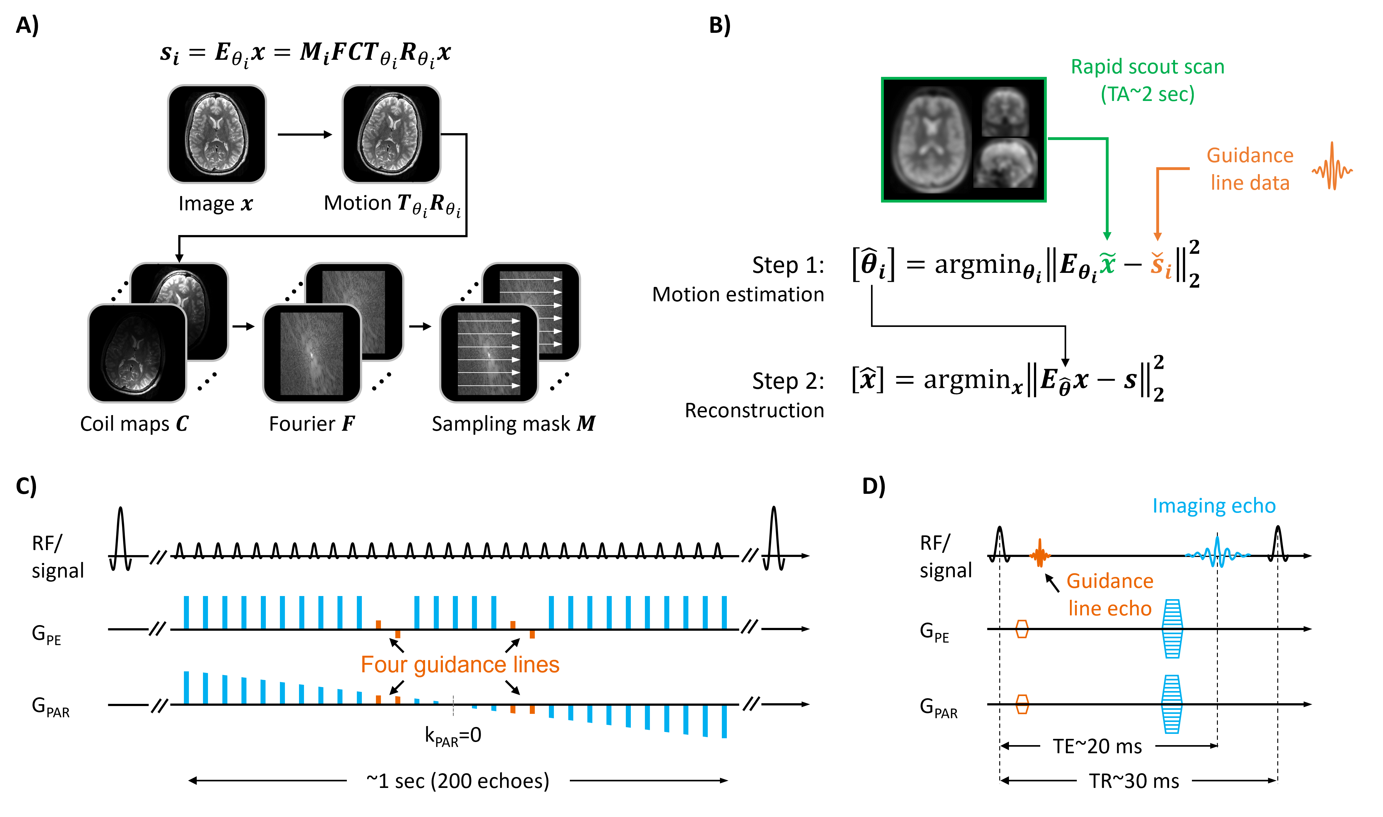
Motion estimation:SAMER is based on SENSE5 parallel imaging, where rigid body motion operators are added to describe motion within the forward model6 (Fig. 1A). Estimating the motion trajectory and the motion-free image is achieved by minimizing the deviation between the physics model prediction and the acquired k-space data. To avoid a computationally costly joint optimization7,8, SAMER acquires additional k-space encoding lines (motion guidance lines) and an ultra-fast scout scan which serves as a prior to guide the motion search (Fig. 1B). This facilitates very rapid trajectory estimation (~1 sec/shot).
In the 3D multi-shot sequences MPRAGE (Fig. 1C) and SPACE, the data collection is divided into echo trains (shots), each containing ~200 phase/partition encoding lines. A limited number of additional guidance lines are added to each echo train which does not affect image quality/contrast or acquisition time. In SWI (Fig. 1D), guidance line information is obtained from an additional gradient-echo played before the imaging-echo. This approach again maintains the original contrast and scan efficiency. The ultra-fast low-resolution scout scans (~2 sec for MPRAGE) match the contrast of the guidance lines and were acquired once before every imaging sequence.
Deep learning reconstruction:
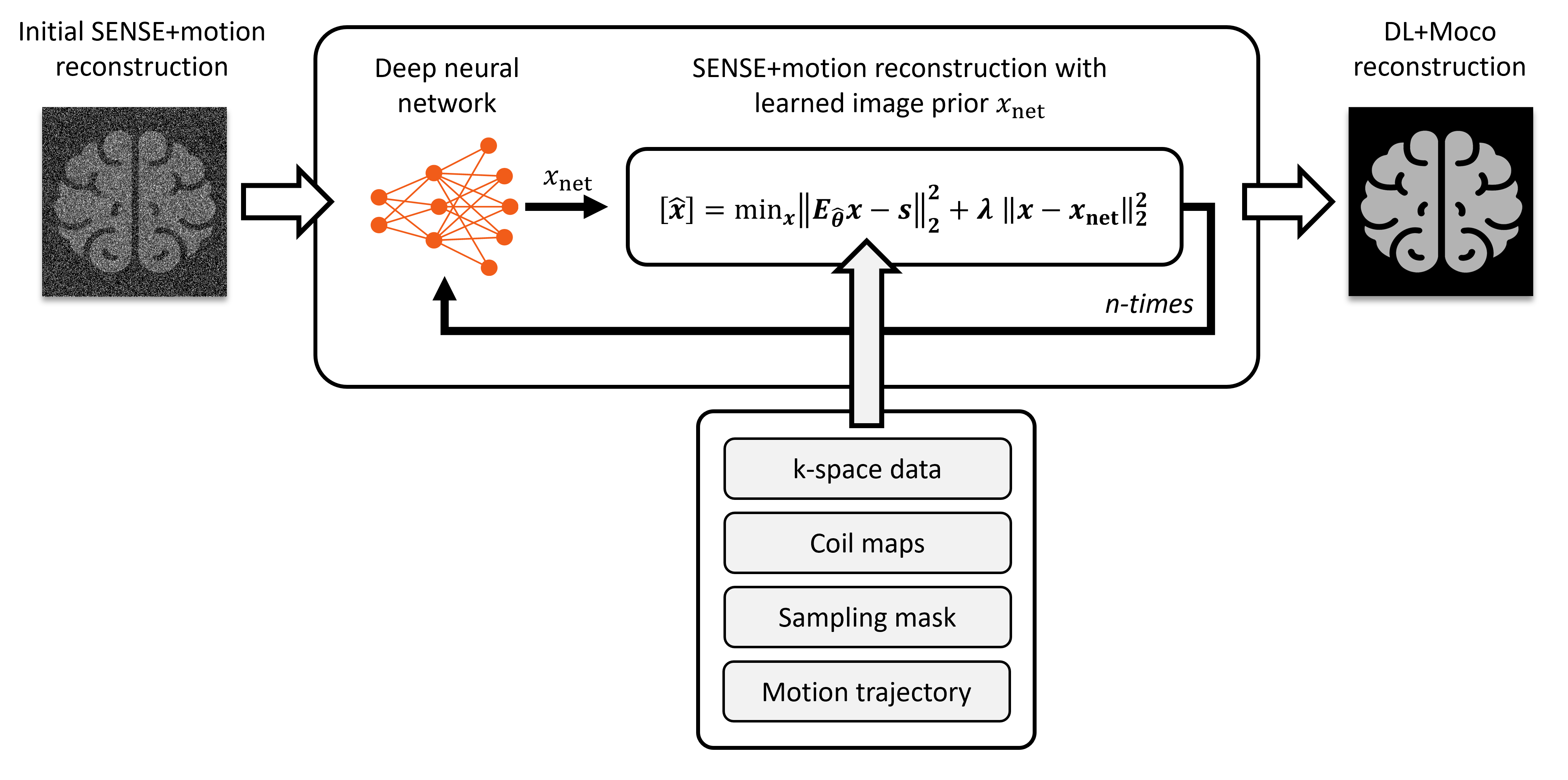
To facilitate highly accelerated motion-robust imaging we integrate deep learning into the SAMER motion-mitigated image reconstruction (Fig. 2). Here, a state-of-the-art unrolled network architecture9 was used which alternates between classical conjugate-gradient SENSE optimization and deep learning based update of the image prior. The final image is obtained after a fixed number of reconstruction instances, each containing a separate neural network regularizer (3D UNet10). The neural networks were pre-trained on motion-free data while motion-correction was performed during inference. Specifically, the data fidelity term (Fig. 2) was adapted to use the SENSE+motion model which is dependent on the estimated motion trajectories.
In vivo experiments:
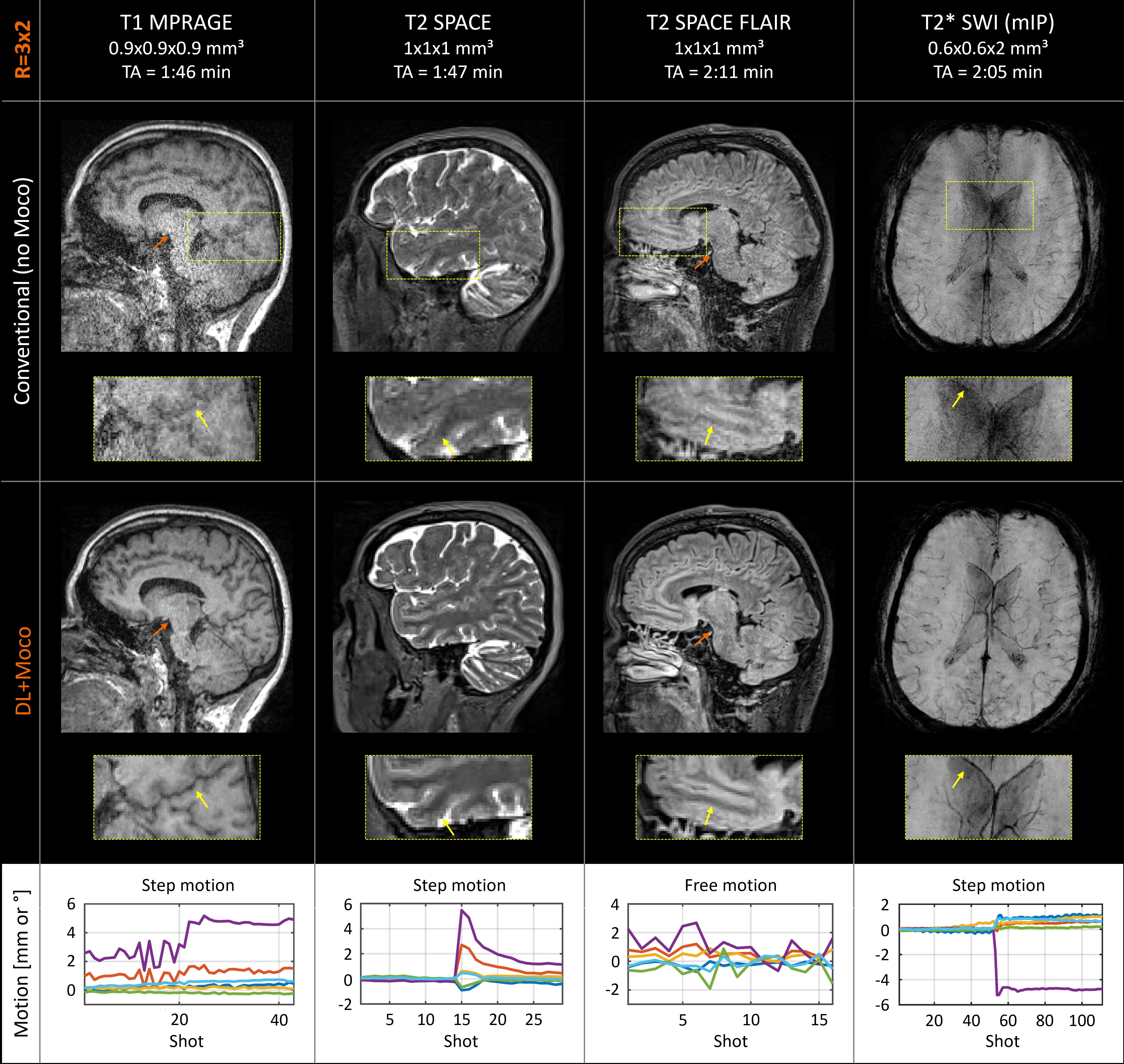
At 3T (MAGNETOM Vida; Siemens Healthineers, Erlangen, Germany), high-resolution MPRAGE, SPACE and SWI data were collected at R=2x2 and R=3x2 acceleration using a 20-channel head/neck coil. With written consent, in vivo measurements with instructed step, nodding and unsupervised free motion were conducted on three healthy volunteers.
Results
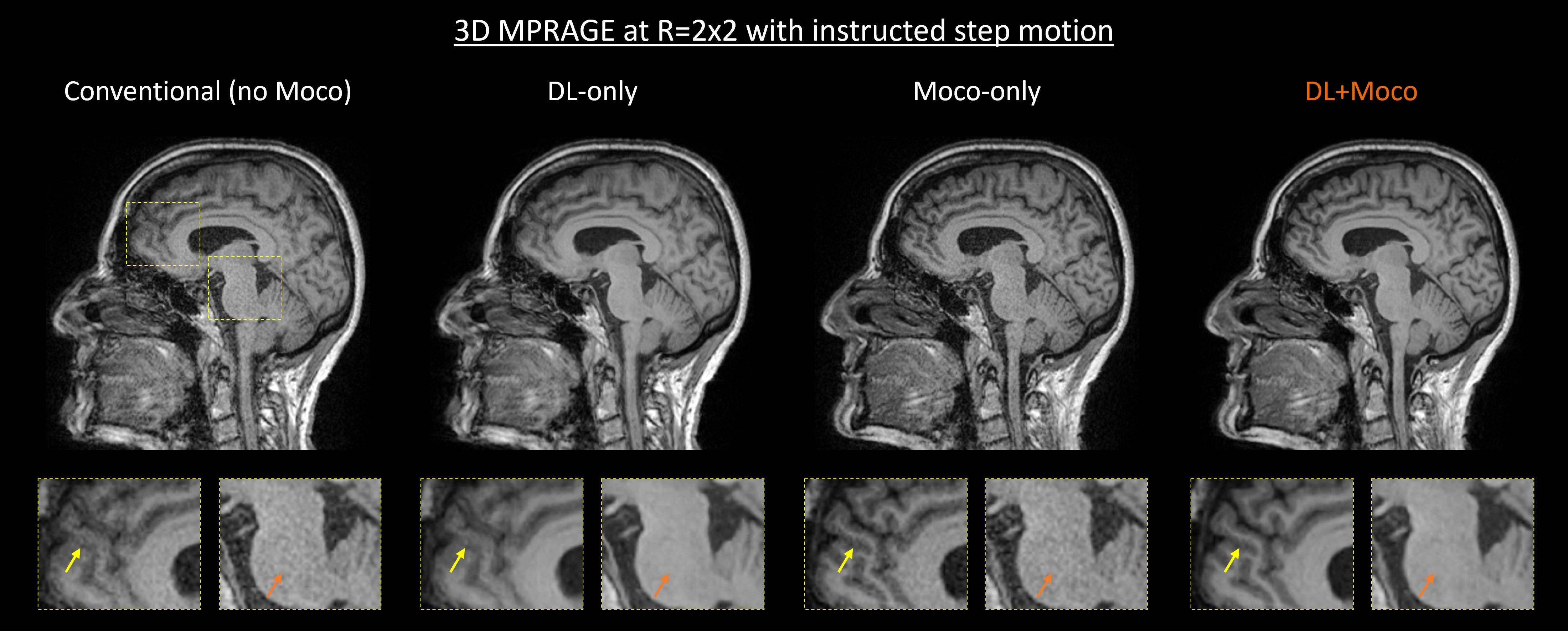
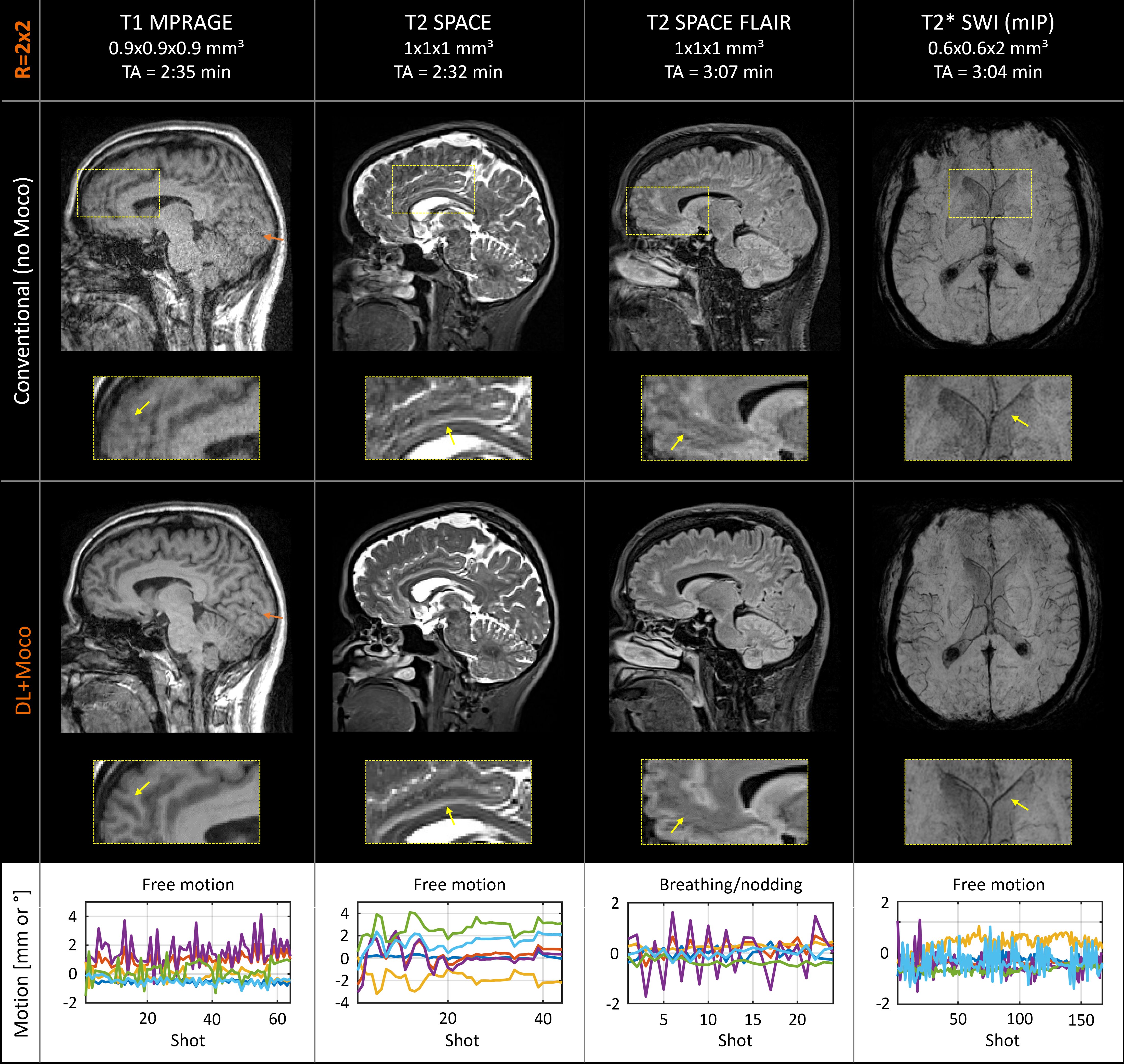
Figure 3 compares the individual image quality improvements afforded by different components of the image reconstruction. We compare the benefits of “DL-only”, “Moco-only” and “DL+Moco” using R=2x2 accelerated MPRAGE data with instructed step motion. Improved SNR (“DL-only”) and increased image sharpness (“Moco-only”) were both obtained with the proposed “DL+Moco” reconstruction.Figures 4 and 5 show the combined “DL+Moco” reconstructions of R=2x2 and R=3x2 accelerated acquisitions under various instructed motion patterns. Across all contrasts, “DL+Moco” consistently reduced noise amplification and mitigated motion artifacts leading to improved image quality.
Conclusions
We integrated SAMER retrospective motion correction into a data-consistency driven neural network reconstruction and demonstrated highly accelerated motion-robust brain imaging. In this work, the scout and guidance line-based motion estimation framework4 was extended to both the SPACE and SWI sequences. This enables retrospective motion correction across the principal clinical contrasts T1, T2, T2 FLAIR and T2*/SWI. Integration into a deep learning reconstruction allowed up to 6-fold acceleration with the standard vendor 20-channel head/neck coil, while retaining high quality and good SNR. Our suite of fast and motion-robust sequences should add clinical value to routine brain exams which typically contain all imaging contrasts from this work. Moreover, they directly align with emerging neuro-degenerative disease screening protocols, e.g., detection of ARIA (amyloid related imaging abnormalities) in Alzheimer’s patients.Acknowledgements
No acknowledgement found.References
1. Cogswell PM, Barakos JA, Barkhof F, et al. Amyloid-Related Imaging Abnormalities with Emerging Alzheimer Disease Therapeutics: Detection and Reporting Recommendations for Clinical Practice. AJNR Am J Neuroradiol. 2022;43(9):E19-E35. doi:10.3174/ajnr.A7586
2. Zaitsev M, Maclaren J, Herbst M. Motion artifacts in MRI: A complex problem with many partial solutions. J Magn Reson Imaging. 2015;42(4):887-901. doi:10.1002/jmri.24850
3. Polak D, Splitthoff DN, Clifford B, et al. Scout accelerated motion estimation and reduction (SAMER). Magn Reson Med. 2022;87(1):163-178. doi:10.1002/mrm.28971
4. Polak D, Hossbach J, Splitthoff DN, et al. Motion guidance lines for robust data consistency–based retrospective motion correction in 2D and 3D MRI. Magn Reson Med. 2023;89(5):1777-1790. doi:10.1002/mrm.29534
5. Pruessmann KP, Weiger M, Börnert P, Boesiger P, Klaas P. Pruessmann. Advances in sensitivity encoding with arbitrary k-space trajectories. Magn Reson Med. 2001;46(4):638-651. doi:10.1002/mrm.1241
6. Batchelor PG, Atkinson D, Irarrazaval P, Hill DLG, Hajnal J, Larkman D. Matrix description of general motion correction applied to multishot images. Magn Reson Med. 2005;54(5):1273-1280. doi:10.1002/mrm.20656
7. Haskell MW, Cauley SF, Wald LL. TArgeted Motion Estimation and Reduction (TAMER): Data consistency based motion mitigation for mri using a reduced model joint optimization. IEEE Trans Med Imaging. 2018;37(5):1253-1265. doi:10.1109/TMI.2018.2791482
8. Cordero-Grande, L., Teixeira, R., Hughes, E., Hutter, J., Price, A., & Hajnal J, Cordero-Grande L, Teixeira RPAG, et al. Sensitivity Encoding for Aligned Multishot Magnetic Resonance Reconstruction. IEEE Trans Comput Imaging. 2016;2(3):266-280. doi:10.1109/tci.2016.2557069
9. Hammernik K, Klatzer T, Kobler E, et al. Learning a variational network for reconstruction of accelerated MRI data. Magn Reson Med. 2018;79(6):3055-3071. doi:10.1002/mrm.26977
10. Ronneberger O, Fischer P, Brox T. U-net: Convolutional networks for biomedical image segmentation. In: Lecture Notes in Computer Science (Including Subseries Lecture Notes in Artificial Intelligence and Lecture Notes in Bioinformatics). Vol 9351. ; 2015:234-241. doi:10.1007/978-3-319-24574-4_28
Figures