0380
qDiMo: Domain-conditioned Diffusion Modeling for Accelerated qMRI Reconstruction1Athinoula A. Martinos Center for Biomedical Imaging, Massachusetts General Hospital, Charlestown, MA, United States, 2Harvard Medical School, Boston, MA, United States
Synopsis
Keywords: AI Diffusion Models, Machine Learning/Artificial Intelligence, Rapid MRI, Quantitative MRI, knee, brain
Motivation: Quantitative MRI (qMRI) is time-consuming and requires substantial efforts for acceleration to cut down the acquisition time.
Goal(s): This paper proposes a novel generative AI approach for image reconstruction based on diffusion modeling conditioned on the native data domain.
Approach: Our method is applied to multi-coil quantitative MRI reconstruction, leveraging the domain-conditioned diffusion model within the tissue parameter domain.
Results: The proposed method demonstrates a significant promise for reconstructing quantitative maps at high acceleration factors. Notably, it maintains excellent reconstruction accuracy and efficiency for MR parameter maps across diverse anatomical structures.
Impact: This work demonstrates the feasibility of a new generative AI method for rapid qMRI. Beyond its immediate applications, this method provides potential generalization capability, making it adaptable to inverse problems across various domains.
INTRODUCTION
Generative AI using diffusion models has been shown as a promising approach for robust and efficient MRI applications. Denoising Diffusion Probabilistic Models (DDPMs) have emerged as a notable advancement in this area and have quickly gained interest in MRI reconstruction [1],[2]. DDPMs for MRI reconstruction generally start in the image domain, where the unknown data distribution from training images goes through repeated Gaussian diffusion processes, which can be reversed by learned transition kernels. However, applying the diffusion process in the image domain overlooks the underlying MRI physical model (i.e., k-space encoding and coil sensitivity), which is embedded in the measured k-space data, leading to suboptimal reconstruction performance [3],[4]. In addition, minimal studies have shown that generative AI can be applied to quantitative MRI due to the additional complexity needed to solve MR signal modeling. This study proposes a novel method that applies domain-conditioned diffusion modeling to accelerated quantitative MRI, which we denote as quantitative Diffusion Modeling (qDiMo).THEORY
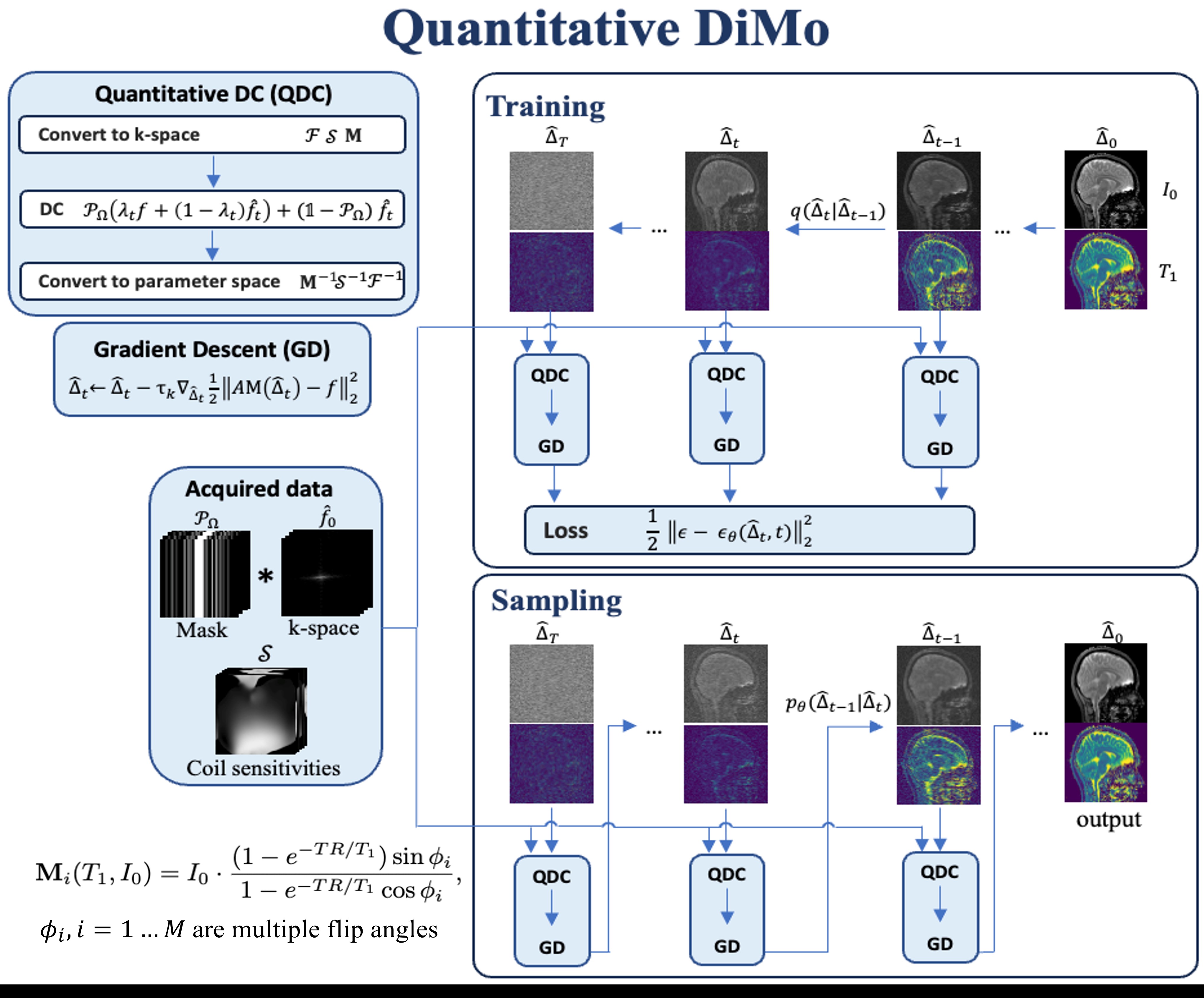
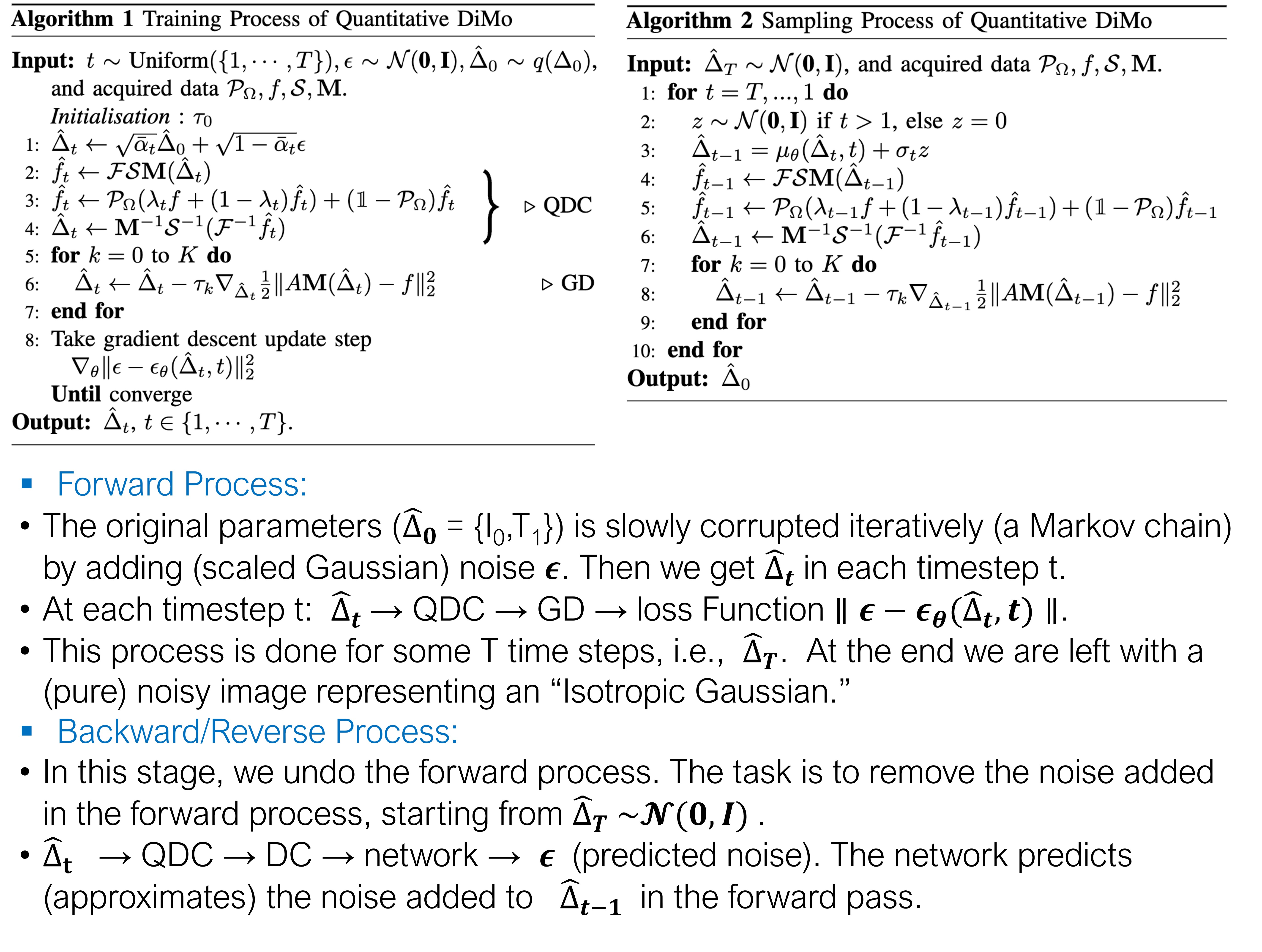
Since qMRI focuses on the quantification of tissue parameters, it can be advantageous to define the diffusion model conditioned on the parameter domain for qMRI (Fig. 1). We propose a novel accelerated qMRI reconstruction method that enables reconstruction of rapid MR parameter mapping from undersampled k-space data $$$f$$$. Define the MR parameter maps as $$$\Delta=\{\delta_i\}_{i=1}^N$$$ where $$$\delta_i$$$ symbolizes each MR parameter. Our qDiMo focuses on estimating the data distribution that is characterized by density $$$q(\Delta|P_\Omega, f, \mathcal{S}, \mathbf{M})$$$ which is conditioned on undersampling mask $$$P_\Omega$$$, partially scanned measurement $$$f$$$, coil sensitivity maps $$$\mathcal{S}$$$, and the MR signal model $$$\mathbf{M}$$$. Using variable flip angle (vFA) T1 mapping [5] as an example, given multiple flip angles $$$\phi_i$$$, the signal model is: $$$\mathbf{M}_i(T_1,I_0)=I_0\cdot\frac{(1-e^{-TR/T_1})sin\phi_i}{1-e^{-TR//T_1}cos\phi_i}$$$. Compared to general DDPMs, qDiMo introduces two additional layers that provide the prior knowledge for the conditions/guidance of diffusion model (Algorithm) in the tissue parameter domain: 1) Quantitative Data Consistency (QDC) Layer to perturb quantitative tissue parameters and guide the parameter space denoising and generation process meanwhile preserving k-space data with a linear combination of partially scanned data $$$f$$$ to enforce data consistency. 2) Gradient Descent (GD) Layer to further fine-tune the QDC layer and optimize perturbed $$$\Delta_t$$$ during each diffusion step using gradient descent optimization.METHODS
The five subjects fully sampled vFA brain data was acquired along the sagittal plane at a Siemens 3T Prisma scanner equipped with a 20-channel head coil using a spoiled gradient echo sequence with imaging parameters TE/TR = 12/40 ms, FA = 5, 10, 20, 40$$$^\circ$$$, 3D matrix size = 176×176×48. Leave-one-out cross-validation was used for all five subjects to train and test our method. Two undersampling schemes were retrospectively used: 1) 1D variable density Cartesian undersampling with acceleration factor AF= 4× and 2) 2D Poisson disk undersampling at AF= 4×. We compared qDiMo with two advanced non-deep learning qMRI reconstruction techniques: Locally Low Rank (LLR) [6], Model-TGV [7], and a self-supervised deep learning method, RELAX [8].RESULTS AND DISCUSSION
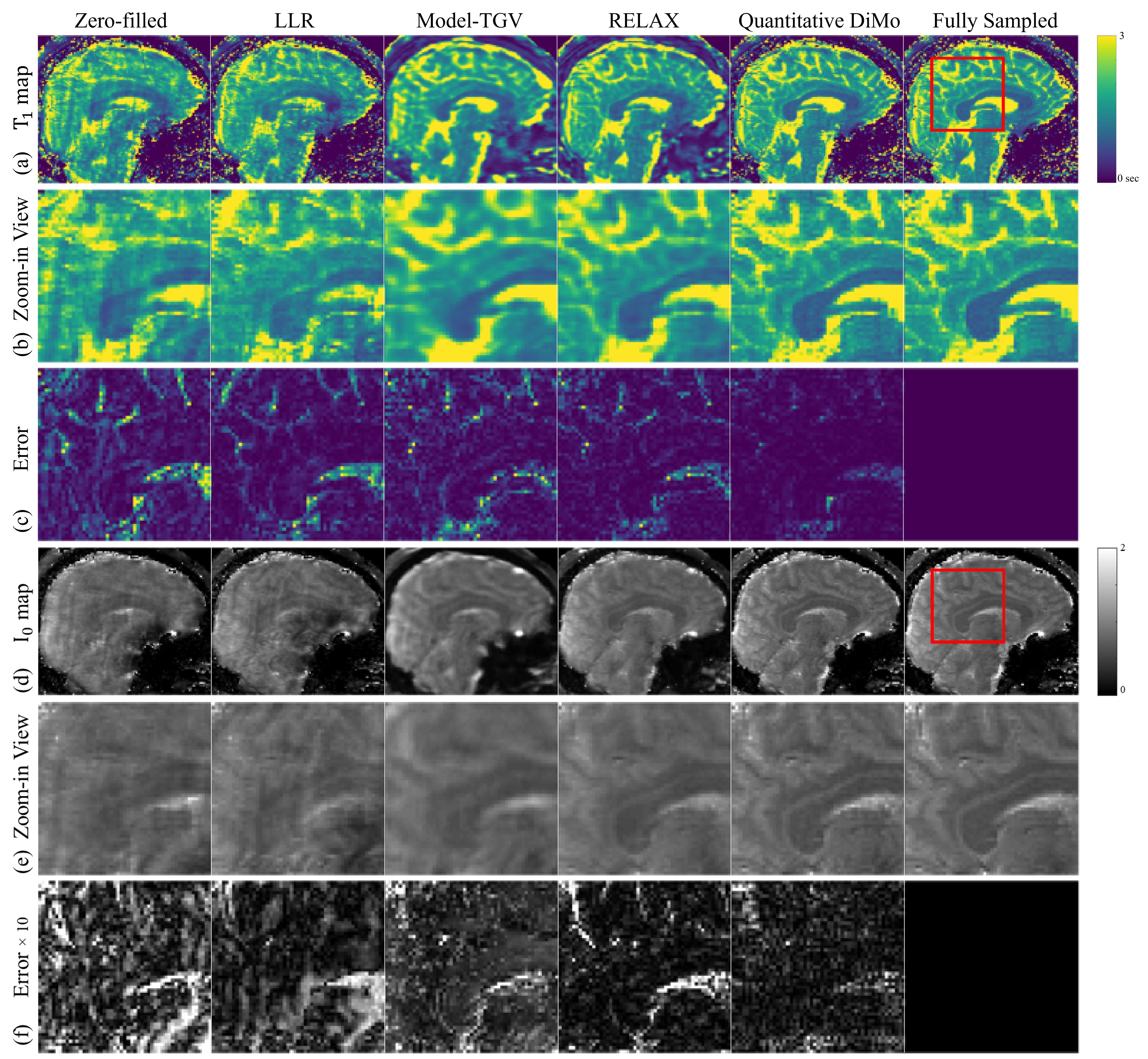
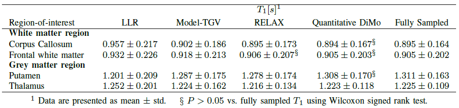
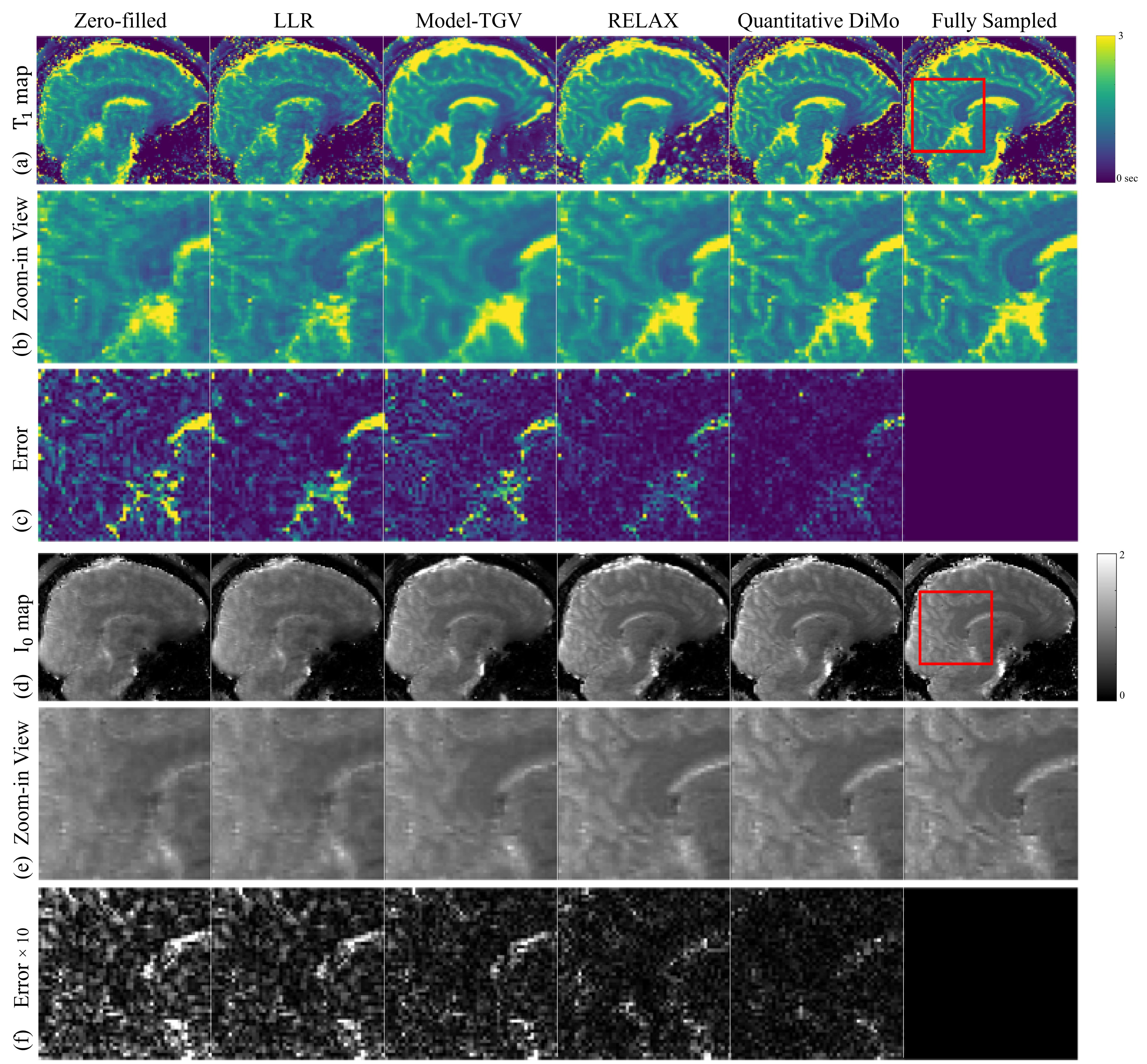
T1 and I0 maps estimated from 4× 1D variable density Cartesian undersampling are shown in Fig. 2. Compared with other methods, qDiMo generates clear and sharp T1 maps. Its proficiency in removing noise translates to its superior map quality in appearance and contrast. This is further witnessed in the zoom-ins (Fig. 2(b)), which show that qDiMo clearly distinguishes the boundary between white matter and grey matter, appearing nearly identical to the fully sampled reference map. This is quantitatively confirmed in the error maps (Fig. 2(c)), where the zero-filled incurs the largest error, followed by LLR, Model-TGV, and RELAX. Mean T1 values obtained from representative white matter and grey matter regions are presented in Table for all subjects. Overall, as shown by the Wilcoxon signed rank test results, qDiMo shows the best similarity with the reference. The I0 maps in Fig 2(d-f) exhibit a similar signature in reconstruction quality as the T1 maps. The T1 and I0 maps estimated from 4× 2D Poisson disk undersampling are shown in Fig. 3. Again, qDiMo produces artifact-free T1 and I0 maps with superior performance compared to other methods. This is also confirmed by the error maps (Fig. 3(c) and Fig. 3(f)), with qDiMo showing the least error. This is likely achieved through integrating the unrolling gradient descent algorithm and diffusion denoising network, prioritizing noise suppression without compromising the fidelity and clarity of the underlying tissue structure.CONCLUSION
This paper proposed a diffusion model conditioned on the native data domain for reconstructing quantitative MRI. The reconstruction shows promising results and great potential for translating rapid quantitative MRI into clinical applications.Acknowledgements
We thank the funding support from NIBIB R21EB031185, NIAMS R01AR079442, R01AR081344, and R56AR081017.References
[1] Chung, H., Medical image analysis, vol. 80, p. 102479, 2022. [2] Gungor, A., Medical Image Analysis, p. 102872, 2023. [3] Ozbey, M., IEEE Transactions on Medical Imaging, 2023. [4] Luo, G., Magnetic Resonance in Medicine, vol. 90, no. 1, pp. 295–311, 2023. [5] Fram, E. K., Magnetic resonance imaging, 1987, 5(3), 201-208. [6] Zhang, T., Magnetic resonance in medicine, 2015, 73(2), 655-661. [7] Maier, O., Magnetic resonance in medicine, 2019, 81(3), 2072-2089. [8] Liu, F., Magnetic resonance in medicine, 2021, 85(6), 3211-3226.Figures