0376
Imaging tumor habitats with PET-MRI HYPERDIrect maps to decode the spatial heterogeneity of malignant gliomas1Università Vita-Salute San Raffaele, Milano, Italy, 2Neuroradiology Unit and CERMAC, IRCCS Ospedale San Raffaele, Milano, Italy, 3Department of Neurosurgery and Gamma Knife Radiosurgery, IRCCS Ospedale San Raffaele, Milan, Italy, 4Department of Nuclear Medicine, IRCCS Ospedale San Raffaele, Milan, Italy, 5Pathology Unit, IRCCS Ospedale San Raffaele, Milan, Italy
Synopsis
Keywords: Tumors (Pre-Treatment), Radiomics, Malignant gliomas, Habitat imaging
Motivation: Malignant gliomas are characterized by considerable intra-tumor heterogeneity directly related to treatment failure. Habitat imaging allows to visualize in vivo such heterogeneity.
Goal(s): Investigating a novel approach based on 3T PET/MRI acquisitions for assessing HYpoxia, PErfusion, DIffusion, and methionine-PET for tissue metabolism in malignant gliomas.
Approach: Quantitative data from PET/MRI are combined in a unique HYPERDIrect map to identify discrete radiomic clusters or `habitats', possibly reflecting diverse genetic and biological features, to be proven by image-guided tissue sampling.
Results: Preliminary analysis showed high habitat imaging reproducibility and a reliable correlation between the expected microenvironment of the different habitats and the actual histopathological characteristics.
Impact: The HYPERDIrect map may represent a tool for decoding pattern of glioma heterogeneity and a new biomarker for stratifying prognosis and selecting patients for personalized approaches.
Introduction
Malignant gliomas are characterized by considerable intra-tumor heterogeneity directly related to treatment failure. A novel method for cancer detection involves identifying regions or habitats within tumors by assessing shared imaging characteristics. This approach utilizes quantitative analysis of conventional and advanced imaging data through algorithms, which effectively partition the tumor into voxel-based subregions exhibiting similar radiological features1. The integration of multiple images further refines the creation of distinct tumor habitats. In this study, habitat analysis has been applied to hybrid PET/MR images of HYpoxia, PERfusion and DIffusion Imaging to map tumor oxygen extraction, neoangiogenesis, cellularity, and metabolism.Materials and Methods
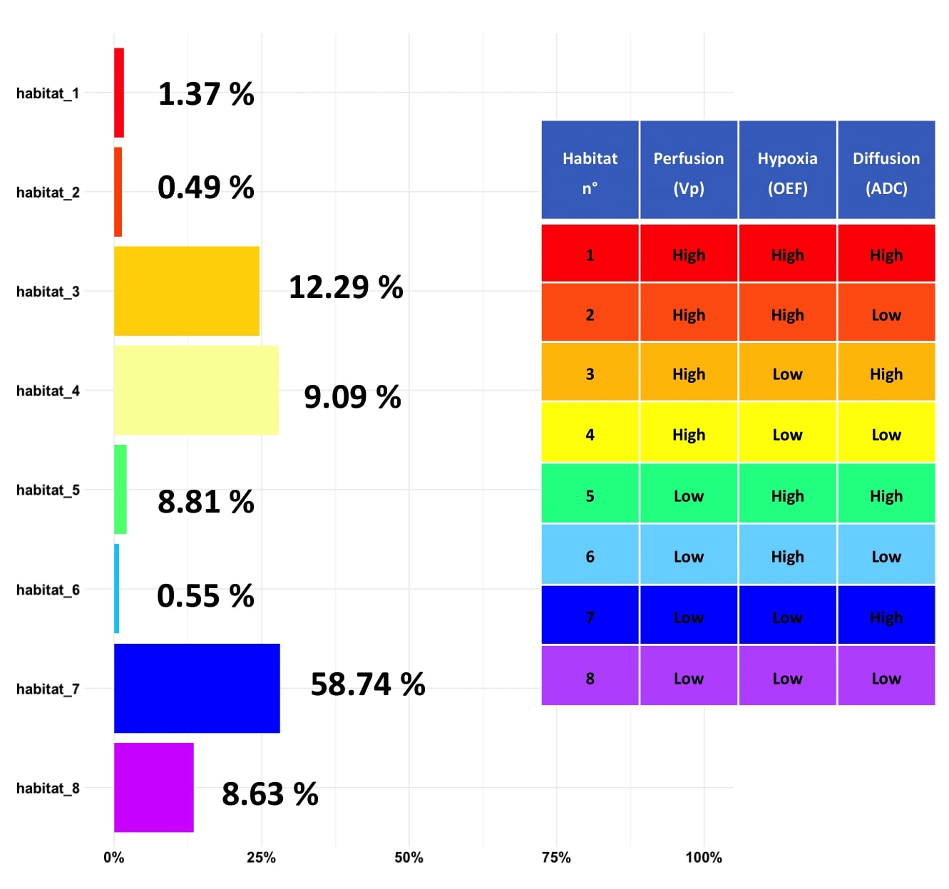
20 patients with radiologically suspected high-grade glioma and eligible for surgical resection or biopsy were enrolled. Patients underwent pre-operative acquisition on a hybrid 3T PET/MRI scan (Signa PET/MR, GE Healthcare) for assessing HYpoxia (using the oxygen extraction map [OEF] from quantitative blood oxygenation level-dependent [q-BOLD] imaging), PERfusion (using the Plasma Volume [Vp] map derived from Dynamic Contrast-Enhanced [DCE] acquisitions), DIffusion (using the Apparent Diffusion Coefficient [ADC] map from DWI acquisitions), and methionine-PET scans for tissue metabolism. Tumor volumes were segmented on FLAIR images. Within these FLAIR-derived regions of interest, we employed the Otsu algorithm to classify tumoral voxels of each quantitative imaging map into two clusters with high and low voxel intensity. ADC-tumoral voxels were split on the basis of a fixed threshold at 1200. Subsequently, the ADC map and methionine-PET data were merged to identify areas of high cellularity with elevated metabolic activity (low ADC and high methionine-PET). By following a voxel-wise clustering procedure outlined in a prior study2, the combination of clusters from Vp, OEF, and the combined ADC and methionine-PET data enabled the identification of eight distinct habitats within the tumor (Figure 1). Enrolled patients can either undergo stereotactic biopsy or open surgical exeresis. The HYPERDIrect map is integrated into the biopsy planning software or into the neuronavigation system to perform image-guided sampling to correlate imaging findings with histopathological and immunohistochemical results.Results
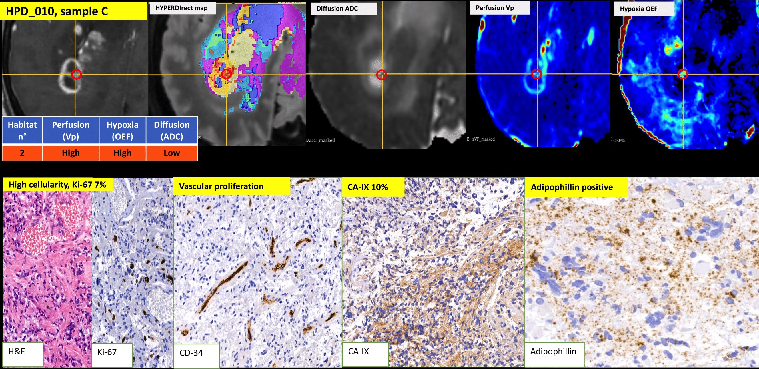
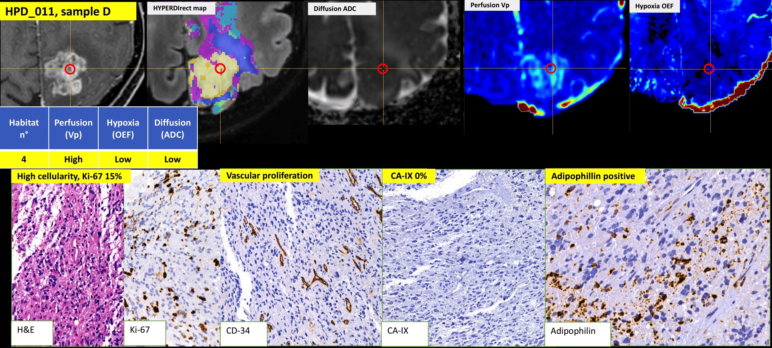
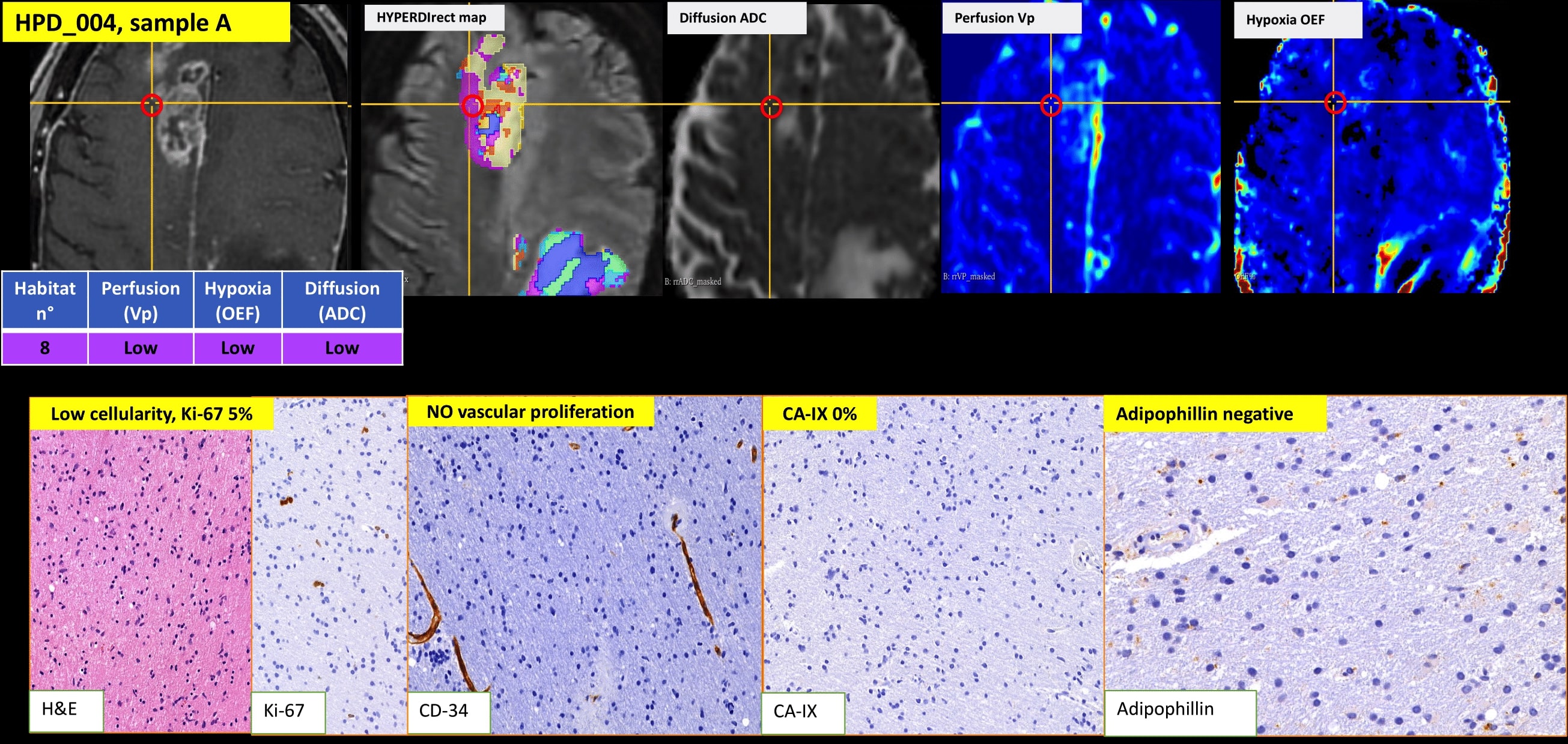
The mean spatial distance between habitats’ centroids was 20.81 mm for the eight habitats for all subjects, representative of good spatial disjunction between them, thus allowing multiple guided sampling from different habitats. Habitat 7 (with low perfusion, low hypoxia, and low cellularity without active metabolism, see Figure 1) and habitat 8 (with low perfusion, low hypoxia, and high metabolic active cellularity) were the most represented in the whole FLAIR tumor volume. Habitat 4 (with high perfusion, low hypoxia, and low cellularity without active metabolism) and habitat 5 (with high perfusion, low hypoxia, and high metabolic active cellularity) were the most found in the enhancing rim. Preliminary findings demonstrated a highly reproducible habitats distribution pattern with common histopathological features. Particularly, samples taken from, hypothetically, more aggressive habitats (with reduced diffusivity, high perfusion, and low hypoxia) histologically correspond to areas of high-grade glial neoplasia with high cellularity and microvascular proliferation. Samples from habitats with less aggressive imaging features (high diffusivity, low perfusion, and low hypoxia) corresponded to glioma infiltrative areas. The association between areas with high radiologically predicted hypoxia and CA-IX immunohistochemical staining reached statistical significance (p = 0.02).Discussion
Malignant gliomas are characterized by extreme biological complexity with considerable intra-tumoral spatio-temporal heterogeneity, driving their dismal prognosis. Histological and molecular diagnosis rely on surgical specimens that could not represent the whole neoplasm; as such, there is an unmet need for non-invasive biomarkers able to provide holistic information regarding tumor microenvironment. Habitat imaging is a subfield of radiomics that could provide disease-wide assessment and possibly a dynamic evaluation of the tumor tissue during therapy. We previously proposed a method for clustering intratumoral heterogeneity by using PET with an experimental tracer to map hypoxia, and separate MRI acquisitions to estimate tissue diffusion and perfusion2. In this study, we have applied this method on fully hybrid PET/MRI images using a clinically feasible MRI protocol with a largely available aminoacidic tracer. The preliminary results demonstrate the feasibility of the integrated PET/MRI approach for assessing HYpoxia, PErfusion, DIffusion, and methionine-PET for tissue metabolism. The good correlation found between predicted imaging habitat characteristics and histological and immunohistochemical features proposes the HYPERDirect map as a valuable tool to obtain a priori knowledge about tumor microenvironment, possibly helping to stratify patients for tailored treatments.Conclusion
Habitat imaging using the HYPERDIrect map approach might serve as a potential biomarker for non-invasively characterizing tumor heterogeneity in vivo.Acknowledgements
This research was funded by the Italian Ministry of Health, grant number GR-2018-12365670. The study was approved by the Ethics Committee of Ospedale San Raffaele on March 9th, 2022 (code 19/INT/2022).References
1. Waqar M, et al. Visualising spatial heterogeneity in glioblastoma using imaging habitats. Front Oncol 12, 1037896 (2022).
2. Bailo M, et al. Decoding the Heterogeneity of Malignant Gliomas by PET and MRI for Spatial Habitat Analysis of Hypoxia, Perfusion, and Diffusion Imaging: A Preliminary Study. Front Neurosci 16, 885291 (2022).
Figures