0375
Deep learning enables robust quantification of cerebral blood flow using ASL in the presence of pathology: Application to treated gliomas1Department of Computer Science, University College London, London, United Kingdom, 2Queen Square Institute of Neurology, University College London, London, United Kingdom, 3Imaging, University College London Hospitals, London, United Kingdom, 4Medical Physics and Biomedical Engineering, University College London Hospitals, London, United Kingdom
Synopsis
Keywords: Tumors (Post-Treatment), Arterial spin labelling, Machine Learning/Artificial Intelligence, Tumor
Motivation: The accuracy of cerebral blood flow (CBF) quantification in arterial spin labelling (ASL) may reduce in regions exhibiting pathology-induced signal abnormalities in proton density (PD) images.
Goal(s): To develop an algorithm for improved CBF quantification by correcting signal abnormalities in PD images due to pathology.
Approach: To correct signal abnormalities, an image-inpainting algorithm based on deep learning (DL) was developed using healthy subject data. The algorithm was demonstrated with an application to patients post tumour treatment.
Results: The developed DL algorithm was able to effectively correct signal abnormalities, resulting in improved CBF maps.
Impact: The improvement in CBF accuracy through DL-corrected PD images may aid clinicians in their assessment of patients. This study demonstrates the potential benefit of the proposed method in an example application of monitoring tumour recurrence post treatment with ASL.
Introduction
Arterial spin labelling (ASL) MRI allows the mapping of cerebral blood flow (CBF) in tissue without the need for contrast agents, which can be valuable in clinical applications, such as the imaging of brain tumours1. As part of the typical processing pipeline to generate CBF maps2, a separately acquired proton density (PD) image is used to provide a calibration factor on a voxel-by-voxel basis3. However, this calibration method may be inappropriate in the presence of pathology. For example, recent studies have reported that higher water content in both untreated and treated gliomas causes signal abnormalities in PD images that result in biased CBF maps4,5. Sokolska et al attempted to mitigate this bias with region-wise calibration correction, i.e., estimating the appropriate calibration factor for a region with signal abnormalities from its normal appearing contralateral counterpart5. However, this approach does not support voxel-wise correction. To enable such fine-grained correction, we propose an image-inpainting algorithm powered by deep learning (DL).Methods
The proposed PD image correction takes as input a PD image with the pathological regions masked out, thus translating the image correction task into a familiar image-inpainting task. The task for the image-inpainting algorithm is to predict normal appearing PD intensities within the masked-out regions. To create such an algorithm, we choose a DL image-inpainting model with a U-Net-like architecture6 for this initial proof-of-concept, which will be referred to as the DL method henceforth. The essential point is that the model was trained using PD images of healthy subjects. By randomly simulating regions to be masked out, we can efficiently produce a large set of labelled examples, with each consisting of a masked-out image paired with the original image. Here, PD images of 49 healthy subjects were split into the training (80%) and test (20%) sets.Evaluation
First, we assessed the performance of the trained DL model for inpainting normal appearing PD intensities. Using the unseen test set, we evaluated the mean-pixel-value differences between the predicted intensities and the ground truth values.Second, the proposed method was compared against the region-wise calibration correction5, which will be referred to as the contralateral mean method henceforth. For this comparison, a set of 57 patients who received standard treatment for gliomas were used. For each subject, ASL data was acquired with a pseudo-continuous ASL sequence, along with PD images for calibration. Masks defining pathological regions (enhancing tumour, non-enhancing tumour, oedema) were automatically produced using an established DL pipeline7. We compared CBF values in pathological regions calibrated using 1) the uncorrected PD intensities, 2) the contralateral mean method, and 3) the proposed DL method.
Results
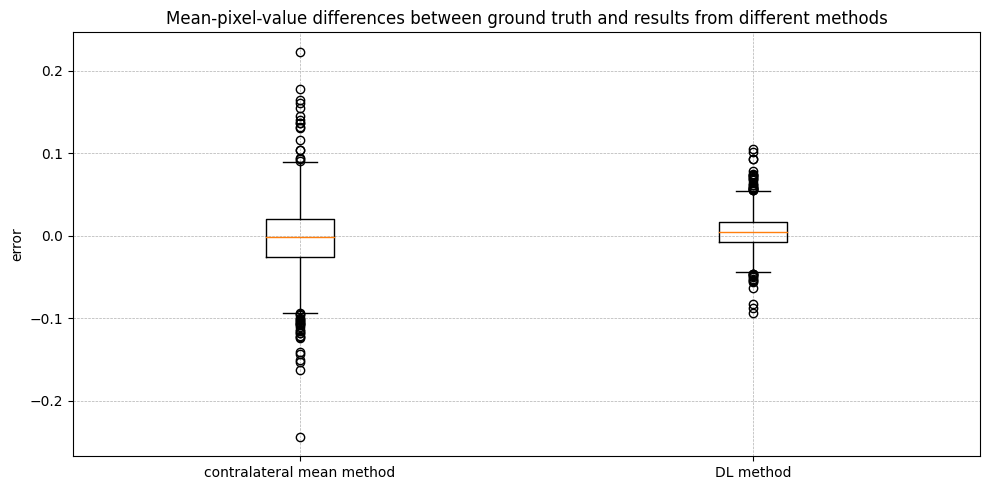
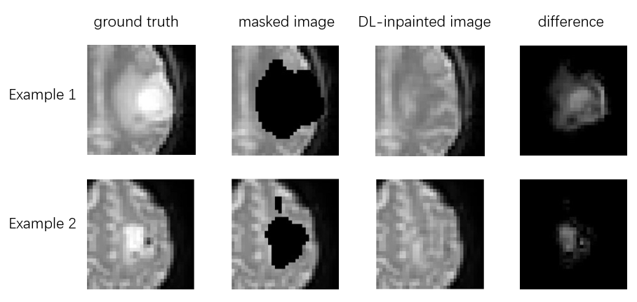
Figure 1 illustrates the inpainting performance of the DL model using two examples from the test set. The inpainted images and the original images (serving as the ground truth) are essentially indistinguishable.Figure 2 demonstrates the inpainting performance quantitatively with the mean-pixel-value differences. Results obtained using inpainting exhibit substantially narrower error spread and fewer outliers compared to the contralateral mean method.
Figure 3 illustrates the inpainting performance of the DL method in the patient dataset. Although there is no ground truth, the inpainted images appear realistic.
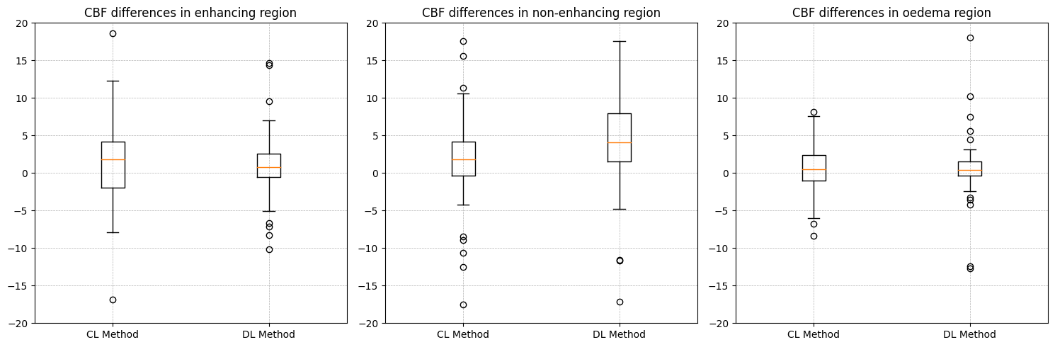
Figure 4 reports the CBF differences between using corrected calibration (either with the contralateral mean method or the DL method) and using uncorrected calibration. Consistent with previous studies4,5, CBF values from uncorrected calibration are on average lower than those from corrected calibrations. The distinction between the two corrected calibrations underscores the value in adopting the proposed approach that enables more fine-grained voxel-wise calibration.
Discussion & Conclusion
The proposed DL method based on image-inpainting enables voxel-wise calibration correction, overcoming the limitations of the contralateral mean method which only supports region-wise correction. The contralateral mean method may also lead to suboptimal results when regions with signal abnormalities lie in the medial part of the brain. More importantly, the proposed DL method can capture spatial variations of intensities in PD images which may be due to anatomical variations between the left and right hemispheres and variations in imaging factors (e.g., coil inhomogeneity). Such variations will render contralateral mean method to be biased.Acknowledgements
University College London Hospitals Biomedical Research CentreReferences
1. Razek, Ahmed Abdel Khalek Abdel, et al. "Clinical applications of arterial spin labeling in brain tumors." Journal of computer assisted tomography 43.4 (2019): 525-532.
2. Telischak, Nicholas A., John A. Detre, and Greg Zaharchuk. "Arterial spin labeling MRI: clinical applications in the brain." Journal of Magnetic Resonance Imaging 41.5 (2015): 1165-1180.
3. Alsop, David C., et al. "Recommended implementation of arterial spin‐labeled perfusion MRI for clinical applications: a consensus of the ISMRM perfusion study group and the European consortium for ASL in dementia." Magnetic resonance in medicine 73.1 (2015): 102-116.
4. Croal, Paula L., et al. "Quantification of cerebral blood flow using arterial spin labeling in glioblastoma multiforme; challenges of calibration in the presence of oedema." Proceedings of the ISMRM 27th Annual meeting & exhibition. Vol. 2315. 2019.
5. Sokolska, Magdalena., et al. "Effects of peritumoral T2 hyperintensities in treated gliomas on the quantification of tumour blood flow with arterial spin labeling." Proceedings of the 2023 ISMRM & ISMRT Annual Meeting & Exhibition. Vol. 1744. 2023.
6. Ronneberger, Olaf, Philipp Fischer, and Thomas Brox. "U-net: Convolutional networks for biomedical image segmentation." Medical Image Computing and Computer-Assisted Intervention–MICCAI 2015: 18th International Conference, Munich, Germany, October 5-9, 2015, Proceedings, Part III 18. Springer International Publishing, 2015.
7. Ruffle, James K., et al. "Brain tumour segmentation with incomplete imaging data." Brain Communications 5.2 (2023): fcad118.
8. Zhang, Hui, et al. "Deformable registration of diffusion tensor MR images with explicit orientation optimization." Medical image analysis 10.5 (2006): 764-785.
9. Liu, Guilin, et al. "Image inpainting for irregular holes using partial convolutions." Proceedings of the European conference on computer vision (ECCV). 2018.
Figures
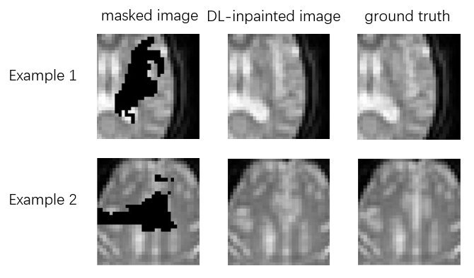
Figure 1: Examples of inpainting applied to the test set (healthy subjects).