0370
Relaxation-compensated CEST imaging of the APT can predict response to radiotherapy and progression-free survival in patients with glioma at 3T1Division of Radiology, German Cancer Research Center, Heidelberg, Germany, 2Division of Medical Physics in Radiology, German Cancer Research Center, Heidelberg, Germany, 3Faculty of Physics and Astronomy, University of Heidelberg, Heidelberg, Germany, 4Department of Radiation Oncology, Heidelberg University Hospital, Heidelberg, Germany, 5Clinical Cooperation Unit Radiation Oncology, German Cancer Research Center, Heidelberg, Germany, 6Faculty of Medicine, University of Heidelberg, Heidelberg, Germany, 7Department of Neuroradiology, Bonn University Hospital, Bonn, Germany
Synopsis
Keywords: Tumors (Pre-Treatment), CEST & MT, Asymmetry-based and Lorentzian-fit-based CEST contrast reconstruction
Motivation: There is a scarcity of studies comparing the clinical value of asymmetry- and different Lorentzian-fit-based CEST contrasts of the amide proton transfer (APT) and semi-solid magnetization transfer (ssMT) in patients with glioma.
Goal(s): To assess and compare the potential of asymmetry-based (APTwasym) and Lorentzian-fit-based CEST imaging with (MTRRexAPT and MTRRexMT) and without (MTconst) relaxation compensation for the prediction of therapy response and survival in patients with glioma.
Approach: 78 study participants prospectively underwent CEST MRI at baseline before radiotherapy.
Results: Imaging of the MTRRexAPT and MTRRexMT predicted response to radiotherapy, whilst the MTRRexAPT was also associated with progression-free survival.
Impact: Here we demonstrate for the first time that Lorentzian-fit-based CEST imaging of the APT and ssMT with relaxation compensation can predict therapy response and progression-free survival of patients with glioma at baseline before radiotherapy, at 3T.
Introduction
Chemical exchange saturation transfer (CEST) MRI can be applied to use peptides and sub-cellular macromolecules as endogenous imaging contrasts via the amide proton transfer (APT) and semi-solid magnetization transfer (ssMT) [1]. Several groups have previously demonstrated the diagnostic and predictive value of CEST imaging in patients with glioma before and after radiotherapy [2-5]. However, CEST contrasts are heavily dependent on the metrics applied for their reconstruction from the Z-spectrum, as well as the magnetic field strength [6-8]. Furthermore, there is a scarcity of cross sectional studies comparing the clinical value of asymmetry-based and different Lorentzian-fit-based CEST contrasts. Therefore, the objective of this study was to assess the potential of asymmetry-based imaging of the APT (APTwasym), as well as Lorentzian-fit-based imaging of the APT and ssMT with (MTRRexAPT and MTRRexMT) and without (MTconst) relaxation compensation for the prediction of therapy response and survival in patients with glioma at baseline before radiotherapy, at a clinical field strength of 3T.Methods
From July 2018 to December 2022, 78 study participants with diffuse glioma underwent 3T CEST MRI (Prisma®, Siemens) at baseline before radiotherapy. Imaging of the APTwasym was performed according to Zhou J, et al. using four rectangular B1 pulses of 2μT [9]. The MTRRexAPT, MTRRexMT and MTconst were acquired with two low-power B1 pulses of 0.6 and 0.9 μT according to Goerke et al. [7] and Mehrabian et al. [8]. Tumor volumes included glioma-associated contrast enhancement (CE) and whole tumor (WT), and were segmented on contrast-enhanced T1w (T1wCE) and T2w TIRM (T2w) images in Matlab® (Mathworks). Therapy response and progression-free survival (PFS) were assessed according to the response assessment in neuro-oncology (RANO) criteria, based on longitudinal follow-up. Median overall survival was not reached at data cut-off on August 6th, 2023. Statistical testing included receiver operating characteristic (ROC) and Kaplan-Meier analyses.Results
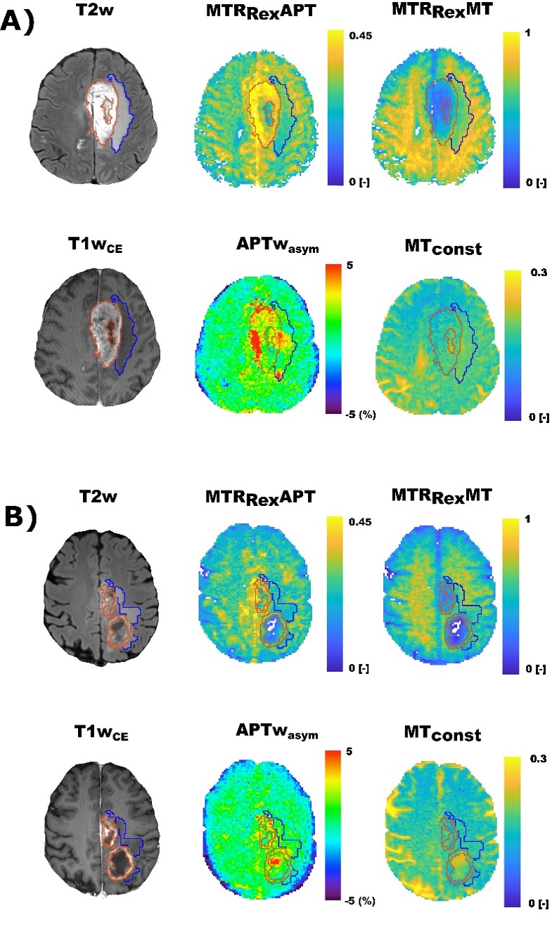
Exemplary contrast maps of two study participants with the diagnosis of glioblastoma (GBM), of whom one was assessed as having progressive disease and the other one as having pseudoprogression after the completion of radiotherapy, are shown in Fig. 1.Therapy response was available for 73 participants. Of those 57 were assessed as having stable disease (SD) and 16 as having progressive disease (PD). Of the participants with SD, 11 were assessed as having pseudoprogression (PP). Median PFS was 4.9 months (min. 0.6 and max. 32.0 months), with a median follow-up of 12.0 months (min. 0.6 and max. 54.5 months). Nine participants were lost to follow-up.
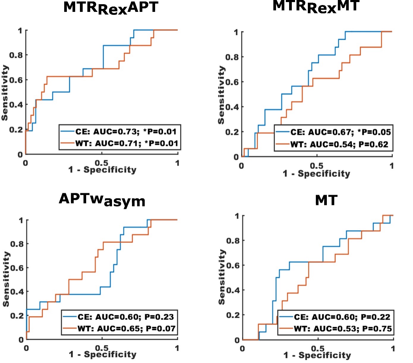
The MTRRexAPT (CE: AUC=0.73, p=0.007; WT: 0.71, p=0.01) and MTRRexMT (CE: AUC=0.67, p=0.05) were associated with therapy response and showed higher mean contrast values for participants with PD compared to those with SD. For WT, the MTRRexAPT (AUC=0.72, p=0.04) showed also higher mean contrast values for participants with PD compared to those with PP.
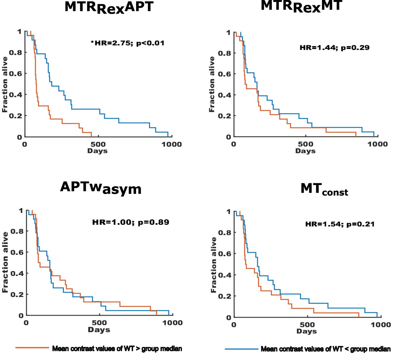
The MTRRexAPT was furthermore the only contrast that was associated with PFS (HR=2.76, p=0.004), and showed shorter survival for participants with mean contrast values above the cohort median (2.6 vs. 6.0 months) for WT.
The APTwasym and the MTconst were not associated with any of the assessed clinical outcomes.
ROC curves indicating the association of the investigated CEST contrasts for CE and WT with therapy response are displayed in Fig. 2. Kaplan-Meier plots showing the association of all contrasts for WT with PFS are displayed in Fig. 3.
Discussion
In this study, we observed a moderate association of the MTRRexAPT with therapy response and PFS in 78 participants with glioma at baseline before radiotherapy and a weaker association of the MTRRexMT with therapy response. However, whilst earlier interim analyses of the same clinical cohort demonstrated greater clinical value for the APTwasym and MTconst at the first follow-up after the completion of radiotherapy[4, 5], no predictive value was observed for these contrasts in this study. The APTwasym and MTconst have stronger contributions from the relayed nuclear Overhauser effect (rNOE), T1, and direct water saturation [6, 8]. Therefore, the clinical value of the APTwasym and MTconst in the early post-radiotherapy interval could be related to radiation-induced tissue necrosis and edema, at least to some extent. The MTRRexAPT, on the other hand, could reflect viable and proliferative tumor tissue in this cohort more accurately, given its higher specificity for the APT [6]. However, further studies comparing the association of asymmetry-based and different Lorentzian-fit-based contrasts of the APT and ssMT with histopathological features of previously untreated gliomas are needed to elucidate this matter further.Conclusion
Relaxation-compensated CEST imaging of the APT demonstrates promising predictive value in patients with glioma at baseline before radiotherapy, at a clinical field strength of 3T.Acknowledgements
We would like to thank all contributing clinicians, technicians and study nurses who made this work possible.References
1. van Zijl, P.C.M., et al., Magnetization Transfer Contrast and Chemical Exchange Saturation Transfer MRI. Features and analysis of the field-dependent saturation spectrum. Neuroimage, 2018. 168: p. 222-241.
2. Paech, D., et al., Relaxation-compensated amide proton transfer (APT) MRI signal intensity is associated with survival and progression in high-grade glioma patients. Eur Radiol, 2019. 29(9): p. 4957-4967.
3. Park, J.E., et al., Pre- and Posttreatment Glioma: Comparison of Amide Proton Transfer Imaging with MR Spectroscopy for Biomarkers of Tumor Proliferation. Radiology, 2016. 278(2): p. 514-23.
4. von Knebel Doeberitz, N., et al., CEST imaging of the APT and ssMT predict the overall survival of patients with glioma at the first follow-up after completion of radiotherapy at 3T. Radiother Oncol, 2023. 184: p. 109694.
5. Kroh, F., et al., Semi-solid MT and APTw CEST-MRI predict clinical outcome of patients with glioma early after radiotherapy. Magn Reson Med, 2023.
6. Zaiss, M., et al., Relaxation-compensated CEST-MRI of the human brain at 7T: Unbiased insight into NOE and amide signal changes in human glioblastoma. Neuroimage, 2015. 112: p. 180-188.
7. Goerke, S., et al., Relaxation-compensated APT and rNOE CEST-MRI of human brain tumors at 3 T. Magn Reson Med, 2019. 82(2): p. 622-632.
8. Mehrabian, H., et al., Evaluation of Glioblastoma Response to Therapy With Chemical Exchange Saturation Transfer. Int J Radiat Oncol Biol Phys, 2018. 101(3): p. 713-723.
9. Zhou, J., et al., APT-weighted MRI: Techniques, current neuro applications, and challenging issues. J Magn Reson Imaging, 2019. 50(2): p. 347-364.
Figures