0364
Improved MR Multitasking-based Dynamic Imaging for Cerebrovascular Evaluation (MT-DICE): Towards Multiparametric Brain Tumor Evaluation1Department of Biomedical Engineering, University of Southern California, Los Angeles, CA, United States, 2Department of Radiology, Keck School of Medicine, University of Southern California, Los Angeles, CA, United States, 3Department of Radiological Sciences, University of California, Los Angeles, Los Angeles, CA, United States, 4Biomedical Imaging Research Institute, Cedars-Sinai Medical Center, Los Angeles, CA, United States, 5Department of Neuro-oncology, Keck School of Medicine, University of Southern California, Los Angeles, CA, United States, 6Department of Neurosurgery, Keck School of Medicine, University of Southern California, Los Angeles, CA, United States, 7Department of Radiation Oncology, Keck School of Medicine, University of Southern California, Los Angeles, CA, United States
Synopsis
Keywords: Tumors (Post-Treatment), DSC & DCE Perfusion, multiparametric brain tumor evaluation
Motivation: The lack of pathophysiologically relevant quantitative information hinders the precision management of brain tumors. Specifically, the characterization of intra-tumor heterogeneity influencing treatment decision-making, remains a critical challenge.
Goal(s): This work aims to optimize a recently developed technique MT-DICE, to provide multiparametric mapping information for more comprehensive brain tumor evaluation.
Approach: The MT-DICE technique was further refined by including 3D flow compensation, dictionary-based mapping, and water exchange quantification. Assessments for the repeatability of MT-DICE parameters and their agreement with routine measurements were performed.
Results: Excellent reproducibility and agreement were achieved. Spatially co-registered multiparametric maps from MT-DICE facilitated comprehensive brain tumor characterization.
Impact: Individuals with brain tumors may benefit from more comprehensive brain tumor evaluation using multiparametric maps derived from our improved MT-DICE technique.
Introduction
MR is an imaging modality of choice for brain tumor evaluation. Routine MR imaging protocols offer valuable qualitative information but lack quantitative information that is highly desired for precision medicine. MR Multitasking-based dynamic imaging for cerebrovascular evaluation (MT-DICE) is a recently developed technique for simultaneous quantification of brain perfusion and permeability with one scan and a single contrast injection1. In addition to these two brain tumor-relevant properties, intra-tumor bleeding and water exchange across the cell membrane have also been shown to be pathologically important and can be quantified by quantitative susceptibility mapping (QSM)2 and dynamic contrast enhanced (DCE) imaging3, respectively. In this work, we further improved the MT-DICE technique to provide multiparametric information including perfusion, permeability, water exchange, and susceptibility with a single 8-minute scan.Materials and Methods
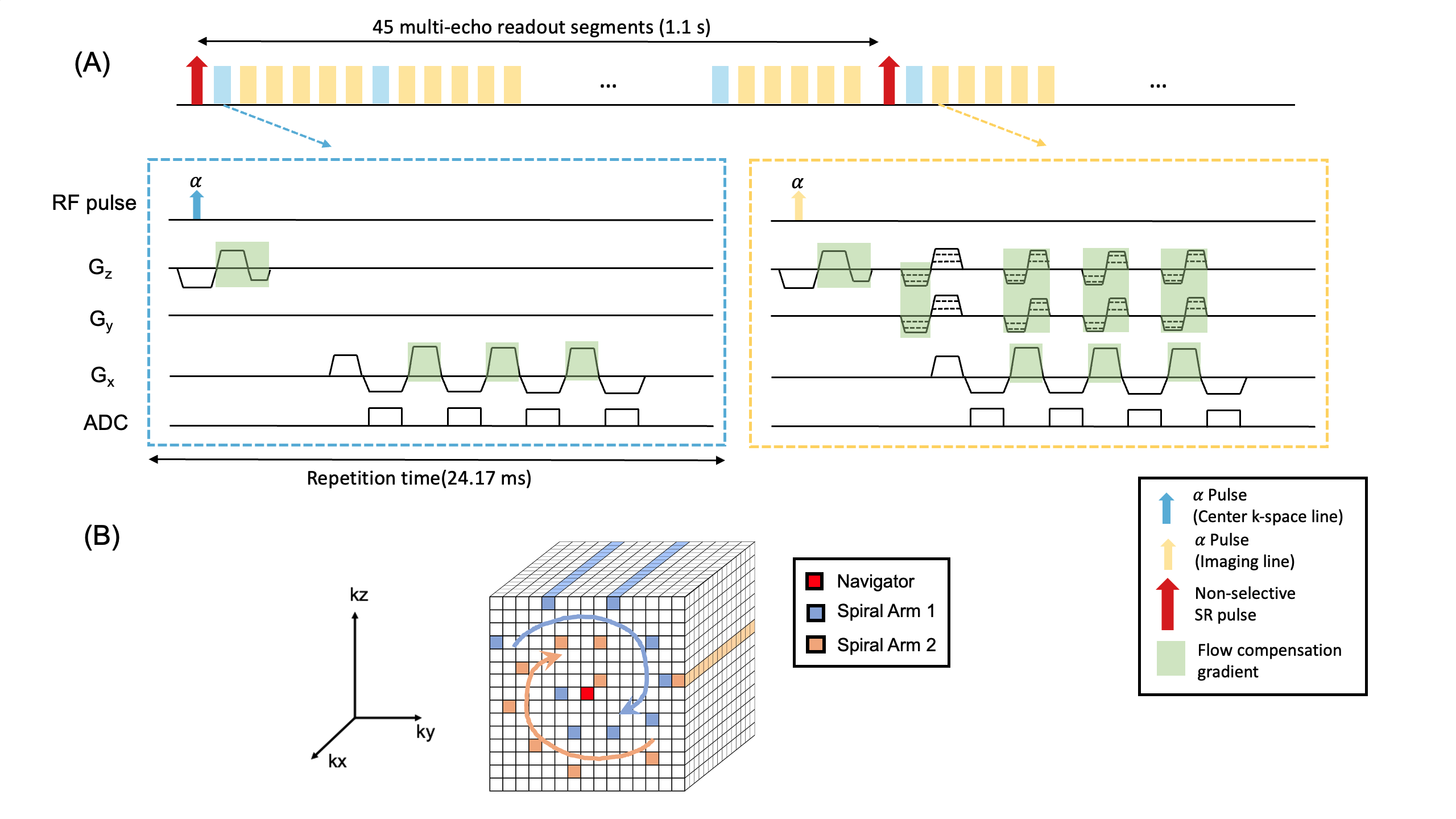
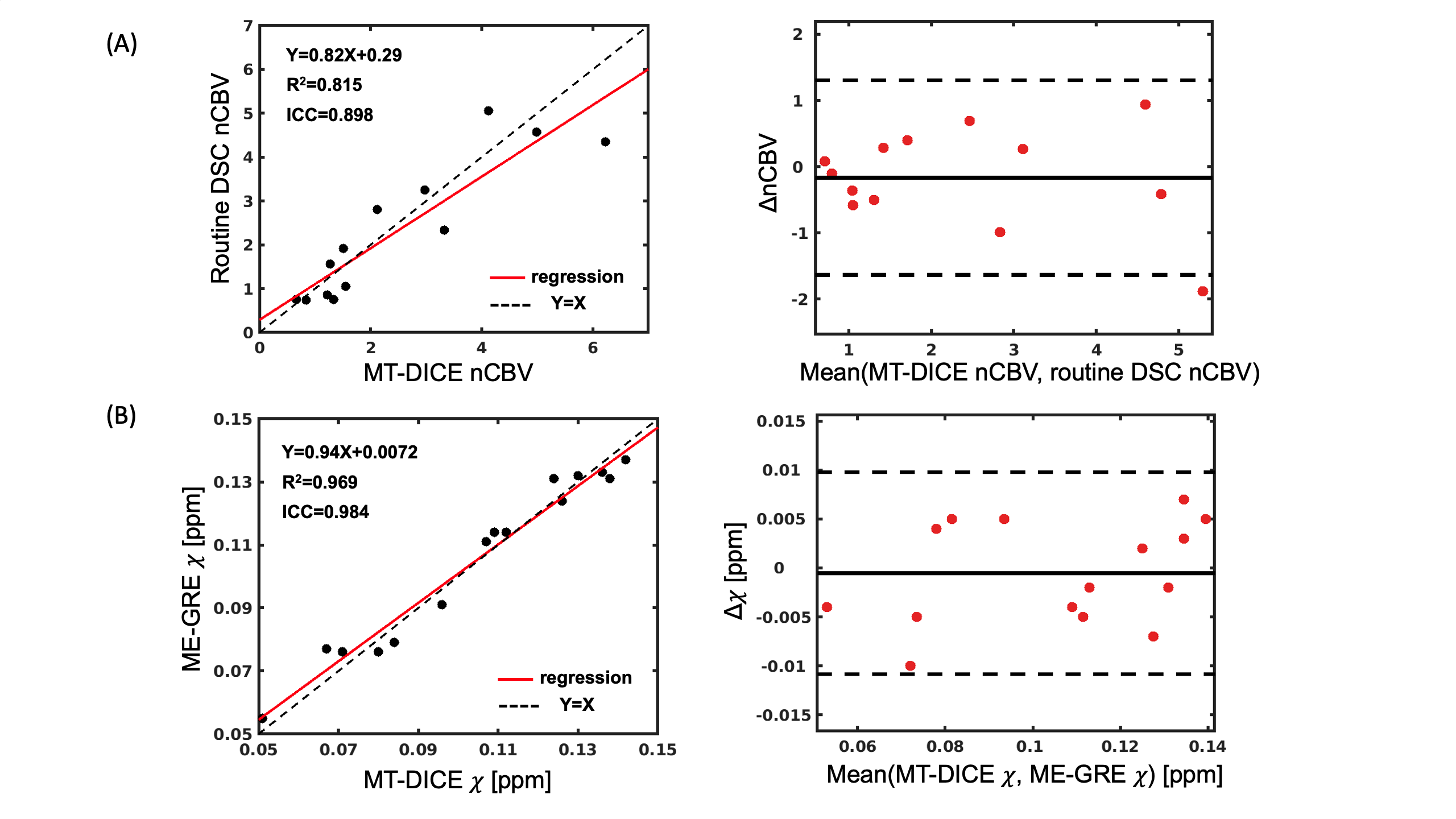
Technical refinements: Three-dimensional flow compensation gradients were added, allowing for more reliable measurement of arterial input function and quantification of susceptibility. Cartesian spiral-in sampling pattern was utilized to lower the gradient variation before each training line acquisition to minimize the eddy current effect. The sequence diagram of the optimized MT-DICE sequence and corresponding k-space sampling pattern are shown in Fig. 1. In addition, a dictionary-based mapping method was implemented to accelerate the dynamic T1/T2* mapping. Moreover, a previously published shutter-speed model4 (SSM) was used to quantify the water exchange between the interstitium and cell. In this model, the transmembrane water exchange rate is treated as finite, whereas other conventional models typically assume it to be infinitely large. Regions without contrast agent leakage, where accurate measurement of water exchange across cell membranes cannot be achieved, were excluded from the final map, due to the SSM model's reliance on the longitudinal relaxation rate difference between distinct compartments. Experiment: Ten healthy subjects were scanned at 3T (Siemens Vida). Five of them were recruited to undergo repetitive MT-DICE scans in two visits one week apart, the other 5 were scanned once with both MT-DICE and multi-echo gradient-echo (GRE) sequence (as the reference for QSM quantification). Ten brain tumor patients (three GBM and seven brain metastases) were recruited to undergo an MT-DICE scan during their clinical studies. Routine dynamic susceptibility contrast (DSC) imaging was also acquired following the MT-DICE scan with a second dose of contrast. Image analysis: Six-dimensional images were reconstructed with MT-DICE reconstruction framework, followed by the dictionary-based dynamic T1/T2* mapping. Based on dynamic T1 maps, contrast agent leakage rate constant, Ktrans, was estimated with the extended Tofts model5 and intracellular-to-extracelluar water-efflux-rate constant, Kio, was estimated with the SSM model mentioned above. Leakage-insensitive CBV and QSM maps were generated with dynamic T2* maps and pre-contrast multi-echo images respectively. The T1 enhancement map was generated by subtracting the pre-contrast image from the post-contrast image, both reconstructed using MT-DICE. Statistical analysis: Bland-Altman plot and intraclass correlation coefficient (ICC) were used to assess the repeatability of MT-DICE derived parameters. The correlation of nCBV (absolute CBV normalized to contralateral normal appearing white matter) between MT-DICE and routine DSC was evaluated with linear regression analysis, ICC, and Bland-Altman plot. The agreement of susceptibility quantification from MT-DICE and multi-echo GRE was also assessed with same analysis methods from three different brain regions including globus pallidus (GP), Putamen (Pt), and substantia nigra (SN).Results
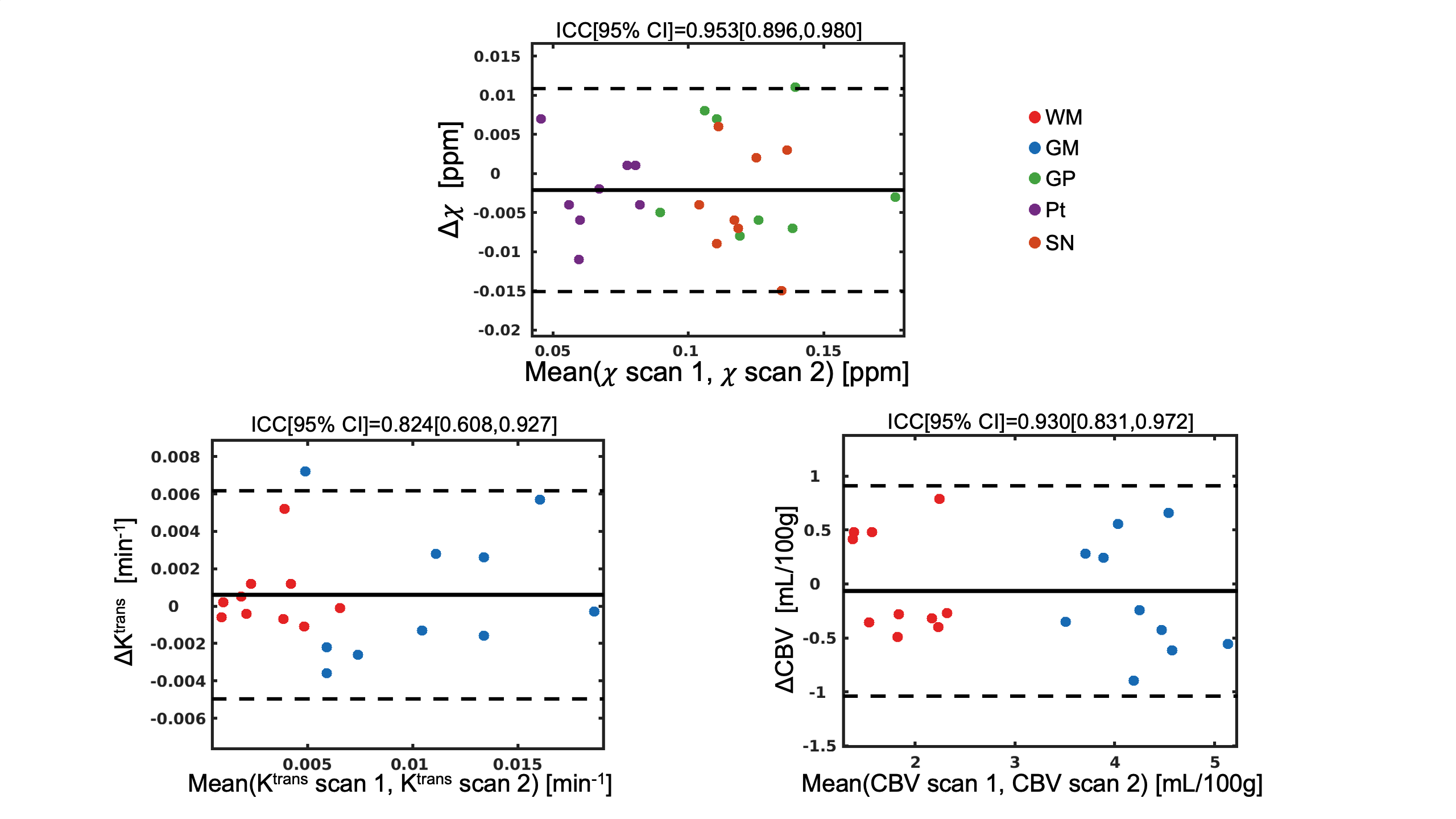
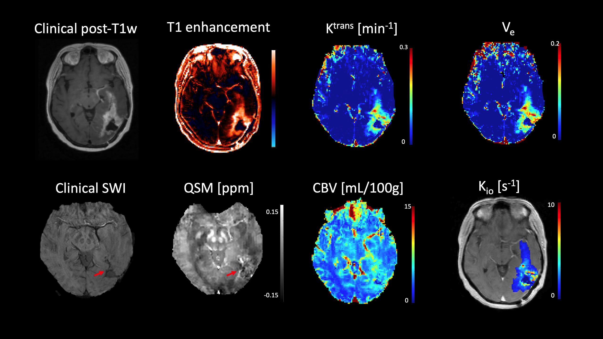
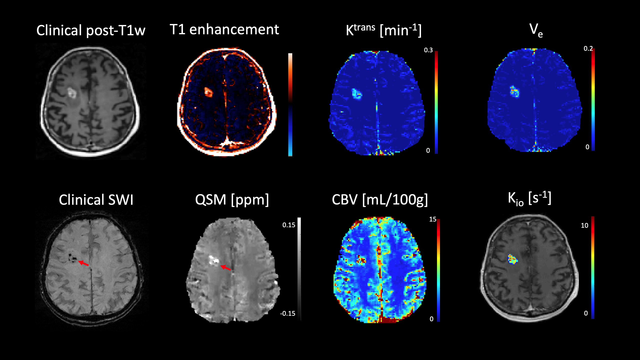
Fig. 2 shows excellent repeatability for CBV (ICC=0.930) and susceptibility (ICC=0.953) and good repeatability for Ktrans (ICC=0.824). The linear regression plots and Bland-Altman plots analyzing correlation of MT-DICE parameters with routine measurements are displayed in Fig. 3. The nCBVs and susceptibility quantified by MT-DICE were in excellent agreement with those measured by clinical DSC measurements (R2=0.815, ICC=0.898) (Fig. 3A) and multi-echo GRE (R2=0.969, ICC=0.984) (Fig. 3B), respectively. Representative maps derived from MT-DICE for in a GBM patient, and a brain metastasis patient are shown in Fig. 4 and Fig. 5 respectively. Intrinsically co-registered multiparametric maps facilitated the visualization of abnormalities within the tumor region.Discussion and conclusion
With the single scan and single contrast dose injection, the improved MT-DICE technique can simultaneously quantify brain perfusion, permeability, water exchange, and susceptibility. In addition to distinguishing the tumor abnormalities from normal tissues, the heterogeneity within the tumor area can be observed from permeability (Ktrans, Ve), water exchange (Kio), and leakage-insensitive perfusion (CBV) maps. The hemosiderin deposition within the surgical bed and intra-tumor micro bleeding are clearly visualized in QSM maps. These valuable multiparametric information obtained from MT-DICE potentially allows for more comprehensive characterization of brain tumors in a time-efficient manner, which has not been achievable feasible using clinical standard protocols. Further clinical evaluation is currently underway.Acknowledgements
No acknowledgement found.References
1. Hu Z, Christodoulou AG, Wang N, et al. MR multitasking-based dynamic imaging for cerebrovascular evaluation (MT-DICE): Simultaneous quantification of permeability and leakage-insensitive perfusion by dynamic T1/T2* mapping. Magnet Reson Med. 2022. doi:10.1002/mrm.29431
2. Deistung A, Schweser F, Wiestler B, et al. Quantitative Susceptibility Mapping Differentiates between Blood Depositions and Calcifications in Patients with Glioblastoma. Plos One. 2013;8(3):e57924. doi:10.1371/journal.pone.0057924
3. Jia Y, Xu S, Han G, et al. Transmembrane water-efflux rate measured by magnetic resonance imaging as a biomarker of the expression of aquaporin-4 in gliomas. Nat Biomed Eng. 2023;7(3):236-252. doi:10.1038/s41551-022-00960-9
4. Tofts PS, Brix G, Buckley DL, et al. Estimating kinetic parameters from dynamic contrast-enhanced t1-weighted MRI of a diffusable tracer: Standardized quantities and symbols. J Magn Reson Imaging. 1999;10(3):223-232. doi:10.1002/(sici)1522-2586(199909)10:3<223::aid-jmri2>3.0.co;2-s
5. Bai R, Wang B, Jia Y, et al. Shutter-Speed DCE-MRI Analyses of Human Glioblastoma Multiforme (GBM) Data. J Magn Reson Imaging. 2020;52(3):850-863. doi:10.1002/jmri.27118
Figures