0363
Evaluating the Clinical Utility and Diagnostic Value of High-Resolution Deuterium MRS Imaging (DMRSI) in Patients with Brain Tumor1CMRR, Department of Radiology, University of Minnesota, Minneapolis, MN, United States, 2Beckman Institute for Advanced Science and Technology, University of Illinois at Urbana-Champaign, Urbana, IL, United States, 3National Center for Supercomputing Applications, University of Illinois at Urbana-Champaign, Urbana, IL, United States, 4Deptartment of Laboratory Medicine and Pathology, University of Minnesota, Minneapolis, MN, United States, 5Department of Electrical and Computer Engineering, University of Illinois at Urbana-Champaign, Urbana, IL, United States, 6Department of Neurosurgery, University of Minnesota, Minneapolis, MN, United States
Synopsis
Keywords: Tumors (Post-Treatment), Cancer, Deuterium metabolic Imaging
Motivation: Deuterium MRS imaging (DMRSI) can detect Warburg Effect in brain tumors; however, its clinical utility and value in brain tumor diagnosis and treatment has not been investigated.
Goal(s): To perform a preliminary investigation in human patients with brain tumor.
Approach: High-resolution dynamic DMRSI (HR-DMRSI) study was conducted in brain tumor patients on a 7T clinical scanner with an oral D66-glucose administration; biospecimens taken from DMRSI positive and negative regions were analyzed and compared with the DMRSI and clinical MRI results.
Results: HR-DMRSI technology enables detection and characterization of glioma infiltration with a level of precision surpassing traditional imaging modalities.
Impact: We clearly demonstrate that 7T high-resolution dynamic deuterium (2H) MRS imaging is able to detect and characterize glioma infiltration in individual human patients with high accuracy and specificity that exceeds conventional imaging modalities available in standard clinical setting.
INTRODUCTION
Glioblastoma (GBM) is the most common primary brain tumor in adults and is associated with very high mortality and intra-tumoral heterogeneity, which poses significant challenges to clinical diagnosis and treatment management [1,2]. A molecular hallmark of primary brain tumor is upregulated glycolysis accompanying with inhibited mitochondrial oxidation, i.e., the “Warburg effect” [3,4]. Recently developed in vivo deuterium (2H) MRS imaging (DMRSI) technique can simultaneously measure the dynamic changes of deuterated glucose substrate and downstream metabolites involved in glucose metabolism, namely, 2H-labeled water (HDO), glucose (Glc), mixed glutamate/glutamine (Glx) and lactate (Lac), thus, has a potential to map the “Warburg effect” and metabolic reprograming in animal and human brain tumors [5-8]. However, the clinical utility of this novel metabolic imaging technique has not been rigorously evaluated. In this study, we conduct such an investigation by comparing DMRSI results with the histological findings of biospecimens from human brain tumor patients for the first time.METHODS
Three brain tumor patients (1 female/2 male, 32-51 years old) participated in this study approved by UMN IRB. A 7T-Terra clinical scanner (Siemens, Germany) and a novel 2H/1H array head coil [9] were used. High-resolution dynamic 3D DMRSI data with 0.7cc nominal voxel and 2.5min/volume were acquired before and after the oral administration of the D66-glucose (Cambridge Isotope Lab, 0.75 g/kg dose) solution [10], and were reconstructed using a SPICE-based processing scheme to reduce the spectral and temporal fluctuations [11]. After SPICE processing, the 2H signals of each metabolites of interest at different spatial and time points were converted to the molar concentrations using the corresponding natural abundance water signal as an internal reference after correcting the saturation effect. Patients also underwent 3T clinical scans and neurosurgery or biopsies as part of standard care; sample location for biospecimens was guided by the individual patient's DMRSI results, and standard neuropathological and immunohistochemical analysis were performed on these samples.RESULTS
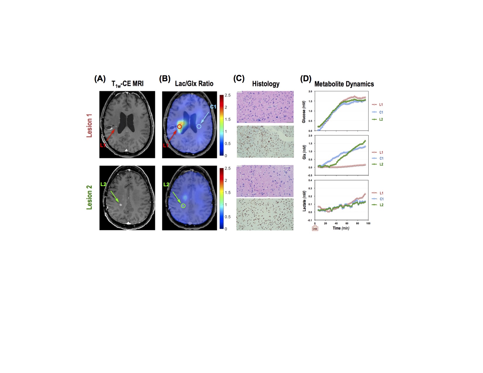
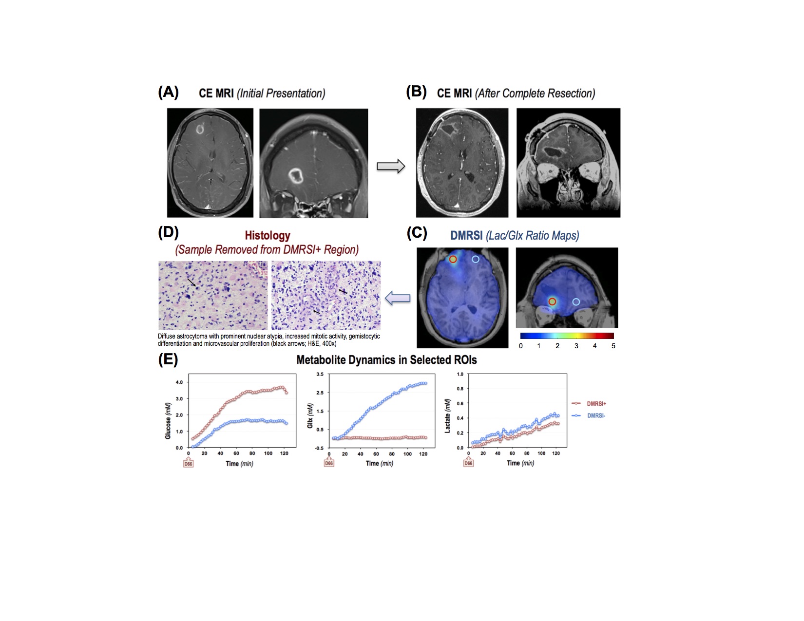
As illustrated in Fig. 1, two distinct contrast-enhancing (CE) lesions were detected in the brain of a glioblastoma patient whose tumor had been removed during a previous surgery. The DMRSI results exhibited elevated Lac/Glx ratio in Lesion 1 but not in Lesion 2. Histological analysis of the biopsy samples confirmed the presence of tumor cells in Lesion 1 (L1, DMRSI+ region with a higher value of [Lac]/[Glx] or 1/[Glx]), while no evidence of tumor in Lesion 2 (L2, DMRSI- region). The dynamic changes of deuterated glucose, Glx and lactate concentrations in the two lesions (L1 & L2) and a corresponding control region (C1) are also displayed, lower Glx and higher Lac were observed in Lesion 1, reflecting a tumor recurrence in this brain region.Another interesting case (see Fig. 2) involved a glioblastoma patient who had undergone a gross total resection of a contrast-enhancing lesion. Surprisingly, DMRSI still indicated significant abnormal signals in the vicinity of the resection cavity. Subsequent investigations led to a repeat resection procedure, with samples from the DMRS+ region revealing clear evidence of tumor infiltration.
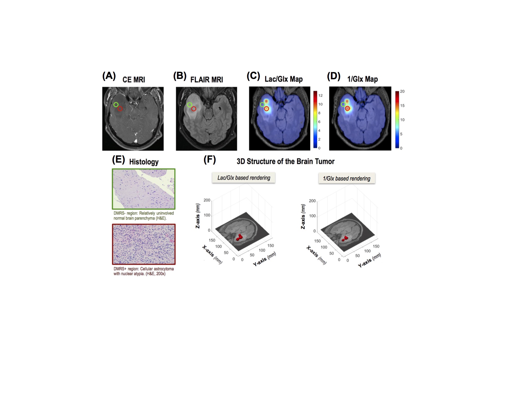
In Fig. 3, a third patient diagnosed with a low-grade glioma showed abnormal DMRSI signals; biopsies taken from DMRS+ regions indicated the presence of tumor infiltration, in contrast to samples taken from DMRS- regions, which exhibited normal brain tissue characteristics. 3D structure of the brain tumor were generated based on the elevated DMRSI signals of Lac/Glx or 1/Glx.
DISCUSSION and CONCLUSION
In previous work, we established technical capability to achieve very high spatial (0.7 cc nominal voxel) and temporal (2.5 min per 3D volume) resolution for dynamic whole-brain DMRSI in healthy human at 7T with oral intake of D66-glucose [10]. In this work, we evaluate the clinical utilities of the HR-DMRSI technology in brain tumor patients. Our results clearly demonstrate that HR-DMRSI is capable of detecting brain regions infiltrated by glioblastoma in each patient, beyond the capabilities offered by conventional gadolinium-enhanced MRI or FLAIR (Fluid-Attenuated Inversion Recovery) imaging. These findings highlight the clinical value and sensitivity of HR-DMRS in identifying and characterizing glioma infiltration with a precision that exceeds traditional imaging modalities. Furthermore, through additional work that combines kinetic modeling with metabolites dynamics (e.g., Figs. 1D & 2E), we expect that metabolic rates of glucose consumption, lactate production and TCA cycle activity can be determined simultaneously in brain tumor and normal appearing tissues.In conclusion, high-resolution dynamic DMRSI technique as utilized in this work can provide a powerful neuroimaging tool for improving clinical diagnosis and treatment management of brain tumor patients.
Acknowledgements
NIH Grants: R01 CA240953, NS118330 and NS133006, U01 EB026978, P41 EB027061 and S10 OD025256.
Technical support from Siemens.
References
1. Louis DN. Molecular pathology of malignant gliomas. Annu Rev Pathol 1:97-117 (2006).
2. Sottoriva A et al. Intratumor heterogeneity in human glioblastoma reflects cancer evolutionary dynamics. Proc Natl Acad Sci U S A 110:4009-4014 (2013).
3. Warburg O. On the origin of cancer cells. Science 123:309-314 (1956).
4. Vander Heiden MG, Cantley LC & Thompson CB. Understanding the Warburg effect: the metabolic requirements of cell proliferation. Science 324:1029-1033 (2009).
5. Lu M, Zhu XH, Zhang Y, Mateescu G and Chen W. Quantitative assessment of brain glucose metabolic rates using in vivo deuterium magnetic resonance spectroscopy. J Cereb Blood Flow Metab, 37:3518-3530 (2017).
6. Lu M, Zhu XH, Zhang Y, Low W and Chen W. Simultaneous assessment of abnormal glycolysis and oxidative metabolisms in brain tumor using in vivo deuterium MRS imaging. Proc. Intl. Soc. Mag. Reson. Med., 24:3962 (2016).
7. Lu M, Zhu XH, Zhang Y, Low W and Chen W. High-resolution deuterium MR spectroscopic imaging of the Warburg effect in brain tumor. Proc. Intl. Soc. Mag. Reson. Med., 26:4852 (2018).
8. De Feyter HM et al. Deuterium metabolic imaging (DMI) for MRI-based 3D mapping of metabolism in vivo. Sci. Adv. 4, eaat7314 (2018).
9. Li X et al. A multinuclear 4-channel 2H loop and 4-channel 1H microstrip array coil for human head MRS/MRI at 7T. Proc. Intl. Soc. Mag. Reson. Med., 31:3727 (2023).
10. Li X et al. Whole brain deuterium MRS imaging (DMRSI) with high spatiotemporal resolution to map and differentiate grey and white matter metabolic dynamics in human brain at 7T. Proc. Intl. Soc. Mag. Reson. Med., 31:3868 (2023).
11. Li Y et al. Machine Learning-Enabled High-Resolution Dynamic Deuterium MR Spectroscopic Imaging. IEEE Trans Med Imaging doi:10.1109/TMI.2021.3101149 (2021).
Figures