0359
Comparison of MUSE and ssEPI for diffusion-weighted imaging in meningioma: imaging quality and grading accuracy1Department of Radiology, Fujian Medical University Union Hospital, Fuzhou, Fujian, China, 2School of Medical Imaging, Fujian Medical University, Fuzhou, Fujian, China, 3MR Research, GE Healthcare, Beijing, China
Synopsis
Keywords: Tumors (Pre-Treatment), Neuro
Motivation: DWI is of key importance in evaluating biological behavior of meningioma, but image quality of conventional ssEPI-DWI is unsatisfactory due to susceptibility artifact near the skull.
Goal(s): Our goal was to compare the image quality of ssEPI-DWI and MUSE-DWI in meningiomas, and to compare diagnostic accuracy of them in meningiomas grading.
Approach: We used a 5-point Likert scale to assess image quality of DWI, and calculated SNR and CNR for quantitative evaluation. Combined models were constructed by using ADC histogram parameters extracted from whole tumor.
Results: MUSE-DWI significantly improved imaging quality of DWI, and showed a significantly higher diagnostic accuracy in meningioma grading.
Impact: Meningioma is the most commom intracranial tumours. This study revealed that MUSE-DWI, compared with ssEPI-DWI, can improve the imaging quality and grading accuracy of meningiomas, contributing to better clinical evaluation of meningiomas.
Introduction
Meningioma is a common kind of primary intracranial tumour, accounting for about one-third of all primary intracranial tumours [1]. Meningiomas can be classified into low-grade and high-grade tumours, which corresponds to differently biological behaviors such as invasiveness, recurrence risk and proliferation activity. Previous studies showed that DWI was important for assessing the biological behavior of meningiomas [2]. However, meningiomas often occur near the skull and even at the base of the skull, where the tumour imaging is prone to distortion and susceptibility artifact on conventional single-shot echo-planar diffusion-weighted imaging (ssEPI-DWI). This makes it difficult to delineate tumor regions and obtain precise ADC values on diffusion images. Multiplexed Sensitivity Encoding diffusion-weighted imaging (MUSE-DWI) has been proposed to reduce image artifacts and geometric distortion. MUSE-DWI has been proven to improve image quality of endometrial cancer, Crohn's disease, liver tumours, etc [3-6].So far, no study has applied MUSE-DWI to the evaluation of meningiomas. Whole-tumour histogram analysis based on ADC can provide more accurate evaluation of tumour heterogeneity [7], and it is also a more objectively quantitative evaluation of tumour grading. Our study used a 5-point Likert scale to qualitatively assess the image quality of MUSE-DWI and ssEPI-DWI, and calculated SNR and CNR for quantitative assessment. For both diffusion techniques, combined models are rconstructed by using ADC parameters extracted from 3D-VOI based histogram analysis.Methods
Consecutive 73 patients with pathologically proven meningiomas were included in this prospective study. Two neuroradiologists independently assessed the image quality using a 5-point Likert scale, while quantitatively evaluated the image quality by calculating the Signal-to-noise ratio of lesion (SNRlesion), Signal-to-noise ratio of the normal white matter (SNRnormal) and lesion to brain contrast-to-noise ratio (CNR). Kappa and Intra-class correlation coefficients (ICC) were used to measure the consistency of qualitative and quantitative parameters between two raters. Quantitative scores, qualitative SNR values and CNR values of the two diffusion techniques were compared using the Wilcoxon signed-rank test. Histogram metrics including mean, median, maximum, minimum, 10th percentiles (C10), 90th percentiles (C90), interquartile range, kurtosis, skewness and variance of ADC values were extracted from the whole tumour. The Mann-Whitney U test was used to compare the histogram parameters between high- and low-grade meningiomas. ROC curve, Delong test, and logistic regression analyses were performed to evaluate the diagnostic performance of single histogram parameters and combined models for tumour grading.Results
Sharpness, distortion, artifact, lesion conspicuity and overall image quality were significantly better in MUSE-DWI than those in ssEPI-DWI with good agreement between the two raters (p < 0.05; ICC: 0.82–0.91), while there was no significant difference in diagnostic confidence between the two DWI sequences (p>0.05; ICC: 0.85-0.86). The values of SNRlesion, SNRnormal and CNR were all significantly higher in MUSE-DWI than in ssEPI-DWI with good agreement between the two raters (p< 0.05; ICC: 0.82–0.92). The best diagnostic accuracy was obtained by combining the ADC C10 and interquartile range (An AUC of 0.774 for MUSE-DWI; 0.713 for ssEPI-DWI). The Delong test showed significant differences in diagnostic accuracy between the two combined models (p=0.045).Discussion
This study found that MUSE-DWI had better image quality than ssEP-DWI in both qualitative and quantitative assessments, which was consistent with previous MUSE-DWI studies in Crohn's disease, endometrial cancer, liver tumours,etc [3-6]. ADC values are more accurate in MUSE-DWI than in ssEPI-DWI, possibly attributing to the fewer susceptibility artifact in MUSE-DWI. As expected, the combined model based on MUSE-DWI had superior grading accuracy. Thus, MUSE-DWI might provide a convenient and reliable method for preoperative evaluation of meningiomas. In previous studies, conventional diffusion techniques based on ssEPI have shown potential in the grading, subtyping, cellular proliferation and recurrence prediction of meningiomas [8-11].The application of MUSE-DWI might better evaluate microstructural characteristics of meningiomas. Notably, we found that MUSE-DWI can better depict the boundary between lesions and normal tissues, suggesting that MUSE-DWI has a promising future in the evaluation of tumour invasion.Conclusion
Compared with ssEPI-DWI, MUSE-DWI improves the diffusion image quality of meningiomas and enhances the accuracy of tumour grading.Acknowledgements
We acknowledge Jialu Zhang from GE Healthcare for the technical support.
References
[1]Yarabarla V, Mylarapu A, Han TJ, McGovern SL, Raza SM, Beckham TH. Intracranial meningiomas: an update of the 2021 World Health Organization classifications and review of management with a focus on radiation therapy. Front Oncol. 2023 Aug 22;13:1137849.
[2]Tsai YT, Hung KC, Shih YJ, Lim SW, Yang CC, Kuo YT, Chen JH, Ko CC. Preoperative Apparent Diffusion Coefficient Values for Differentiation between Low and High Grade Meningiomas: An Updated Systematic Review and Meta-Analysis. Diagnostics (Basel). 2022 Mar 4;12(3):630.
[3]Ota T, Tsuboyama T, Onishi H, Nakamoto A, Fukui H, Yano K, Honda T, Kiso K, Tatsumi M, Tomiyama N. Diagnostic accuracy of MRI for evaluating myometrial invasion in endometrial cancer: a comparison of MUSE-DWI, rFOV-DWI, and DCE-MRI. Radiol Med. 2023 Jun;128(6):629-643.
[4]Chang HC, Chen G, Chung HW, Wu PY, Liang L, Juan CJ, Liu YJ, Tse MD, Chan A, Zhang S, Chiu KW. Multi-shot Diffusion-Weighted MRI With Multiplexed Sensitivity Encoding (MUSE) in the Assessment of Active Inflammation in Crohn's Disease. J Magn Reson Imaging. 2022 Jan;55(1):126-137.
[5]Kim YY, Kim MJ, Gho SM, Seo N. Comparison of multiplexed sensitivity encoding and single-shot echo-planar imaging for diffusion-weighted imaging of the liver. Eur J Radiol. 2020 Nov;132:109292.
[6]Zhao W, Ju S, Yang H, Wang Q, Fang L, Pylypenko D, Wang W. Improved Value of Multiplexed Sensitivity Encoding DWI with Reversed Polarity Gradients in Diagnosing Prostate Cancer: A Comparison Study with Single-Shot DWI and MUSE DWI. Acad Radiol. 2023 Sep 29:S1076-6332(23)00446-4.
[7]Gao A, Zhang H, Yan X, Wang S, Chen Q, Gao E, Qi J, Bai J, Zhang Y, Cheng J. Whole-Tumor Histogram Analysis of Multiple Diffusion Metrics for Glioma Genotyping. Radiology. 2022 Mar;302(3):652-661.
[8]Cao T, Jiang R, Zheng L, Zhang R, Chen X, Wang Z, Jiang P, Chen Y, Zhong T, Chen H, Wu P, Xue Y, Lin L. T1 and ADC histogram parameters may be an in vivo biomarker for predicting the grade, subtype, and proliferative activity of meningioma. Eur Radiol. 2023 Jan;33(1):258-269.
[9]Miyoshi K, Wada T, Uwano I, Sasaki M, Saura H, Fujiwara S, Takahashi F, Tsushima E, Ogasawara K. Predicting the consistency of intracranial meningiomas using apparent diffusion coefficient maps derived from preoperative diffusion-weighted imaging. J Neurosurg. 2020 Nov 13;135(3):969-976.
[10]Lin L, Bhawana R, Xue Y, Duan Q, Jiang R, Chen H, Chen X, Sun B, Lin H. Comparative Analysis of Diffusional Kurtosis Imaging, Diffusion Tensor Imaging, and Diffusion-Weighted Imaging in Grading and Assessing Cellular Proliferation of Meningiomas. AJNR Am J Neuroradiol. 2018 Jun;39(6):1032-1038.
[11]Hsieh HP, Wu DY, Hung KC, Lim SW, Chen TY, Fan-Chiang Y, Ko CC. Machine Learning for Prediction of Recurrence in Parasagittal and Parafalcine Meningiomas: Combined Clinical and MRI Texture Features. J Pers Med. 2022 Mar 24;12(4):522.
Figures
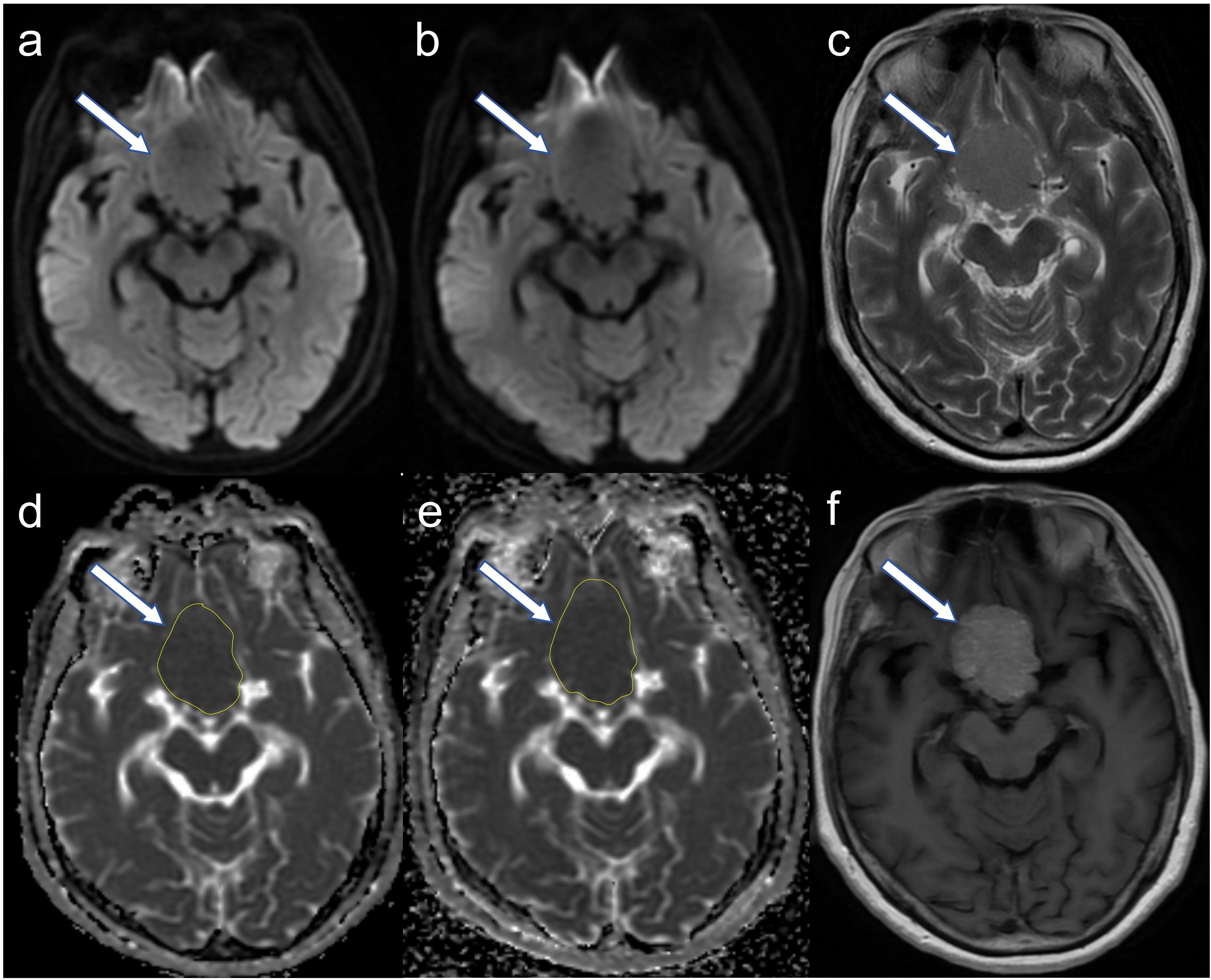
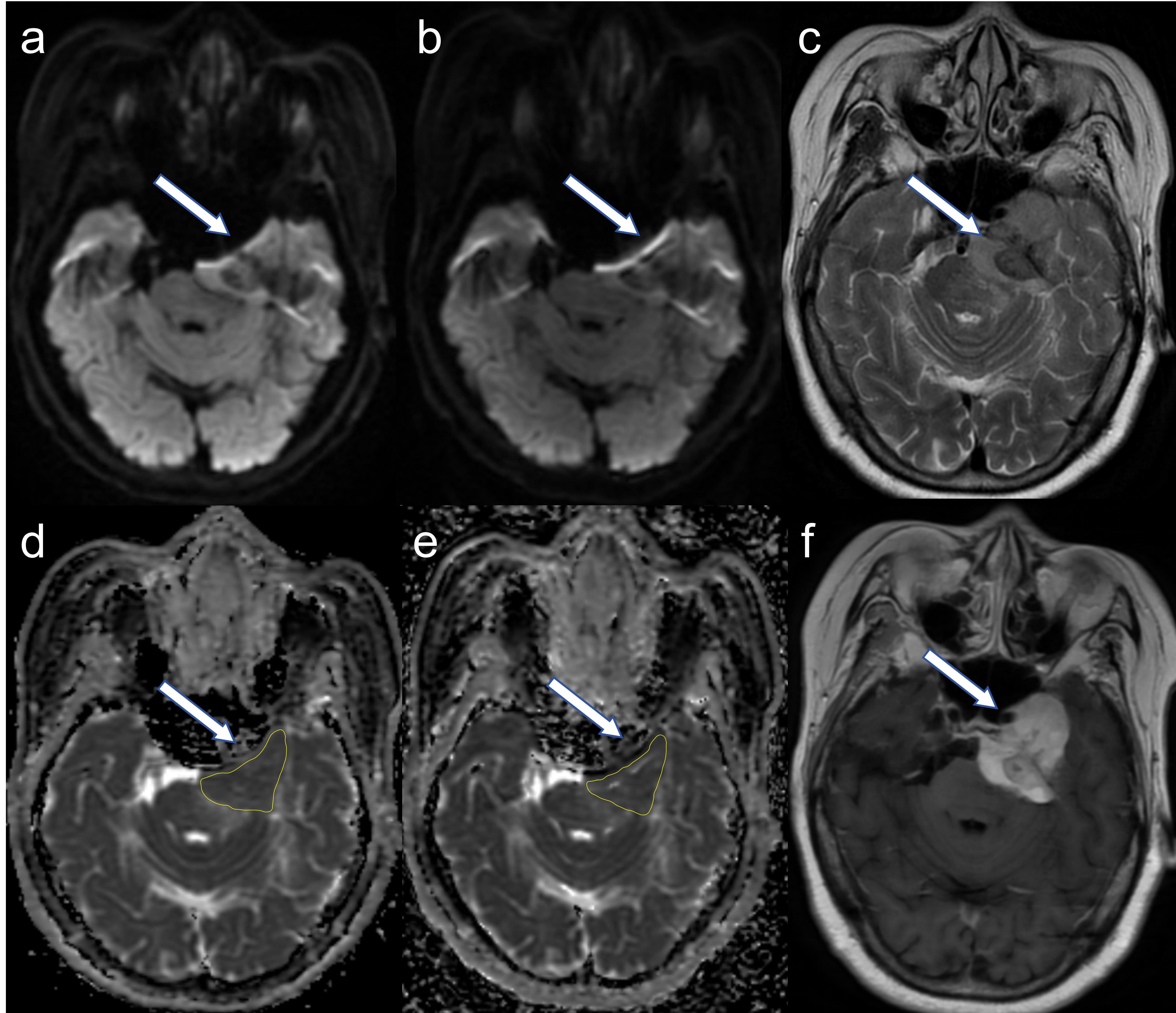
Figure: 2 Images from a 69-year-old woman with histologically proven meningioma, including MUSE-DWI image (a), ssEPI-DWI image (b), T2-weighted image (c), ADC map of MUSE-DWI (d), ADC map of ssEPI-DWI (e), T1-CE image(f ). In this case, the meningioma is located at the petroclival region near the base of posterior fossa (white arrow). Affected by the air in the inferior mastoid air cells of temporal bone, the local magnetic susceptibility varies greatly. The tumor on MUSE-DWI (a) has superior sharpness, less susceptibility artifact and fewer distortion than on ssEPI-DWI (b) .
.
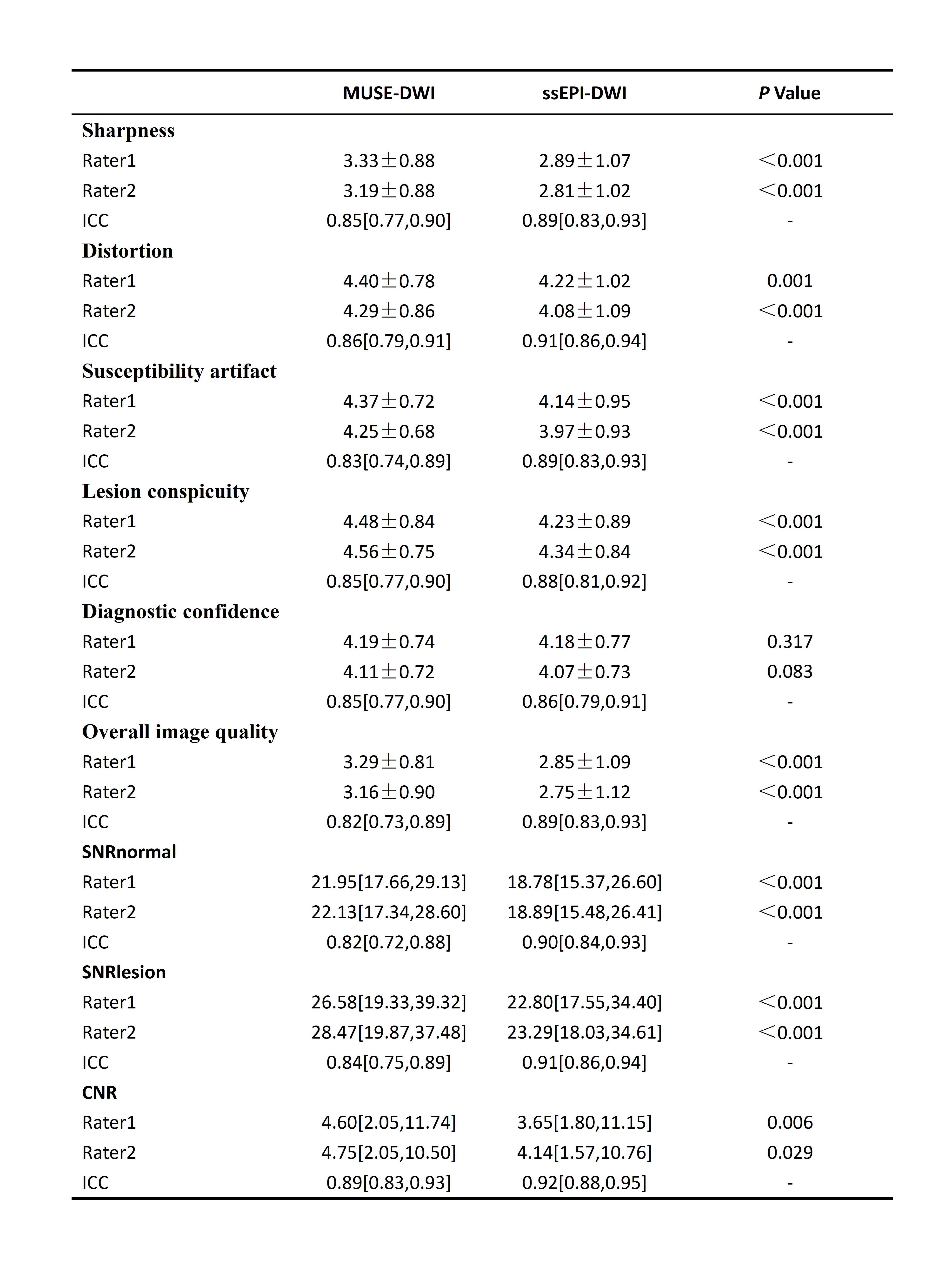
Table: 1 Qualitative and quantitative comparison of image quality between MUSE-DWI and ssEPI-DWI in meningiomas by the two raters.
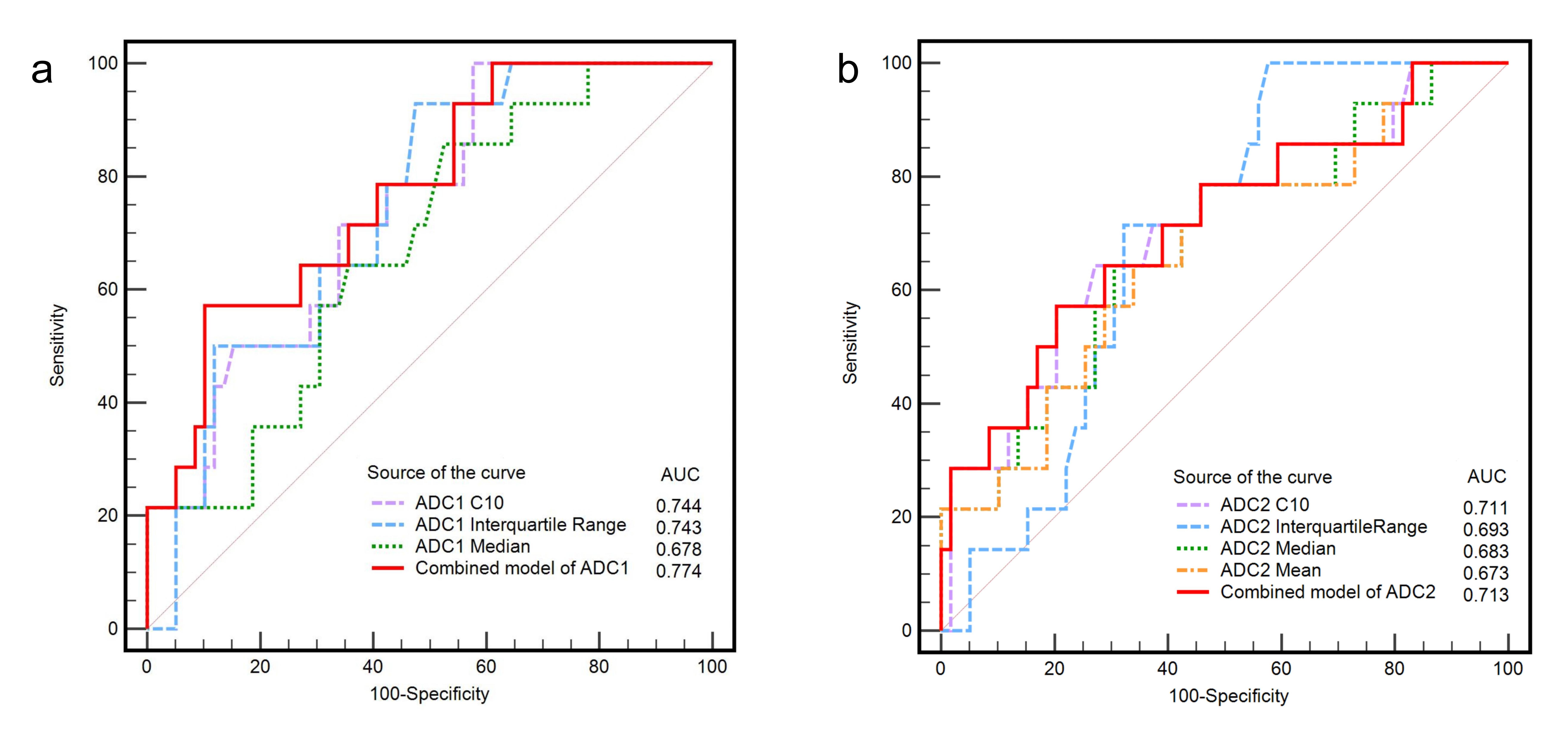
Figure:3 ROC curves of ADC1 (a. ADC of MUSE-DWI) and ADC2 (b. ADC of ssEPI-DWI) histogram parameters in tumours to differentiate low- and high-grade
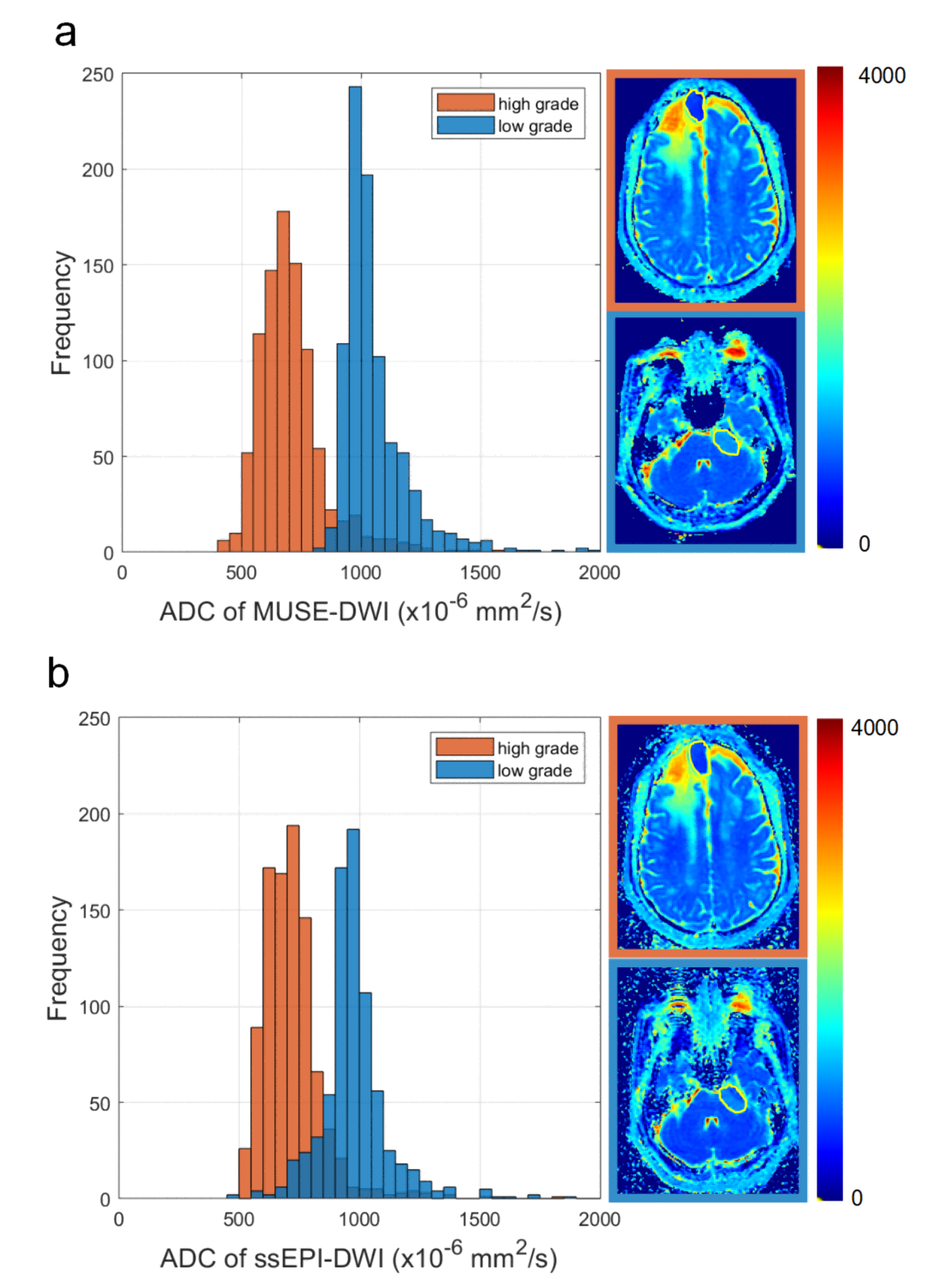
Figure: 4 ADC histograms and maps of MUSE-DWI and SSEPI-DWI. In the orange box at the upper right corner is a case of a 67-year-old male WHO grade 2 atypical meningioma (high grade). In the blue box at the lower right corner is another case of a 54-year-old woman WHO grade 1 angiomatous type meningioma (low grade).The left side of figure4a and figure4b correspond to the respective ADC histograms of the two diffusion techniques for the two cases.