0355
LIPCON: Lipid Identification with Convolutional Neural Network for MR Spectroscopic Imaging1Athinoula A. Martinos Center for Biomedical Imaging, Massachusetts General Hospital, Boston, MA, United States, 2Department of Radiology, Massachusetts General Hospital, Harvard Medical School, Boston, MA, United States, 3Computational Imaging Research Lab - Department of Biomedical Imaging and Image-guided Therapy, Medical University of Vienna, Vienna, Austria, 4High Field MR Center - Department of Biomedical Imaging and Image‐Guided Therapy, Medical University of Vienna, Vienna, Austria, 5Center for Biomedical Imaging (CIBM), Geneva, Switzerland, 6Department of Radiology and Medical Informatics, University of Geneva, Geneva, Switzerland, 7Advanced Clinical Imaging Technology, Siemens Healthineers International AG, Lausanne, Switzerland
Synopsis
Keywords: AI/ML Image Reconstruction, Spectroscopy, Lipid-Suppression, Brain, High-Field MR, Deep Learning
Motivation: Magnetic Resonance Spectroscopic Imaging (MRSI) offers non-invasive metabolic concentration mapping, aiding early pathology detection like brain tumors. However, extracranial lipid signals can compromise neurochemical data.
Goal(s): The potential of supervised neural networks remains unexplored, despite their success in other artifact removal and metabolite quantification tasks. We introduce a deep learning method for robust lipid removal.
Approach: Our approach is compared to a state-of-the-art L2-lipid-regularization using simulated and in-vivo whole-brain MRSI data.
Results: Our supervised deep learning method showed improved performance to the L2-lipid-regularization method by eliminating more lipid signal while preserving metabolic signals and spectral baseline.
Impact: The LIPCON method achieves accurate lipid suppression across whole-brain MRSI datasets without the need for parameter tuning and within a few seconds. This should mark a step in enhancing the reproducibility and efficiency of MRSI pipelines.
Authorship
* Sharing senior authorship.Introduction
Magnetic resonance spectroscopic imaging (MRSI) has gained growing influence as it enables non-invasively visualization of metabolite concentrations across the brain [6], which has great potential for disease investigation [7].However, artifact removal of lipid signal originating from the scalp region remains a persistent challenge [5]. The overlap of low-intensity metabolites with lipid signals that are orders of magnitude greater make this task particularly challenging.
In response to this challenge, we introduce the Lipid Identification with Convolutional Neural Network (LIPCON), a deep learning approach for lipid removal, enhancing the suppression of lipid-contaminated spectra while preserving the metabolic signals.
Method
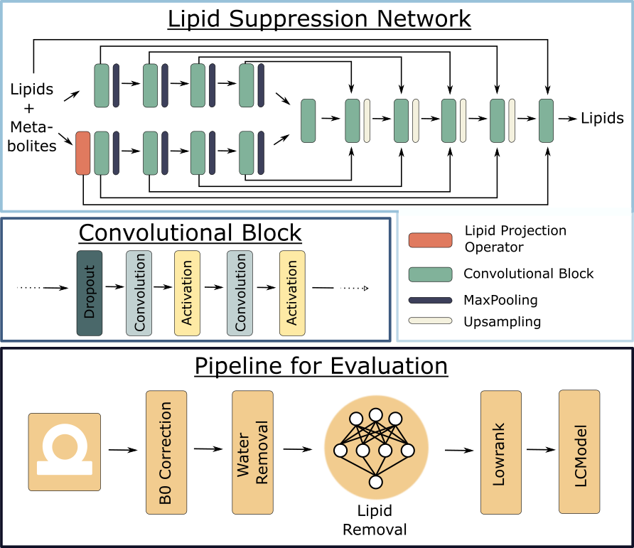
LIPCON employs a Y-Net convolutional neural network structure [2], depicted in Figure 1, utilizing two encoders instead of one. The network takes two distinct inputs: an MRS spectrum containing lipid signal contamination and the same spectrum subjected to a linear projection operator onto the lipid subspace (defined as the (1 - Lipid removal operator) in [1]). The aimed outcome is a spectrum exclusively containing the lipid signal, which is subsequently removed from the contaminated MRS spectra.Architecture:
Each encoder/decoder of the Y-Net comprised four convolutional blocks, each consisting of two convolutional layers, PReLU activation functions [4], a dropout rate of 0.01, and MaxPooling/upsampling with a factor of 2. Skip connections are ussed to connect the encoders and decoder via concatenation. An additional convolutional block is incorporated in the bottleneck region and after the decoder.
Data Simulation and training:
The training spectra were generated through a multi-step process. First, metabolite spectra were simulated using a physical model [10]. This model was tailored to simulate 25 common 1H metabolites across an extensive dataset of 106 spectra. The spectra were generated with a parameter distribution that encompassed a wide range of possibilities, including variations in frequency offset (-100 to 100 Hz), linewidth (4 to 30 Hz), and signal-to-noise ratio (SNR from 1 to 10). Additionally, a random baseline was introduced, characterized by 10 broad gaussian components. Subsequently, we extracted a collection of lipid spectra from voxels within the scalp region obtained from in-vivo datasets (acquired as part of the study [9]). To create the training input spectra, the simulated metabolite and collected lipid spectra were randomly combined. The relative lipid amplitude was varied within a broad range, spanning from 10-2 to 103. The network's reference training output was established by exclusively considering the lipid component of the spectra. The training dataset was generated with lipid signals from 19 subjects, with 2 of them being patients, while the validation dataset includes 2 other subjects.
Test data Acquisition:
2D 1H-FID-MRSI data were acquired on 3 subjects at 7T (MAGNETOM Terra.X, Siemens Healthcare, Erlangen, Germany) with a 1Tx/32Rx head coil (NovaMedical, USA 1Tx/32Rx) and with 0.9ms TE, 275ms TR, a 4x4x10mm3 voxel size, a matrix size of (53x41). The spectral bandwidth was set to 2326Hz with 450 FID points. The data was acquired by a fully sampled elliptical phase encoding.
Results
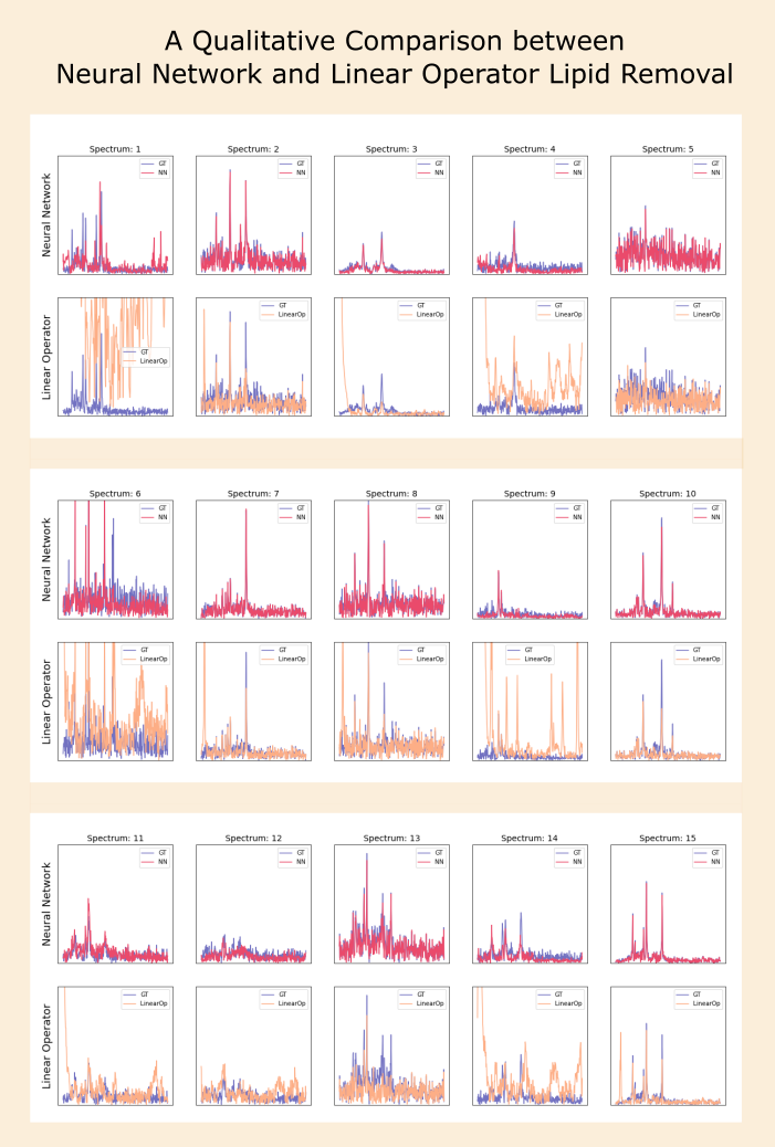
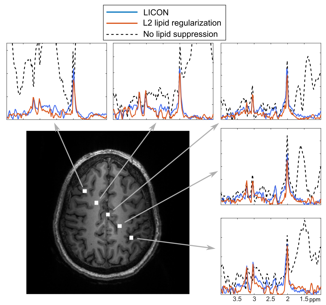
In Figure 2, we present the results of applying the LIPCON and L2-lipid-regularization methods to suppress lipid signals in simulated spectra, with both approaches compared against the ground truth. Generally, the results reveal that the spectra processed with LIPCON exhibit both reduced residual lipid signal and less attenuation of metabolic signals compared to the L2-lipid-regularization method.Figure 3 showcases the in-vivo results of LIPCON's lipid suppression. The data without lipid suppression illustrate the strong signal contamination originating from the skull lipids. Notably, the results obtained with LIPCON demonstrate an improved preservation of the NAA signal (2ppm) and the spectral baseline.
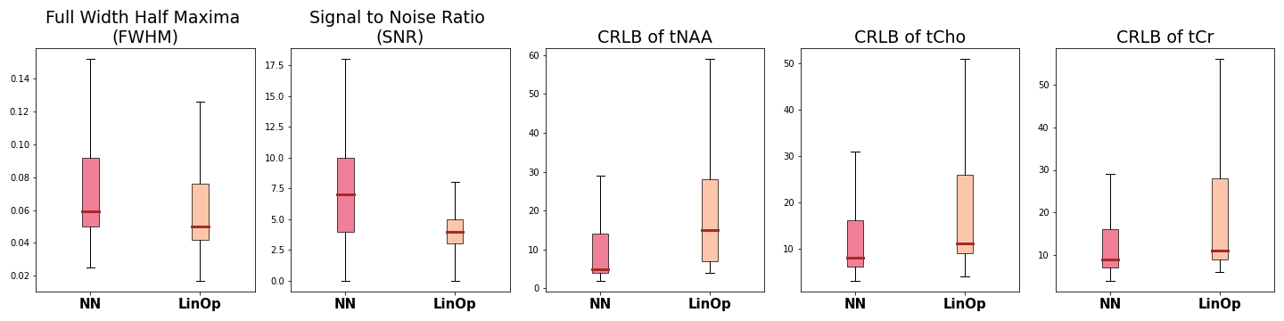
In Figure 4, we display spectral quality metrics evaluated by LCModel for the three test subjects. The results reveal stable values across methods, eliminating any systematic impact on spectral quality.
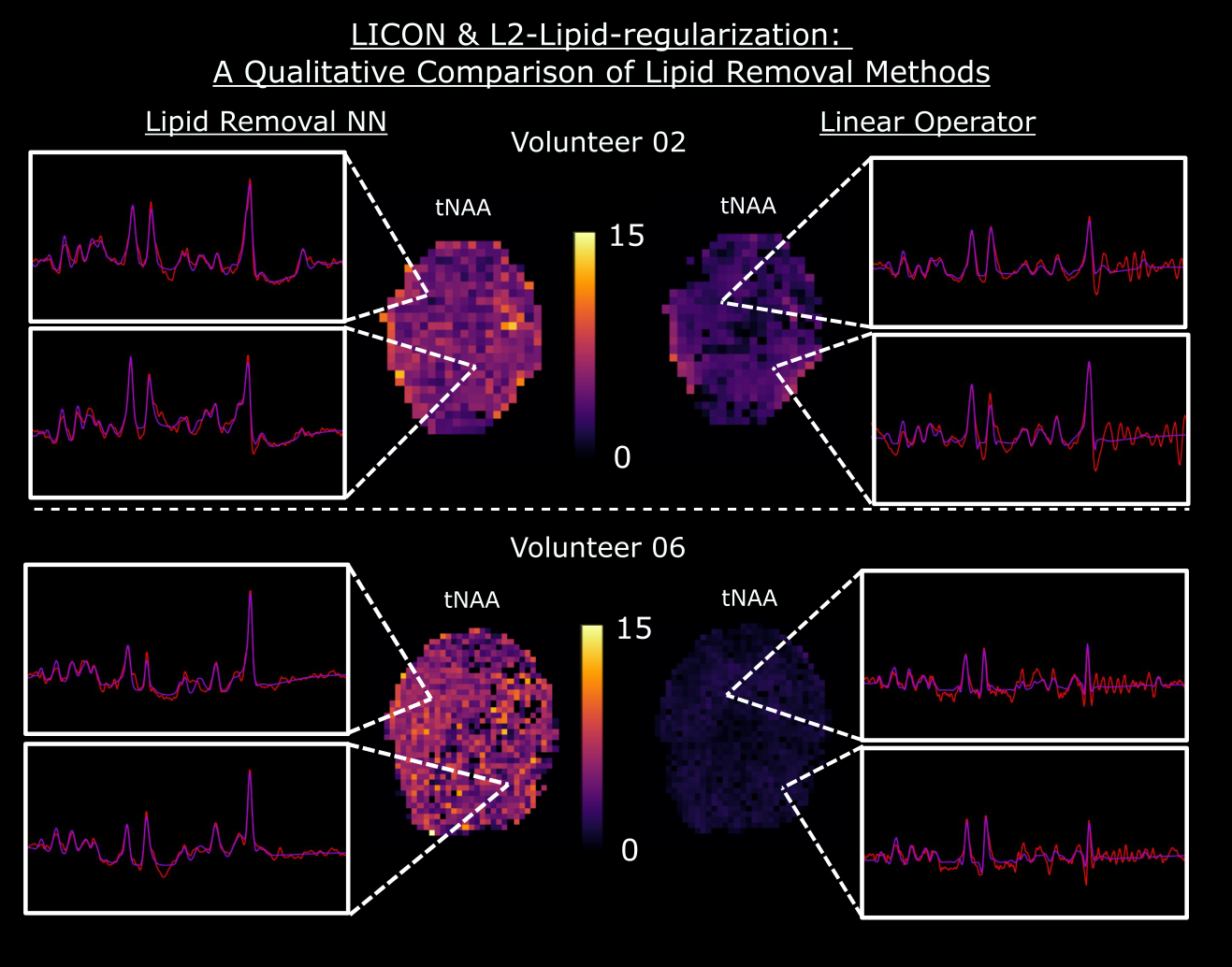
Figure 5 presents a qualitative comparison on NAA maps and selected spectra with LCModel fit of two subjects. When using the L2-lipid-regularization technique, a reduction in the NAA signal is observed with a dip in the baseline around 2 ppm and some remaining lipid signal. In contrast, LIPCON effectively removes even small lipid signals while maintaining the integrity of both the metabolic signal and baseline.
Conclusion
The LIPCON method has demonstrated notable enhancements in the removal of skull lipid signals from whole-brain MRSI data. This pipeline represents a streamlining of the lipid suppression process, as it necessitates no parameter adjustments and operates on data within seconds, highlighting its efficiency and effectiveness.Acknowledgements
No acknowledgement found.References
[1]: Bilgic, Berkin, et al. "Fast image reconstruction with L2‐regularization." Journal of magnetic resonance imaging 40.1 (2014): 181-191.
[2]: Mohammed, Ahmed, et al. "Y-net: A deep convolutional neural network for polyp detection." arXiv preprint arXiv:1806.01907 (2018).
[3]: Ronneberger, Olaf, Philipp Fischer, and Thomas Brox. "U-net: Convolutional networks for biomedical image segmentation." Medical Image Computing and Computer-Assisted Intervention–MICCAI 2015: 18th International Conference, Munich, Germany, October 5-9, 2015, Proceedings, Part III 18. Springer International Publishing, 2015.
[4]: He, Kaiming, et al. "Delving deep into rectifiers: Surpassing human-level performance on imagenet classification." Proceedings of the IEEE international conference on computer vision. 2015
[5]: Tkáč, I., Deelchand, D., Dreher, W., Hetherington, H., Kreis, R., Kumaragamage, C., Považan, M., Spielman, D. M., Strasser, B., & Graaf, R. A. (2021). Water and lipid suppression techniques for advanced 1 H MRS and MRSI of the human brain: Experts’ consensus recommendations. NMR in Biomedicine, 34(5). https://doi.org/10.1002/nbm.4459
[6]: Wolfgang Bogner, Ricardo Otazo, and Anke Henning. “Accelerated MR spectroscopic imaging—a review of current and emerging techniques”. In: NMR in Biomedicine 34.5 (2021), e4314.
[7]: Philipp Kickingereder and Ovidiu Cristian Andronesi. “Radiomics, metabolic, and molecular MRI for brain tumors”. In: Seminars in neurology. Vol. 38.01. Thieme Medical Publishers. 2018, pp. 032–040.
[8]: Lustig M, Pauly JM. SPIRiT: Iterative Self-consistent Parallel Imaging Reconstruction From Arbitrary k-Space. Magnet Reson Med 2010;64(2):457–471.
[9]: Klauser, A., Strasser, B., Bogner, W., Hingerl, L., Courvoisier, S., Schirda, C., Babolin, M., Dietrich, J., Arrillaga-Romany, I., Miller, J., Uhlmann, E., Cahill, D. P., Batchelor, T. T., Lazeyras, F.; Andronesi, O. C. (2023). ECCENTRIC: a fast and unrestrained approach for high-resolution in vivo metabolic imaging at ultra-high field MR. http://arxiv.org/abs/2305.13822
[10]: Songeon, J., Courvoisier, S., Xin, L., Agius, T., Dabrowski, O., Longchamp, A., Lazeyras, F.,; Klauser, A. (2023). “In vivo magnetic resonance 31P‐Spectral Analysis With Neural Networks: 31P‐SPAWNN.” Magnetic Resonance in Medicine, 89(1), 40–53. https://doi.org/10.1002/mrm.29446
Figures