0351
Coil Geometry Effects on Deep-learning-based MR Image Reconstruction1Biomedical Engineering, University of Calgary, Calgary, AB, Canada, 2Seaman Family MR Research Centre, Foothills Medical Centre, Calgary, AB, Canada, 3Hotchkiss Brain Institute, University of Calgary, Calgary, AB, Canada, 4O'Brien Centre for the Health Sciences, Cumming School of Medicine, Calgary, AB, Canada, 5Radiology and Clinical Neuroscience, University of Calgary, Calgary, AB, Canada, 6Electrical and Software Engineering, University of Calgary, Calgary, AB, Canada
Synopsis
Keywords: AI/ML Image Reconstruction, Image Reconstruction
Motivation: Parallel imaging coil constraints can make it difficult to design comfortable coil arrays.
Goal(s): To investigate whether parallel imaging-imposed geometric coil constraints can be relaxed when using a deep learning (DL)-based image reconstruction method as opposed to a traditional non-DL method.
Approach: We synthesized an eight-channel head coil configuration and gradually increased coil overlap making the coils less ideal for parallel imaging. A DL reconstruction method was compared to a traditional non-DL method.
Results: As coil overlap increased, a smaller decrease in reconstruction performance was seen when using a DL method versus a non-DL method.
Impact: Our works suggests parallel imaging geometric coil constraints may be relaxed when using a deep learning reconstruction method. This flexibility would lead to an increased range of coil configurations that allow for improved patient comfort while decreasing scan times.
Introduction
The magnetic resonance (MR) scanner may be a claustrophobic environment to many patients.1,2 Additionally, MR receiver coils are often bulky and uncomfortable when placed on a patient.3 To improve patient tolerance, parallel imaging decreases (accelerates) scan times by a factor of R. Parallel imaging constrains MR coils to specific geometries that ensure coil sensitivities are sufficiently unique from one another.3 We investigate whether and to what extent coil uniqueness constraints can be relaxed when using deep learning (DL)-based MR reconstruction methods through the use of larger, more overlapped coils. Larger coils improve the coil receptive depth,4 uniformity, and potentially accommodate larger head sizes.5 We hypothesize that parallel imaging constraints do not always apply to DL reconstruction methods as these approaches operate in a more flexible, nonlinear manner that employs a priori information, often learned from a training set.Methods
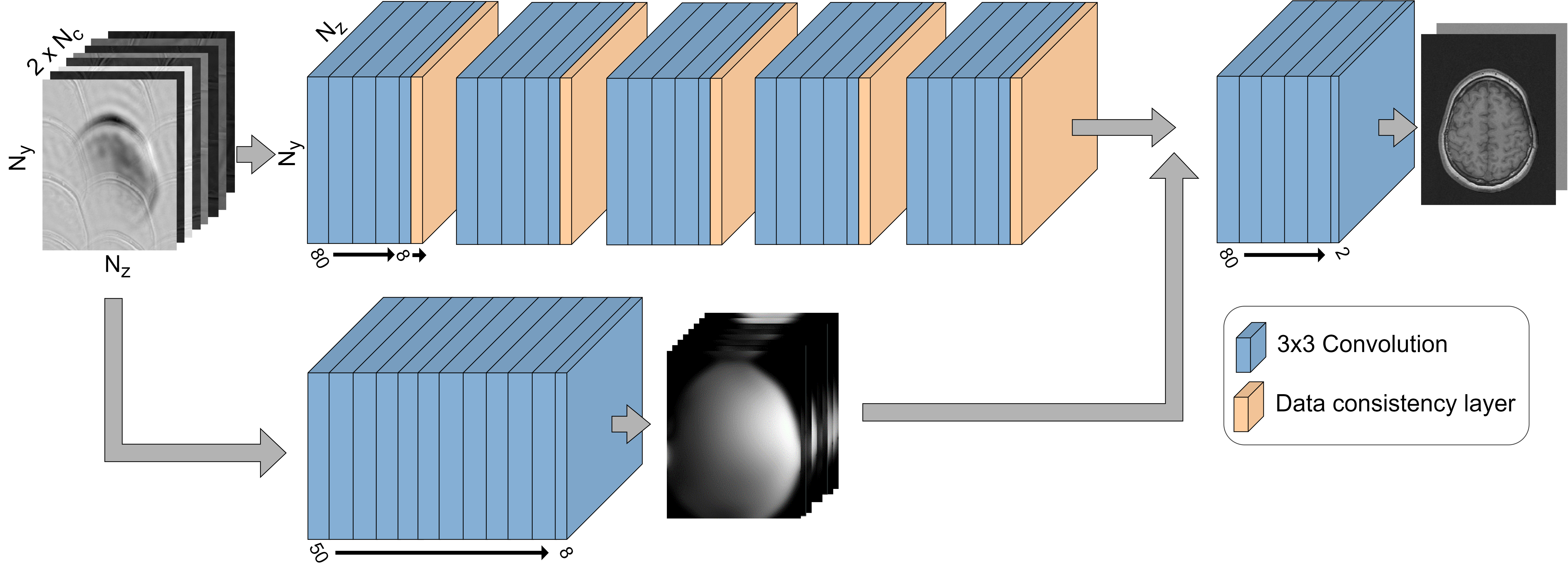
An eight-channel head coil was synthesized with sensitivities computed using the Biot-Savart law (Figure 1).3 The radius of the coil elements was increased from 8 cm to 12 cm in 1 cm increments to increase overlap in the sensitivity profiles. Multi-channel data was generated by multiplying a fully sampled T1-weighted image by sensitivity profiles taken along the central axial plane of the coil. The Calgary-Campinas dataset6 was used which contained 117 volumes from healthy subjects (split into 47/20/50 training/validation/test sets). Reconstruction was applied to 2D axial slices of which the central 100 images were used for testing. Retrospective 2D uniform undersampling was used. The DL model architecture was a modified deep convolutional cascade (Figure 2).7 The DL model was compared to conjugate gradient SENSE8 (CG-SENSE) at R = 6 and 8. Structural similarity index measurement (SSIM) and peak signal to noise ratio (PSNR) were used to assess performance as a function of increasing coil overlap. Significance was analyzed using the non-parametric Friedman test. As appropriate, this test was followed by post-hoc one-tailed Wilcoxon signed-rank tests between adjacent levels of coil overlap. The Holm-Bonferroni method was used to correct for multiple comparisons. The threshold for significance was α = 0.05.Results
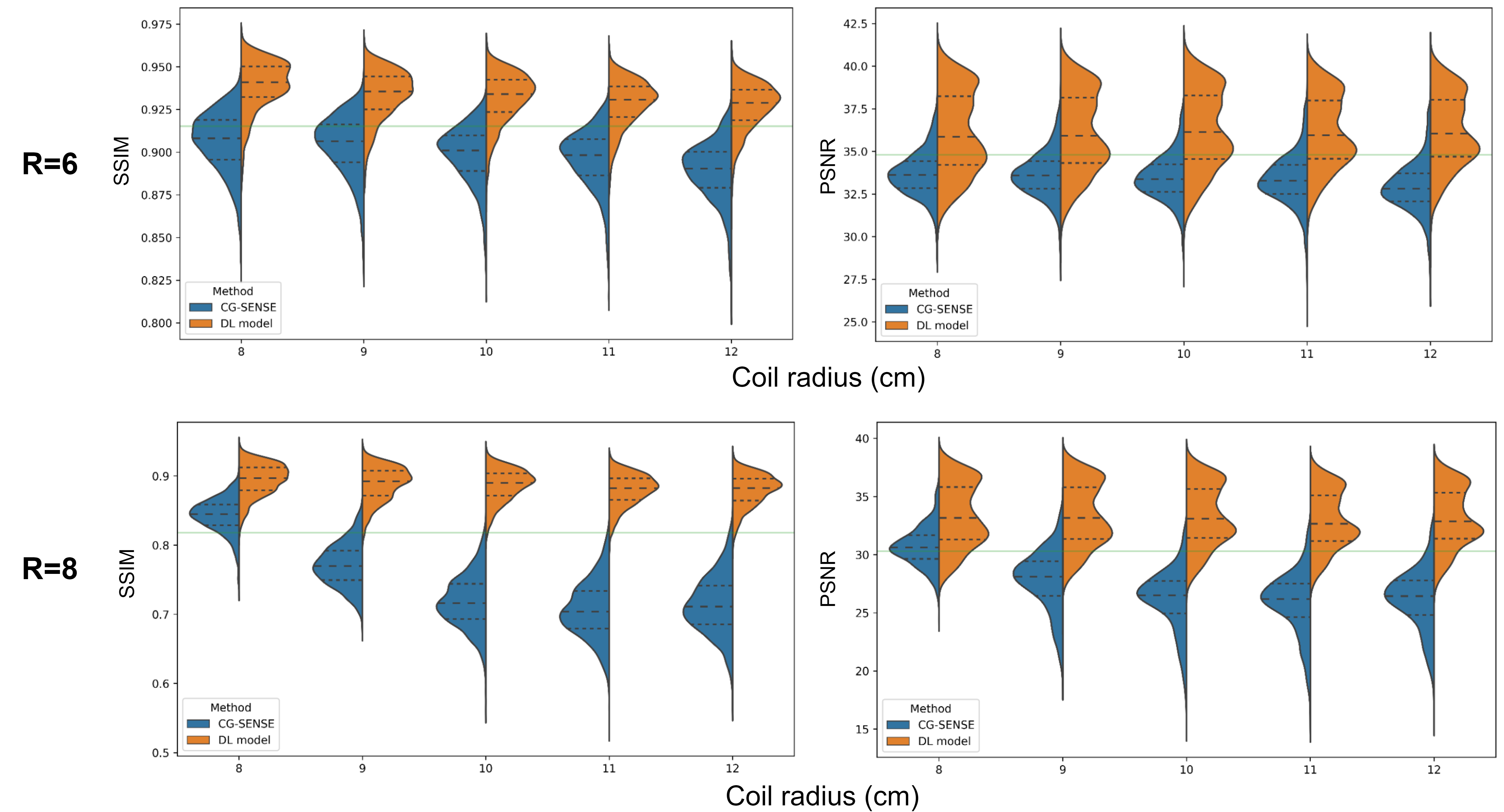
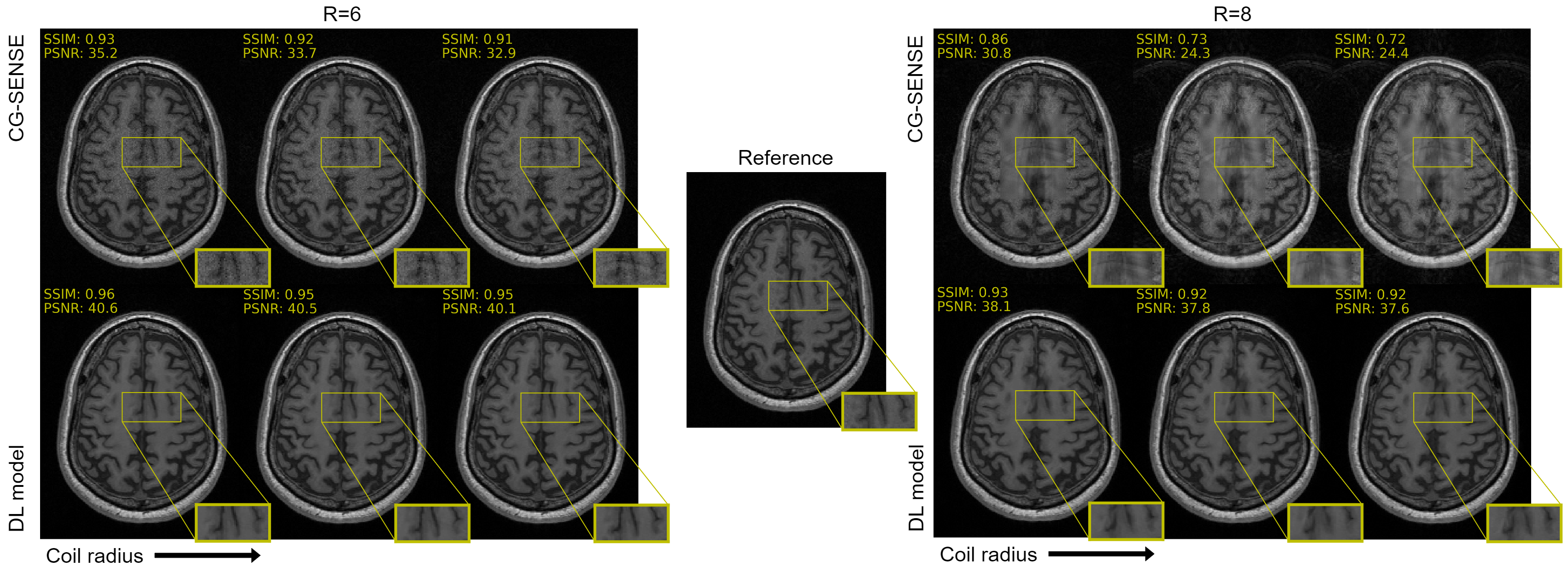
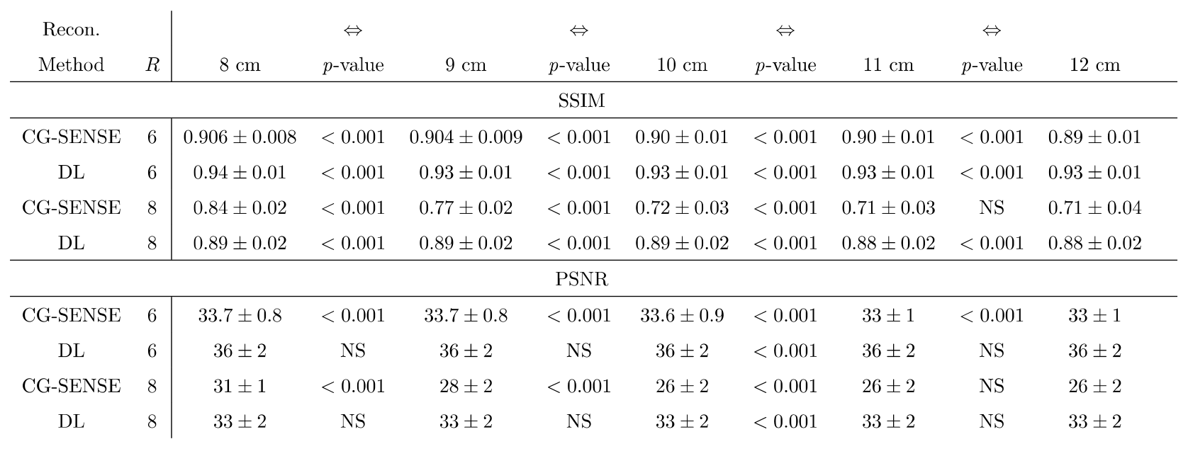
Table 1 summarizes performance results. At R = 6, a larger decrease was observed in CG-SENSE performance compared to the DL model with increasing coil radius (Figure 3). For CG-SENSE, the degree of aliasing increased (Figure 4). However, the introduced artifacts were subtle and masked by noise amplification. Noise amplification was not observed in the DL reconstruction and there were no visual differences across coil radii. At R = 8, CG-SENSE failed to accurately reconstruct the center of the image (Figure 4). This issue was not seen in the DL reconstruction. As with R = 6, as coil radius increased, a larger decrease was observed in CG-SENSE performance compared to DL (Figure 3).Discussion
Our results showed that DL models were less sensitive to coil overlap than parallel imaging approaches, such as CG-SENSE. A smaller decrease in performance was seen using DL compared to CG-SENSE. The performance decrease in CG-SENSE was visually characterized by greater amounts of aliasing (Figure 4). The DL model reconstructed images had little aliasing but tended towards smoothing of fine details. Although only small decreases in DL model performance were measured, these decreases were still significant across radii (adjacent coil configurations). These results suggest that the DL model was less impacted but not completely immune to increasing coil overlap.The DL model had a smoothing effect and thus reduced noise, including in the background. Figure 4 shows CG-SENSE exhibiting noise amplification unlike the DL model. This process perhaps resulted in a less exact reconstruction by one or more performance metrics despite the final image being perceived as potentially better by a human observer. Ultimately, assessments from expert observers are necessary to judge reconstruction quality.
Conclusion
Compared to CG-SENSE, the DL model showed reduced aliasing artifacts and smaller overall changes in performance metrics (SSIM, PSNR) as coil overlap increased. These findings suggest that some geometric constraints currently influencing MR imaging coil arrays could be relaxed when using a DL image reconstruction method, allowing for a more comfortable patient experience.Acknowledgements
We acknowledge Nicola De Zanche, PhD (Cross Cancer Institute, University of Alberta) for his contributions to the early conversation about this project. ND is supported by a Natural Sciences and Engineering Research Council (NSERC) BRAIN CREATE award, an NSERC Canada Graduate Scholarship (CGS-M), and an Alberta Graduate Excellence Scholarship (AGES). RS and RF thank NSERC for ongoing operating support for this project (RGPIN/2021-02858 and RGPIN/2021-02867). RS also acknowledges operational support from the NSERC Alliance–Alberta Innovates Advance Program.References
1 Munn Z, Moola S, Lisy K, Riitano D, Murphy F. Claustrophobia in magnetic resonance imaging: A systematic review and meta-analysis. Radiography. 2015;21(2):e59-e63.
2 Nguyen XV, Tahir Sana, Bresnahan BW, et al. Prevalence and financial impact of claustrophobia, anxiety, patient motion, and other patient events in magnetic resonance imaging. Top Magn Reson. 2020;29(3):125-130.
3 Ohliger MA, Sodickson DK. An introduction to coil array design for parallel MRI. NMR Biomed. 2006;19(3):300-315.
4 Lu M, Gore JC, Yan X. Over-overlapped loop arrays: A numerical study. Magn Reson Imaging. 2020;72:135-142.
5 Stickle Y, Taracila V, Tenley S, et al. Universally Sized, High-Resolution and ASSET Optimized AIR Cervical Coils Combined with a 48-Channel Head Coil for 3T MRI. In: Proc Intl Soc Mag Reson Med 25:1226; 2017.
6 Souza R, Lucena O, Garrafa J, et al. An open, multi-vendor, multi-field-strength brain MR dataset and analysis of publicly available skull stripping methods agreement. NeuroImage. 2018;170:482-494.
7 Schlemper J, Caballero J, Hajnal JV, Price AN, Rueckert D. A deep cascade of convolutional neural networks for dynamic MR image reconstruction. IEEE Trans Med Imaging. 2018;37(2):491-503.
8 Pruessmann KP, Weiger M, Börnert P, Boesiger P. Advances in sensitivity encoding with arbitrary k-space trajectories. Magn Reson Med. 2001;46(4):638-651.
Figures
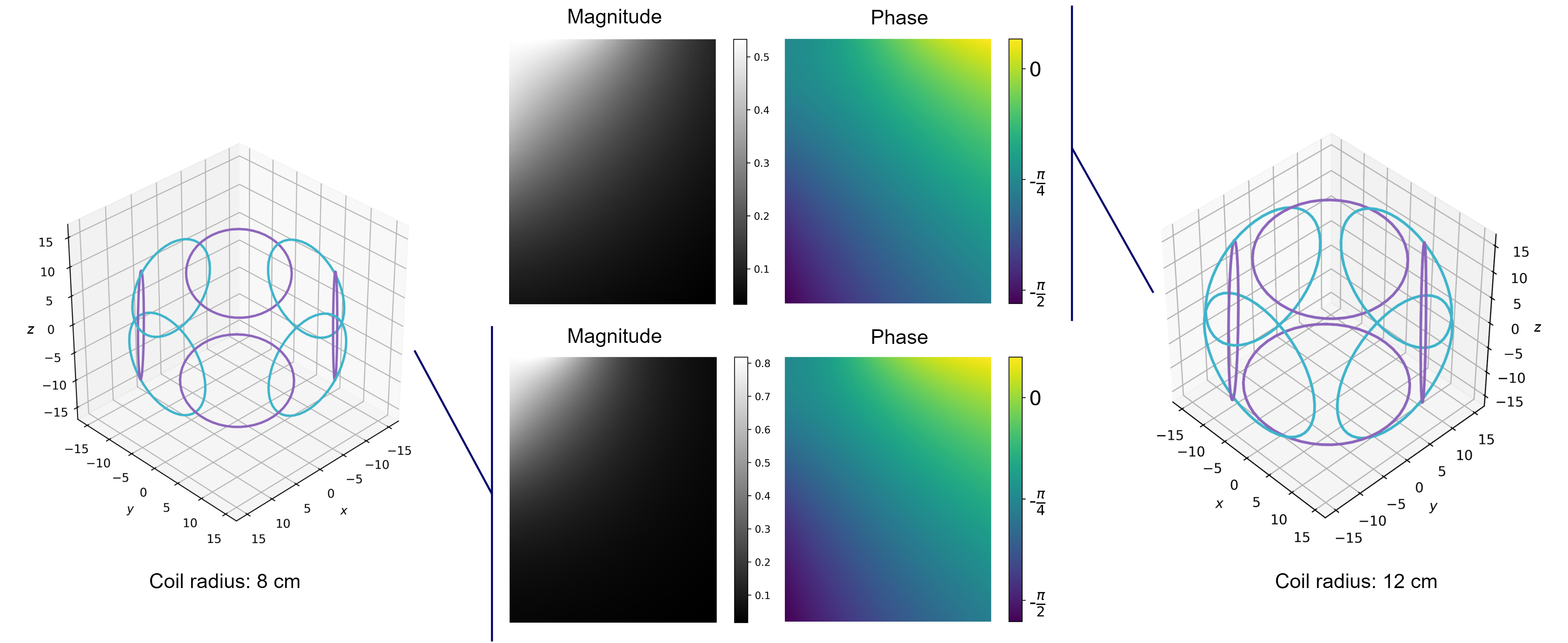
Figure 1: An eight-channel head coil array was simulated using the Biot-Savart law. The radii of the coils were increased from 8 cm to 12 cm to increase overlap. The magnitude and phase sensitivity profiles at z = 0 cm from a single 8 cm [bottom] and 12 cm [top] coil are shown.