0348
Accelerate 3D Coronary Magnetic Resonance Angiography by De-Aliasing Regularization based Compressed Sensing (DARCS)1Institute of Medical Imaging Technology, School of Biomedical Engineering, Shanghai Jiao Tong University, Shanghai, China, 2Shenzhen Institutes of Advanced Technology, Chinese Academy of Sciences, Guangdong, China
Synopsis
Keywords: Machine Learning/Artificial Intelligence, Image Reconstruction, coronary magnetic resonance angiography, compressed sensing, deep learning
Motivation: While classical non-learning reconstruction methods for 3D coronary magnetic resonance angiography (CMRA) lack a task-adaptive image prior, 3D deep unrolling suffers from a low memory efficiency, causing a reduced number of iterations and a compromised image quality.
Goal(s): We aim to combine compressed sensing and deep learning regularization by using a trained de-aliasing network as the sparsifying transform.
Approach: We compared the method with PROST, Plug-and-Play, DAGAN, and MoDL for accelerating CMRA in 20 healthy subjects.
Results: Visual inspections and quantitative comparisons both found a substantially improved reconstruction quality from DARCS relative to the other methods.
Impact: The proposed method overcomes an important limitation of 3D unrolling while maintaining its core advantage of task-adaptive regularization. The method not only can accelerate 3D CMRA, but also has the potential for general 3D image reconstructions.
Introduction
Three-dimensional coronary magnetic resonance angiography (3D CMRA) is a valuable technique to image the coronary arteries1-3, yet its long scan time often requires dedicated reconstruction algorithms for acceleration. Early reconstruction methods often employ hand-crafted regularizations, such as sparsity4-6 and low-rankness7-9, which are not adapted to the artifacts present in CMRA. Deep unrolled networks have shown the state-of-the-art performance by leveraging a data-driven regularizer10-13. However, these 3D unrolled networks require an exceedingly large memory usage, which limits the number of unrollable iterations resulting in a compromised performance. To address the issue, we propose to combine the framework of compressed sensing and the adaptivity of the deep learning regularizer by using a trained de-aliasing network as the sparsifying transform. We validated the method—De-Aliasing Regularization based Compressed Sensing (DARCS)—against a number of other reconstruction methods for 3D CMRA over a cohort of 20 healthy subjects.Methods
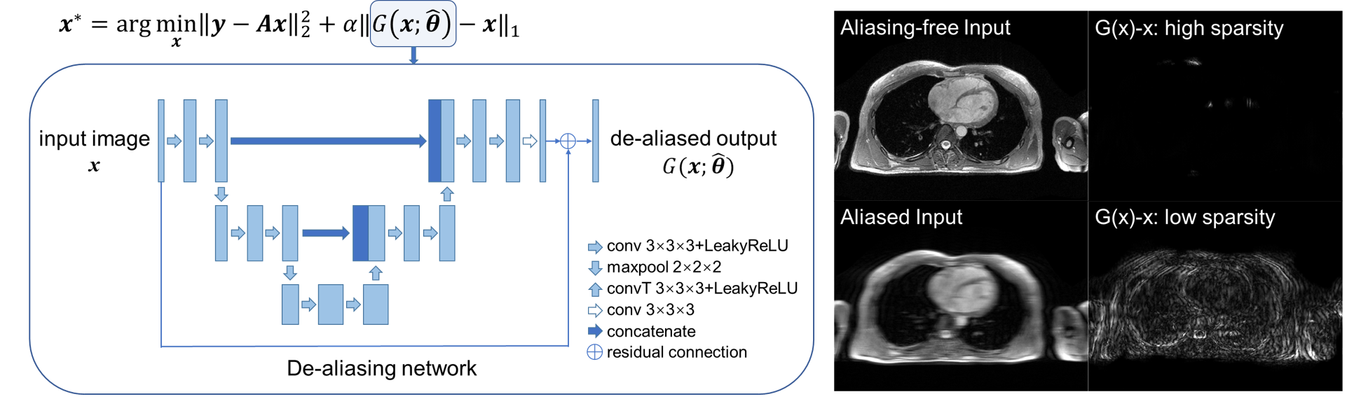
TheoryFigure 1 shows a schematic of DARCS. We start with the standard compressed sensing cost function
$$\Vert \boldsymbol{y}-\boldsymbol{A}\boldsymbol{x}\Vert_2^2 + \alpha\Vert \Phi(\boldsymbol{x})\Vert_1 \tag{1}$$
where $$$\boldsymbol{y}$$$ is the undersampled k-space data, $$$\boldsymbol{A}$$$ the forward imaging model, $$$\boldsymbol{x}$$$ the underlying image, $$$\Phi$$$ the sparsifying transform, and $$$\alpha$$$ the regularization parameter. Let $$$G(\boldsymbol{x};\hat{\boldsymbol{\theta}})$$$ represent a de-aliasing network trained to remove aliasing artifacts in an image and consider the mapping $$$\boldsymbol{x}\rightarrow G(\boldsymbol{x};\hat{\boldsymbol{\theta}})$$$. If the image is aliasing-free, $$$G(\boldsymbol{x};\hat{\boldsymbol{\theta}}) - \boldsymbol{x}$$$ should output a zero image, which intrinsically has the highest sparsity. If the image contains artifacts, $$$G(\boldsymbol{x};\hat{\boldsymbol{\theta}}) - \boldsymbol{x}$$$ should output a recognized artifact map, which has a lower sparsity due to the presence of artifacts. Thus, $$$\Vert G(\boldsymbol{x};\hat{\boldsymbol{\theta}}) - \boldsymbol{x}\Vert_1$$$ acts as an intrinsic sparsity metric and can thus serve as a regularizer. In this work, we used the generator of De-Aliasing Generative Adversarial Network (DAGAN)14 as $$$G(\boldsymbol{x};\hat{\boldsymbol{\theta}})$$$, and trained it with both aliasing-free and aliasing-affected images. The resultant cost function is
$$\Vert \boldsymbol{y}-\boldsymbol{A}\boldsymbol{x}\Vert_2^2 + \alpha\Vert G(\boldsymbol{x};\hat{\boldsymbol{\theta}}) - \boldsymbol{x}\Vert_1 \tag{2}$$
We use the Alternating Direction Method of Multipliers (ADMM) to solve the problem. The data-fidelity sub-problem and the proximal step of the ADMM algorithm were solved by conjugate gradients and gradient descent, respectively.
Experiments
The institutional review board approved the study. We performed CMRA in 20 healthy subjects using an electrocardiogram-triggered, navigator-gated, T2-prepared dual-echo spoiled gradient echo sequence in a 3T scanner (uMR790, United Imaging Healthcare, Shanghai, China) after obtaining written informed consent from each subject. Elliptical sampling with a 2-fold undersampling was employed for the acquisition. Reconstruction was performed by iterative SENSE, whose result was used as the ground truth. Retrospective pseudorandom undersampling of k-space was performed with acceleration rates from 4 to 10. Training was performed in 10 subjects with an acceleration rate of 8. Reconstruction was then performed with patch-based reconstruction(PROST)7, Plug-and-Play(PnP)15, 3D Model-based Deep Learning (MoDL)12, 16 with 3 unrolled iterations, DAGAN14, and DARCS in the other 10 subjects. Evaluation metrics included PSNR, SSIM, NMSE of each method, and the length of the visible coronary arteries.
Results
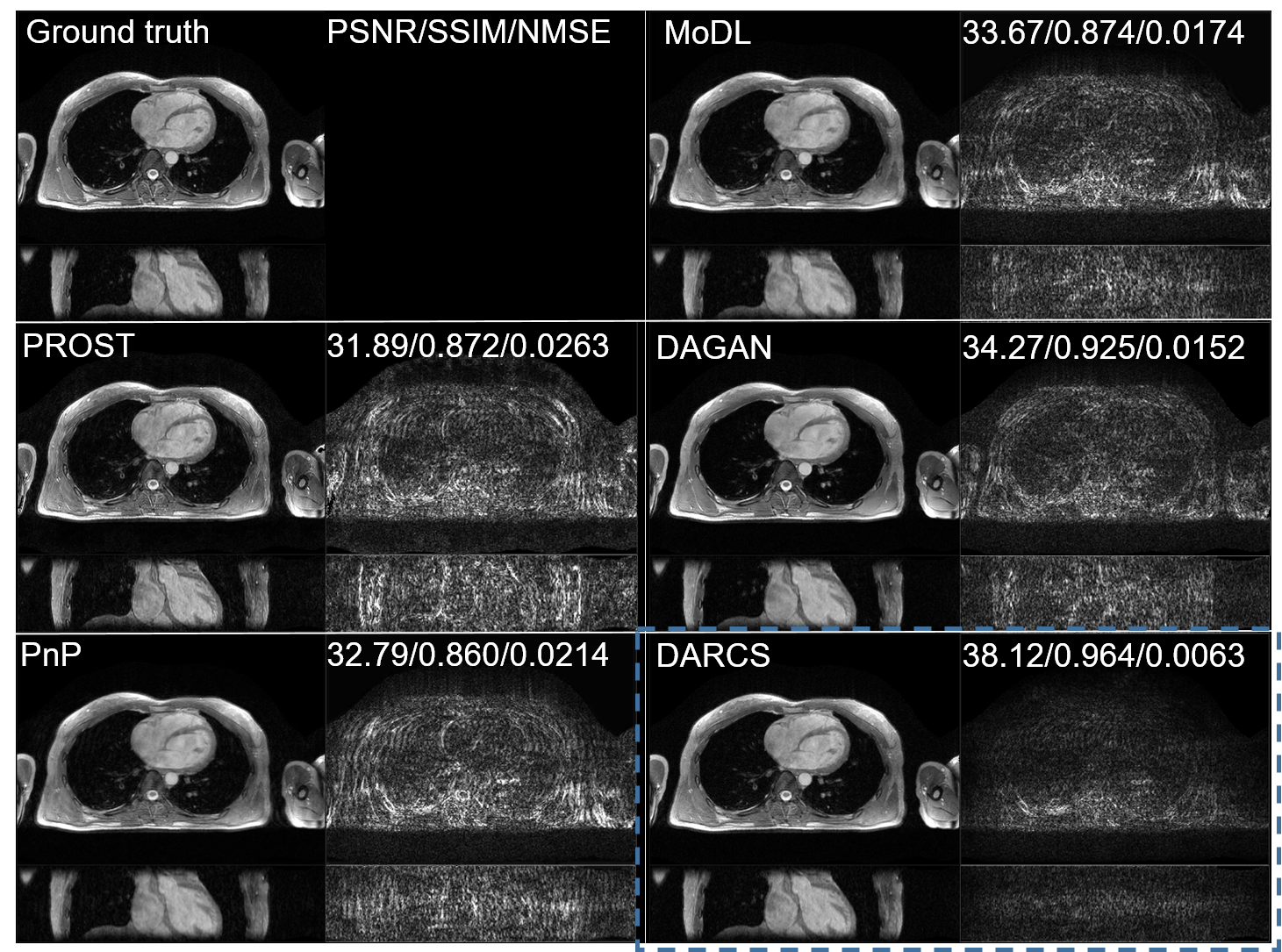
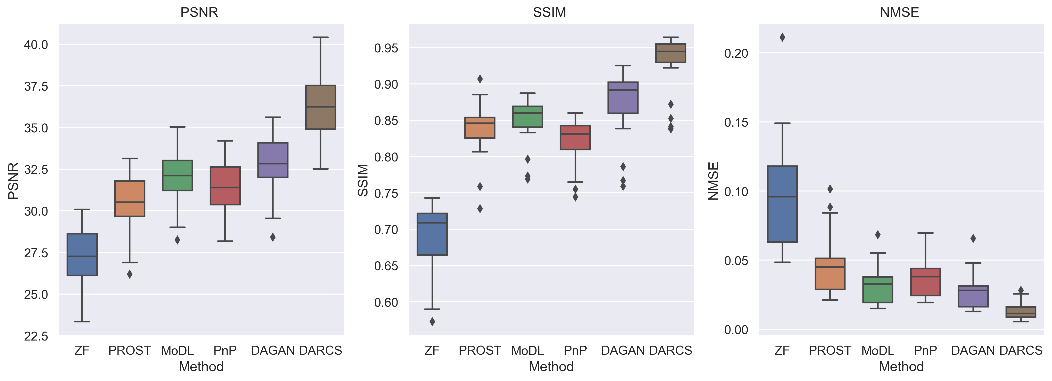
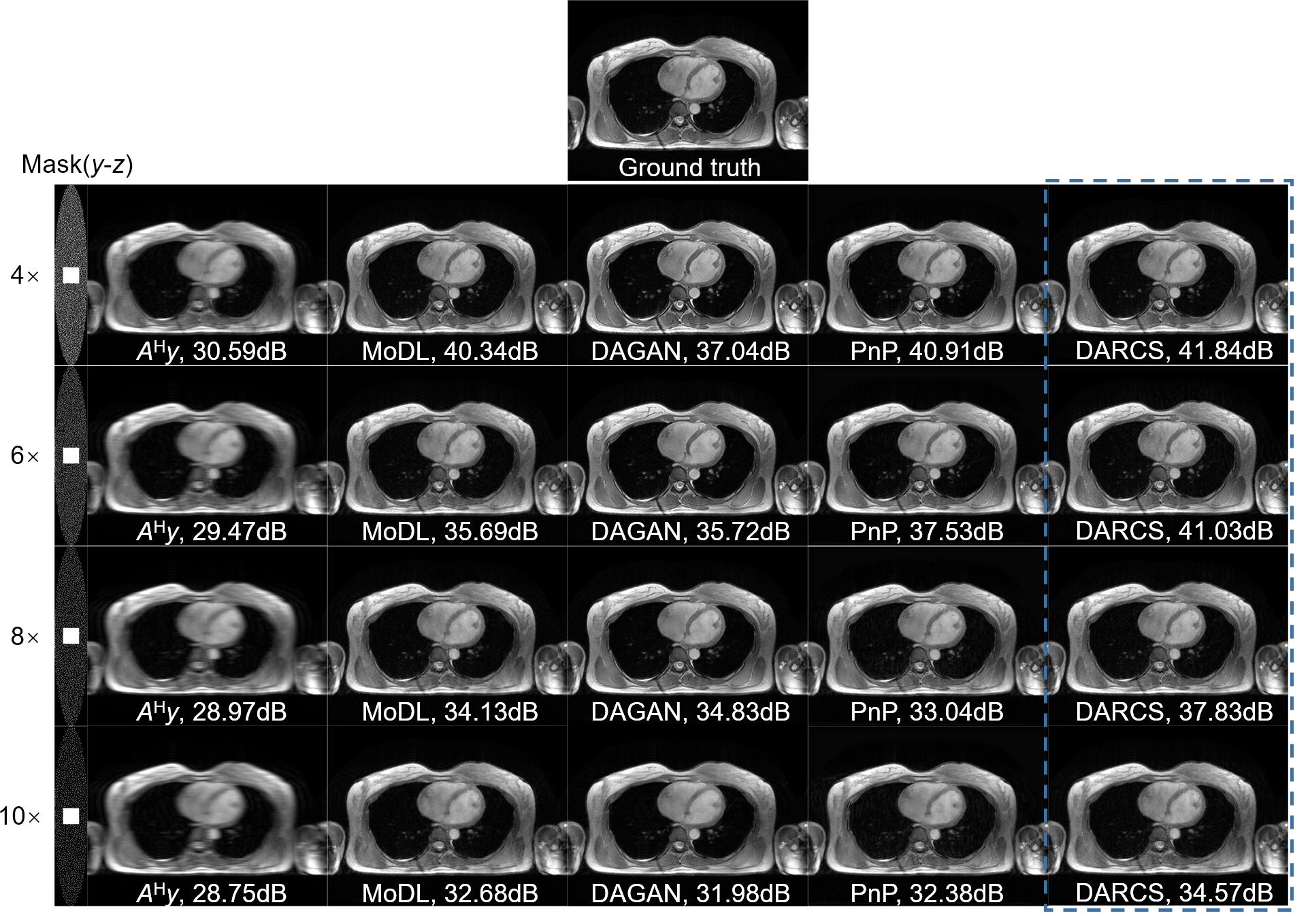
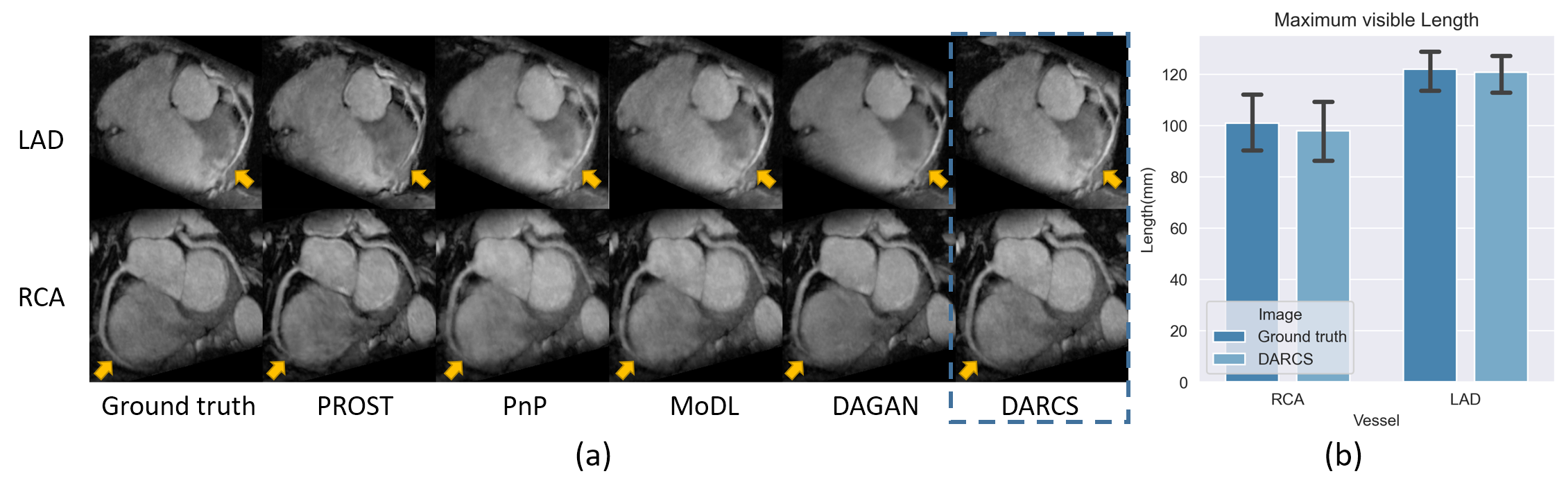
Figure 2 shows representative reconstructions from the five studied methods. DARCS exhibited higher image qualities in both coronal and transversal views than the other methods. Figure 3 shows the statistical comparison between these methods based on the t-test. DARCS significantly improved PSNR, SSIM, and NMSE over the other methods. Figure 4 shows the reconstructions of DARCS, MoDL, PnP, and DAGAN with different sampling rates, despite all of them were trained with data generated by 8-fold undersampling. DARCS consistently provided reconstructions of higher qualities relative to the other methods, suggesting that the method was reasonably generalizable at different accelerate rates. Figure 5 shows reformatted CMRA images and the comparison of visible lengths of the right coronary artery (RCA) and left anterior descending artery (LAD) between DARCS and the ground truth. DARCS exhibited better reconstructions of the coronary arteries than the other methods. The vessel lengths were not significantly different between the ground truth and DARCS reconstructions.Discussion and Conclusions
The improvement achieved by DARCS can be explained by its distinctions from other methods. Firstly, unlike PROST or PnP, DARCS uses a task-adaptive de-aliasing regularizer, which can more effectively suppress the aliasing artifacts for this application. Secondly, although DARCS uses DAGAN to build its regularizer, it also involves the data-fidelity term in the optimization, which is not used by DAGAN alone. Thirdly, unlike MoDL, DARCS does not unroll the iterations or use end-to-end training, thereby embracing an unlimited number of iterations and possibly improved interpretability. In conclusion, we developed a task-adaptive compressed sensing method based on a learned deep de-aliasing regularizer, which showed an improved image quality for accelerating 3D CMRA than both classical and other deep learning methods.Acknowledgements
No acknowledgement found.References
1. Sakuma H. Coronary CT versus MR Angiography: The Role of MR Angiography. Radiology. 2011 2011/02/01;258(2):340-9.
2. Hajhosseiny R, Bustin A, Munoz C, Rashid I, Cruz G, Manning WJ, et al. Coronary Magnetic Resonance Angiography: Technical Innovations Leading Us to the Promised Land? JACC Cardiovasc Imaging. 2020 Dec;13(12):2653-72.
3. Hajhosseiny R, Munoz C, Cruz G, Khamis R, Kim WY, Prieto C, et al. Coronary Magnetic Resonance Angiography in Chronic Coronary Syndromes. Front Cardiovasc Med. 2021;8:682924.
4. Lustig M, Donoho D, Pauly JM. Sparse MRI: The application of compressed sensing for rapid MR imaging. Magn Reson Med. 2007 Dec;58(6):1182-95.
5. Hirai K, Kido T, Kido T, Ogawa R, Tanabe Y, Nakamura M, et al. Feasibility of contrast-enhanced coronary artery magnetic resonance angiography using compressed sensing. Journal of Cardiovascular Magnetic Resonance. 2020;22(1).
6. Lu H, Zhao S, Tian D, Yang S, Ma J, Chen Y, et al. Clinical Application of Non-Contrast-Enhanced Dixon Water-Fat Separation Compressed SENSE Whole-Heart Coronary MR Angiography at 3.0 T With and Without Nitroglycerin. J Magn Reson Imaging. 2022 Feb;55(2):579-91.
7. Bustin A, Ginami G, Cruz G, Correia T, Ismail TF, Rashid I, et al. Five-minute whole-heart coronary MRA with sub-millimeter isotropic resolution, 100% respiratory scan efficiency, and 3D-PROST reconstruction. Magn Reson Med. 2019 Jan;81(1):102-15.
8. Bustin A, Rashid I, Cruz G, Hajhosseiny R, Correia T, Neji R, et al. 3D whole-heart isotropic sub-millimeter resolution coronary magnetic resonance angiography with non-rigid motion-compensated PROST. J Cardiovasc Magn Reson. 2020 Apr 16;22(1):24.
9. Nazir MS, Bustin A, Hajhosseiny R, Yazdani M, Ryan M, Vergani V, et al. High-resolution non-contrast free-breathing coronary cardiovascular magnetic resonance angiography for detection of coronary artery disease: validation against invasive coronary angiography. J Cardiovasc Magn Reson. 2022 Apr 11;24(1):26.
10. Yan Yang, Jian Sun, Huibin Li, Xu Z. ADMM-Net: A Deep Learning Approach for Compressive Sensing MRI. arXiv. 2017-05-19.
11. Zhang J, Ghanem B. ISTA-Net: Interpretable Optimization-Inspired Deep Network for Image Compressive Sensing. IEEE Conference on Computer Vision and Pattern Recognition; 2018. p. 1828-37.
12. Aggarwal HK, Mani MP, Jacob M. MoDL: Model-Based Deep Learning Architecture for Inverse Problems. IEEE Trans Med Imaging. 2019 Feb;38(2):394-405.
13. Hammernik K, Klatzer T, Kobler E, Recht MP, Sodickson DK, Pock T, et al. Learning a variational network for reconstruction of accelerated MRI data. Magn Reson Med. 2018 Jun;79(6):3055-71.
14. Yang G, Yu S, Dong H, Slabaugh G, Dragotti PL, Ye X, et al. DAGAN: Deep De-Aliasing Generative Adversarial Networks for Fast Compressed Sensing MRI Reconstruction. IEEE Trans Med Imaging. 2018 Jun;37(6):1310-21.
15. Ahmad R, Bouman CA, Buzzard GT, Chan S, Liu S, Reehorst ET, et al. Plug-and-Play Methods for Magnetic Resonance Imaging: Using Denoisers for Image Recovery. IEEE Signal Process Mag. 2020 Jan;37(1):105-16.
16. Deng Z, Yaman B, Zhang C, Moeller S, Akcakaya M. Efficient Training of 3D Unrolled Neural Networks for MRI Reconstruction Using Small Databases. 2021 55th Asilomar Conference on Signals, Systems, and Computers; 2021: IEEE; 2021. p. 886-9.
Figures