0346
Unsupervised 4D-Flow MRI reconstruction with Deep Image Prior and Graph Convolution Neural-Network1Tsinghua University, Beijing, China, 2Oden Institute for Computational Engineering and Sciences, University of Texas at Austin, Austin, TX, United States
Synopsis
Keywords: AI/ML Image Reconstruction, Cardiovascular
Motivation: Deep learning reconstruction algorithms offer significant advantages for accelerating 4D-Flow MRI acquisition. However, a large high-quality fully-sampled dataset is usually unavailable for network training.
Goal(s): To propose an unsupervised algorithm for 4D-Flow MRI reconstruction, without the need for any fully-sampled data.
Approach: We use branched CNNs and a Graph-Convolution-Network as the generator. Additionally, we devise an ADMM algorithm to alternately optimize the images and the network parameters. Experiments are conducted on aortic and intracranial 4D-Flow data.
Results: The proposed algorithm demonstrates superior reconstruction results, outperforming even supervised deep-learning method. Moreover, it exhibits good generalization capability when applied to another imaging target.
Impact: The proposed method is a promising algorithm for accelerating MR blood-flow imaging, owing to its exceptional performance and generalization capacity. Furthermore, the algorithm introduces a new model for 4D-flow MRI reconstruction which is valuable for further research.
Introduction
4D-Flow MRI can provide spatio-temporally resolved blood flow quantification[1]. Due to the high dimensionality of 4D-Flow MRI data, k-space undersampling is essential to reduce the acquisition time. In recent years, deep learning-based methods have been demonstrated effective for 4D-Flow MRI reconstruction[2][3]. However, these methods need a fully-sampled dataset for training, which is usually not available for 4D-Flow MRI. Besides, supervised deep-learning algorithms often face challenges in terms of generalization, especially when training on very limited data or transferring to other imaging targets.In this work, we propose an unsupervised 4D-Flow MRI reconstruction algorithm without the need for any fully-sampled data. We utilize the inductive bias of CNN as the spatial regularization[4], and employ the graph convolution neural-network(GCN)[5][6] to further exploit the spatio-temporal redundancy. Additionally, we devise an ADMM algorithm[7] to alternately optimize the images and the network to improve the final reconstruction results. The proposed method was then evaluated using a publicly available aorta 4D-Flow dataset and an in-house intracranial 4D-Flow dataset, and compared with several state-of-art algorithms.
Methods
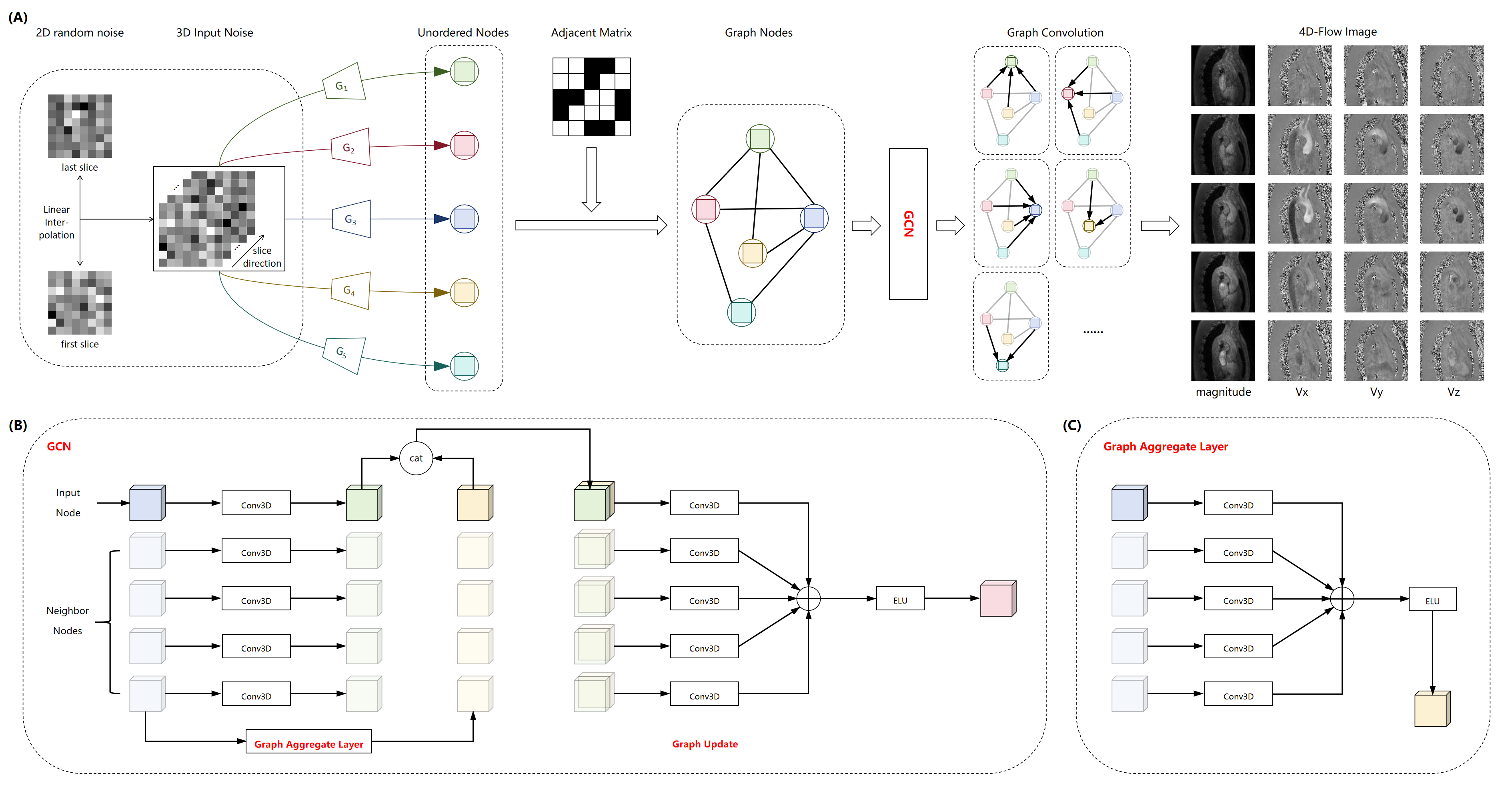
We propose the following objective for 4D-Flow MRI reconstruction:$$\min_{x,\theta}\,\,\frac{1}{2}\left\|Ax-y\right\|_2^2+\lambda\left\|Dx\right\|_1\\s.t.\,\,x=G_{\theta}(z)$$, where $$$x$$$ represents 4D-Flow images, $$$A$$$ is a multi-frame multi-coil MRI encoding matrix, $$$y$$$ denotes k-space measurements, $$$D$$$ is the complex-difference[8] operator along the velocity-encoding direction, $$$G_{\theta}$$$ is the neural-network generator, and $$$z$$$ is a fixed latent variable.The structure of $$$G_{\theta}$$$ structure is illustrated in Fig1. The input of the network is a 3D noise map, which is interpolated from two 2D Gaussian noise variables. Independent small CNNs(G1,......,GN) are used for each frame to recover the image structure. The output frames are considered as graph nodes, and connected by a pre-calculated graph adjacent matrix. Then a Graph-Convolution-Network(GCN) is adopted for updating the graph nodes, finally producing dynamic 4D-Flow MRI images. The detailed structure of GCN is shown in Fig1B and Fig1C. Here, we utilize the Euclidean distance of the ACS data in k-space center as the neighborhood similarity index[9], and apply the K-nearest-neighbor(k-NN) algorithm to calculate the adjacent matrix.
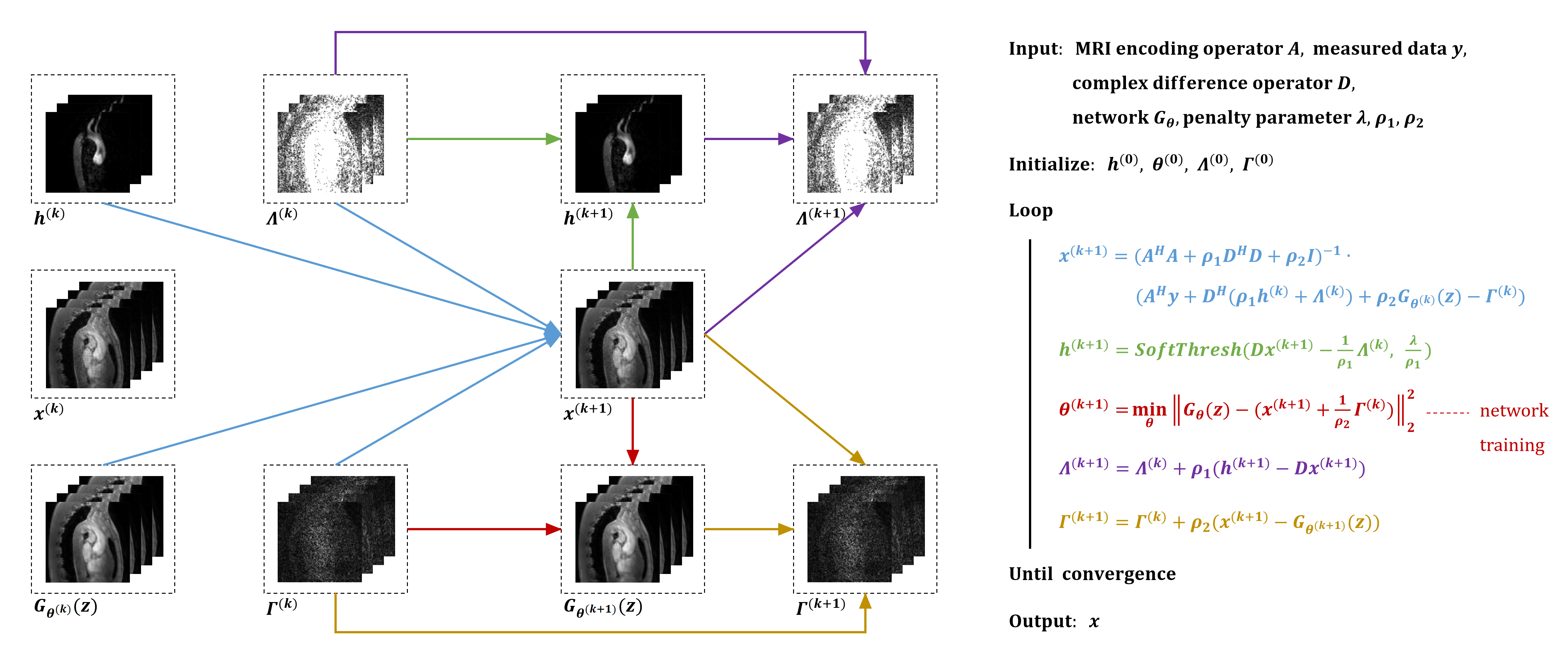
Additionally, we use the augmented Lagrangian method to rewrite the objective as:$$\min_{x,h,\theta,\Lambda,\Gamma}\frac{1}{2}\left \|Ax-y\right\|_2^2+\lambda\left\|h\right\|_1+\frac{\rho_1}{2}\left\|h-Dx\right\|_2^2+Re(\left\langle\Lambda,\,h-Dx\right\rangle)+\frac{\rho_2}{2}\left\|x-G_{\theta}\right\|_2^2+Re(\left \langle\Gamma,\,x-G_{\theta}\right\rangle)$$, where $$$h$$$ is an auxiliary variable, $$$\rho_1$$$ and $$$\rho_2$$$ are relaxation coefficients, $$$\Lambda$$$ and $$$\Gamma$$$ are Lagrangian multipliers. We use the ADMM algorithm to solve this problem:\begin{aligned}x^{(k+1)}&=(A^HA+\rho_1D^HD+\rho_2I)^{-1}(A^Hy+D^H(\rho_1h^{(k)}+\Lambda^{(k)})+\rho_2G_{\theta^{(k)}}(z)-\Gamma^{(k)})\\h^{(k+1)}&=SoftThresh(Dx^{(k+1)}-\frac{1}{\rho_1}\Lambda^{(k)},\,\frac{\lambda}{\rho_1})\\\theta^{(k+1)}&=\min_{\theta}\left\|G_{\theta}(z)-(x^{(k+1)}+\frac{1}{\rho_2}\Gamma^{(k)})\right\|_2^2\\\Lambda^{(k+1)}&=\Lambda^{(k)}+\rho_1(h^{(k+1)}-Dx^{(k+1)})\\\Gamma^{(k+1)}&=\Gamma^{(k)}+\rho_2(x^{(k+1)}-G_{\theta^{(k+1)}}(z))\end{aligned}. The overall optimization algorithm is illustrated in Fig2.
Experiments and Results
We conducted 4D-Flow MRI reconstruction experiments on the aorta and intracranial vessels, respectively. In addition to the proposed algorithm, a traditional algorithm L+S-Hadamard[10] and a supervised deep-learning algorithm FlowVN[2] were also implemented for comparison. These algorithms were all tuned on the aortic dataset, and then directly applied to the intracranial dataset to test their generalization capability.Aortic Experiments
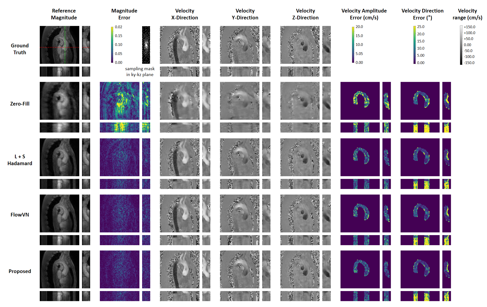
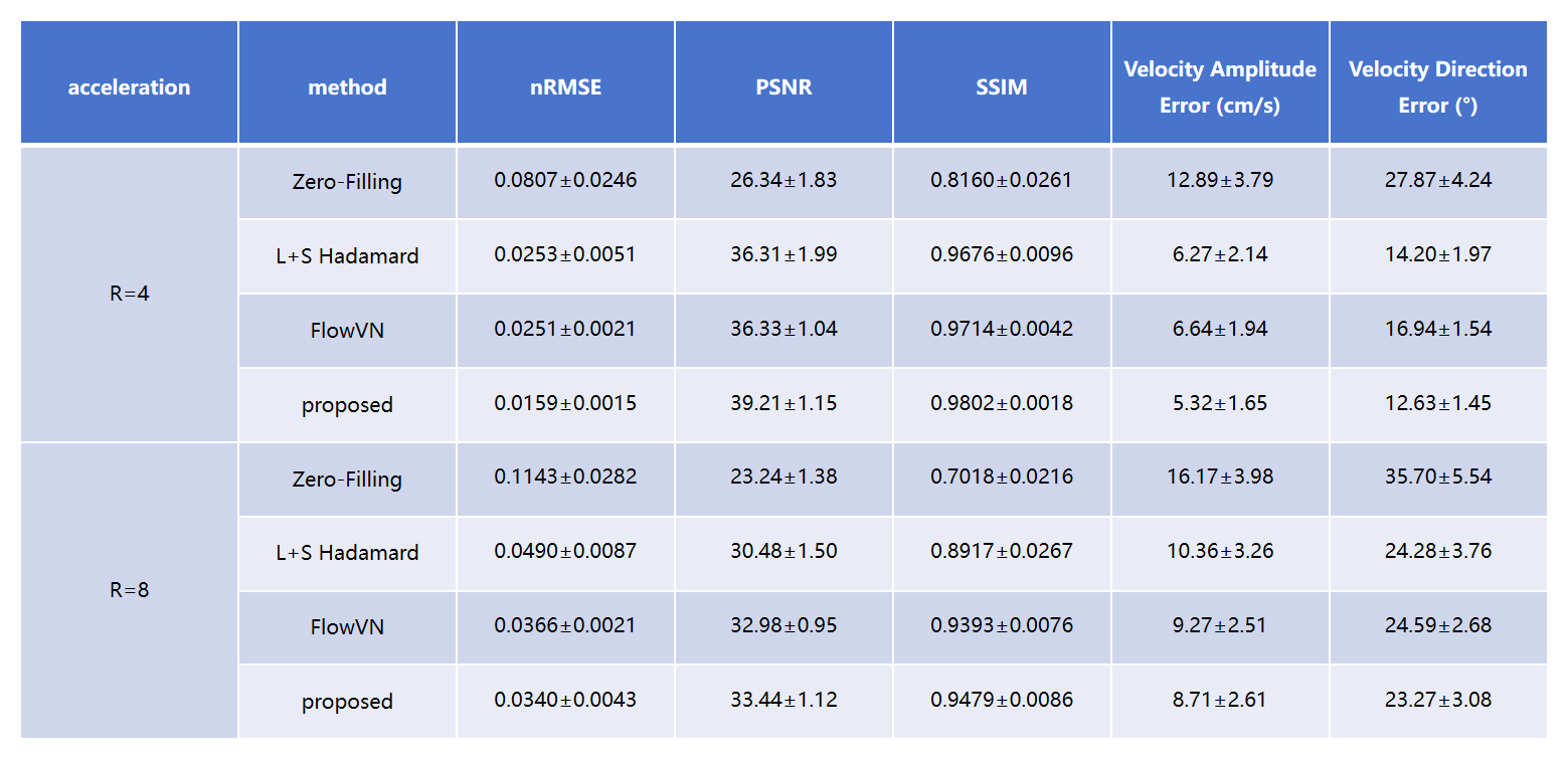
We utilized the public dataset provided by FlowVN[2], which consists of seven fully-sampled aortic 4D-Flow data. 2D Poisson mask is used for sampling in the ky-kz plane. Two acceleration factors R=4.0(ACS=12x5) and R=8.0(ACS=6x4) were tested. To ensure fairness in the comparison, the model of FlowVN was re-trained for each dataset using the leave-one-out method. The reconstructed images are shown in Fig3, where the red dotted line represents the 1D dynamic profile along the time dimension, and the green dotted line represents the slice profile along z-direction. The proposed method achieves the lowest magnitude error and the most accurate quantification of the three-directional flow velocity, compared with the state-of-art traditional and supervised deep-learning algorithms. The quantitative results are summarized in Fig4, which further demonstrates the superiority of the proposed method.
Intracranial Experiments
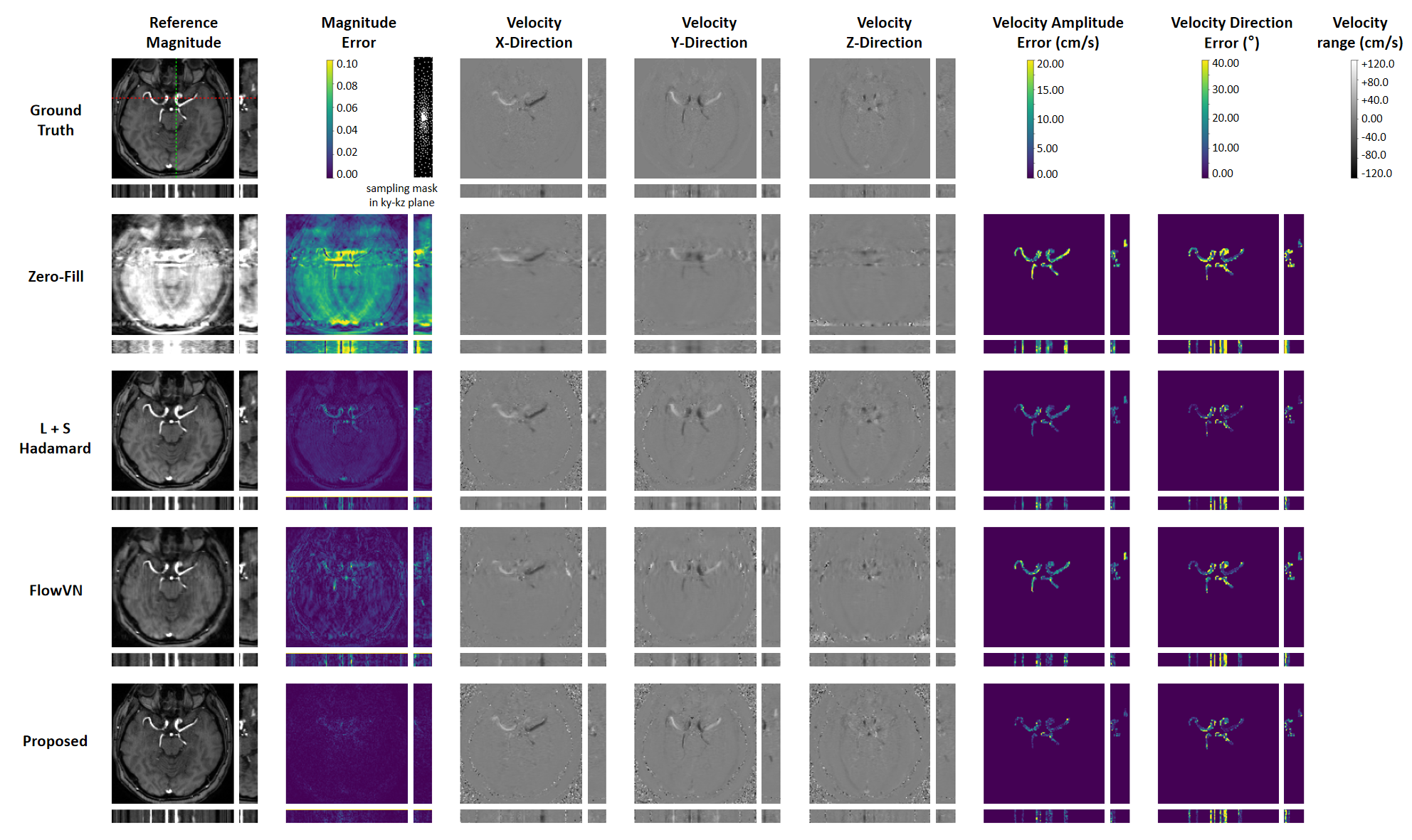
We used an in-house 4D-Flow MRI dataset of intracranial vessels. The multi-coil k-space data were retrospectively generated by Fourier transform and simulated sensitivity maps with five coils. 2D Poisson mask was employed for sampling in the ky-kz plane. An acceleration factor of R=8.0(ACS=6x4) was tested. The algorithms fine-tuned in the aorta dataset were directly applied for reconstruction. Representative reconstructed images are shown in Fig5. It can be seen that the proposed method maintains superior reconstruction performance with the same hyper-parameters, while the supervised deep-learning algorithm FlowVN exhibits even poorer results than the traditional algorithm.
Conclusion
In this work, we propose a new model and unsupervised algorithm for 4D-Flow MRI reconstruction, demonstrating promising results in both aortic and intracranial flow imaging. Given the challenges of obtaining a large high-quality training set for 4D-Flow MRI, the proposed method holds significant value for advancing research and applications in MR flow imaging.Acknowledgements
No acknowledgement found.References
[1]. Markl, M., Frydrychowicz, A., Kozerke, S., Hope, M., & Wieben, O. (2012). 4D flow MRI. Journal of Magnetic Resonance Imaging, 36(5), 1015-1036.
[2]. Vishnevskiy, V., Walheim, J., & Kozerke, S. (2020). Deep variational network for rapid 4D flow MRI reconstruction. Nature Machine Intelligence, 2(4), 228-235.
[3]. Nath, R., Callahan, S., Stoddard, M., & Amini, A. A. (2022). FlowRAU-Net: Accelerated 4D Flow MRI of Aortic Valvular Flows with a Deep 2D Residual Attention Network. IEEE Transactions on Biomedical Engineering, 69(12), 3812-3824.
[4]. Ulyanov, D., Vedaldi, A., & Lempitsky, V. (2018). Deep image prior. In Proceedings of the IEEE conference on computer vision and pattern recognition (pp. 9446-9454).
[5]. Kipf, T. N., & Welling, M. (2016). Semi-supervised classification with graph convolutional networks. arXiv preprint arXiv:1609.02907.
[6]. Han, K., Wang, Y., Guo, J., Tang, Y., & Wu, E. (2022). Vision gnn: An image is worth graph of nodes. Advances in Neural Information Processing Systems, 35, 8291-8303.
[7]. Boyd, S., Parikh, N., Chu, E., Peleato, B., & Eckstein, J. (2011). Distributed optimization and statistical learning via the alternating direction method of multipliers. Foundations and Trends® in Machine learning, 3(1), 1-122.
[8]. Sun, A., Zhao, B., Ma, K., Zhou, Z., He, L., Li, R., & Yuan, C. (2017). Accelerated phase contrast flow imaging with direct complex difference reconstruction. Magnetic resonance in medicine, 77(3), 1036-1048.
[9]. Poddar, S., & Jacob, M. (2015). Dynamic MRI using smoothness regularization on manifolds (SToRM). IEEE transactions on medical imaging, 35(4), 1106-1115.
[10]. Valvano, G., Martini, N., Huber, A., Santelli, C., Binter, C., Chiappino, D., ... & Kozerke, S. (2017). Accelerating 4 D flow MRI by exploiting low‐rank matrix structure and hadamard sparsity. Magnetic resonance in medicine, 78(4), 1330-1341.
Figures