0338
Deep Complex Neural Network for Undersampling Spiral Artefact Removal in Diffusion Tensor Cardiovascular Magnetic Resonance with In-vivo Study1National Heart and Lung Institute, Imperial College London, London, United Kingdom, 2CMR Unit, Royal Brompton Hospital, London, United Kingdom, 3EPSRC Centre for Doctoral Training in Smart Medical Imaging, King’s College London and Imperial College London, London, United Kingdom, 4Bioengineering Department and Imperial-X, Imperial College London, London, United Kingdom
Synopsis
Keywords: AI/ML Image Reconstruction, Diffusion Tensor Imaging
Motivation: Diffusion Tensor Cardiovascular Magnetic Resonance (DT-CMR) is hindered by low resolution and long acquisitions. Spiral trajectories could be efficient with effective removal of artefacts from undersampled images.
Goal(s): To remove artefacts from highly accelerated spiral in-vivo DT-CMR acquisitions using a novel deep learning method.
Approach: We proposed a Residual U-Net based Complex-valued Edge Attention Network (CEAN) to remove undersampling artefacts. Training with and without transfer learning were explored.
Results: CEAN with transfer learning outperformed other networks, achieving the lowest Mean Absolute Error (MAE) for DT-CMR parameters and preserving diffusion encoding information, suggesting future potentials in accelerating clinical DT-CMR studies.
Impact: This work will allow the acquisition and reconstruction of highly accelerated STEAM spiral DT-CMR, aided by the proposed deep Complex-valued Edge Attention Network. Further developments will allow increases in spatial resolution to facilitate clinical translation of DT-CMR.
Introduction
Diffusion Tensor Cardiovascular Magnetic Resonance1-3 (DT-CMR) provides non-invasive insights into in-vivo myocardial microstructure, with proven utility in cardiovascular diseases4. However, it has a limited spatial resolution due to inherent low Signal to Noise Ratio (SNR), and time-consuming5 acquisitions. Spiral sampling is efficient and well-suited to sub-Nyquist acquisitions6,7. It has been implemented with DT-CMR to enhance resolution7, but acceleration is required for future clinical translation.This work aims to demonstrate effective artefact removal from highly accelerated in-vivo spiral DT-CMR, using a novel Residual U-Net based deep Complex-valued Edge Attention Network (CEAN).
Methods
Proposed ModelWe proposed a complex-valued Edge Attention Module (EAM) within a Residual U-Net structure8 to minimise the loss of high-frequency information. The EAM uses Sobel filtering, complex convolution and sigmoid activation function to create edge attention maps. We introduced the complex-valued Spatial Attention Module (SAM) to emphasis the salient regions and further improve the quality of the predicted images. The proposed network architecture is illustrated in Figure 1.
We compared Magnitude Residual U-Net (MRUN) with CEAN. Each network was trained directly on acquired spiral DT-CMR data, and with transfer learning, which is to train on a simulated dataset before fine-tuning on the acquired in-vivo data.
Acquisition
Simulated dataset was calculated from short axis single slice STEAM-EPI DT-CMR3 from 20 controls, in diastole and systole, at 1.5T and 3T (40 datasets). Spatial resolution = 2.8×2.8×8mm3 and Field of View (FOV) = 358×134mm2. Six encoding directions at b = 600s/mm2 (8 averages) and b = 150s/mm2 (2 averages) in addition to “b0”. To simulate spiral acquisitions, we cropped then resampled the STEAM-EPI images along variable density spiral trajectories (central 25% fully-sampled, linearly reducing 30%, outer 45% undersampling x7), equivalent to a uniform four-fold acceleration. Two-fold data augmentation was implemented using random rotations of the spirals.
In-vivo STEAM-spiral DT-CMR was acquired using the same trajectories (Siemens 3T Vida), for 10 subjects in diastole and systole at 3T. Spatial resolution = 2.8×2.8×8mm3, imaging FOV = 134×134mm2 (reduced via in-plane zonal RF excitations), TR=2RR-intervals and TE=11ms. Six encoding directions with b=600s/mm2 (2 averages) and b=150s/mm2 (1 average) were acquired. Data was sampled along 7 interleaves (1 interleave per breath-hold), the ground truth combined all interleaves (after motion-induced phase correction7, interleave registration and coil weighted combination), and undersampled images were each generated from 1 interleave.
The training-validation-testing split by subjects was 14(4319 images)-3(973 images)-3(980 images), and 6(252 images)-1(42 images)-3(126 images) respectively for simulated and in-vivo datasets.
Networks were trained using MAE loss and Stochastic Gradient Descent (SGD) on a NVIDIA GeForce RTX 3090 GPU, and separately on the 1st and 3rd interleaves. DT-CMR parameter maps were calculated using the fully sampled data (21 breath-holds) and predictions from 1st and 3rd interleaves with b=600s/mm2 and b=150s/mm2.
Results
Examples of predicted DT-CMR images are illustrated in Figure 2 and image quality metrics are shown in Table 1. Our proposed method, CEAN, with transfer learning, outperformed other networks across considered measures.Example DT-CMR maps and pixelwise Mean Absolute Error (MAE) for Fractional Anisotropy (FA), Mean Diffusivity (MD), Sheetlet Angle (E2A) and Helix Angle (HA) for the test dataset are presented in Figures 3 and 4. The median MAE of DT-CMR parameters was lowest for the CEAN compared to MRUN and undersampled data.
Discussion
Predictions from CEAN were most similar to the ground truth, and sharper at the myocardial edge with more high-frequency details. CEAN utilises complex MRI information and the complex EAM aims to limit the over-smoothing often presents in convolutions. When trained directly on a limited acquired spiral dataset, CEAN performed adequately, but pre-training the network using a simulated dataset improved the results.DT-CMR parameters from the predicted images of CEAN with transfer learning, were comparable to the ground truth and the results showed the smallest errors relative to the equivalent results from the ground truth, suggesting that the diffusion encoding information is preserved by the CEAN.
While STEAM DT-CMR data would typically be acquired with ~8 averages, for the spiral ground truth we acquired 2 averages, with 7 interleaves per average. Further optimisation of the spiral reconstruction and an increase in the training data set is expected to improve the quality of the ground truth and predicted images.
Conclusion
The proposed CEAN effectively reduces artefacts in undersampled spiral DT-CMR data. Performance of CEAN improved when trained with transfer learning, which helps to overcome the challenges of acquiring large specific training datasets. Our approach should lead to future improvements in DT-CMR efficiency and spatial resolution, which are important steps towards clinical deployment in patient cohorts.Acknowledgements
The authors would like to acknowledge funding from the British Heart Foundation programme grant (RG/19/1/34160) and the EPSRC Centre for Doctoral Training in Smart Medical Imaging, co-funded by the Siemens Healthineers grant (EP/S022104/1).
Guang Yang was supported in part by the ERC IMI (101005122), the H2020 (952172), the MRC (MC/PC/21013), the Royal Society (IEC/NSFC/211235), the NVIDIA Academic Hardware Grant Program, the SABER project supported by Boehringer Ingelheim Ltd, and the UKRI Future Leaders Fellowship (MR/V023799/1).
References
1. Edelman, R.R., Gaa, J., Wedeen, V.J., Loh, E., Hare, J.M., Prasad, P. and Li, W.: In vivo measurement of water diffusion in the human heart. Magnetic resonance in medicine, 32(3), pp.423-428. (1994)2. Reese, T.G., Weisskoff, R.M., Smith, R.N., Rosen, B.R., Dinsmore, R.E. and Wedeen, V.J.: Imaging myocardial fiber architecture in vivo with magnetic resonance. Magnetic Resonance in Medicine, 34(6), pp.786-791. (1995)
3. Nielles‐Vallespin, S., Mekkaoui, C., Gatehouse, P., Reese, T.G., Keegan, J., Ferreira, P.F., Collins, S., Speier, P., Feiweier, T., De Silva, R. and Jackowski, M.P.: In vivo diffusion tensor MRI of the human heart: reproducibility of breath‐hold and navigator‐based approaches. Magnetic resonance in medicine, 70(2), pp.454-465. (2013)
4. Nielles-Vallespin, S., Khalique, Z., Ferreira, P.F., de Silva, R., Scott, A.D., Kilner, P., McGill, L.A., Giannakidis, A., Gatehouse, P.D., Ennis, D. and Aliotta, E.: Assessment of myocardial microstructural dynamics by in vivo diffusion tensor cardiac magnetic resonance. Journal of the American College of Cardiology, 69(6), pp.661-676. (2017)
5. Nielles‐Vallespin, S., Scott, A., Ferreira, P., Khalique, Z., Pennell, D. and Firmin, D.: Cardiac diffusion: technique and practical applications. Journal of Magnetic Resonance Imaging, 52(2), pp.348-368. (2020)
6. Gorodezky, M., Scott, A.D., Ferreira, P.F., Nielles‐Vallespin, S., Pennell, D.J. and Firmin, D.N.: Diffusion tensor cardiovascular magnetic resonance with a spiral trajectory: An in vivo comparison of echo planar and spiral stimulated echo sequences. Magnetic resonance in medicine, 80(2), pp.648-654. (2018)
7. Gorodezky, M., Ferreira, P.F., Nielles‐Vallespin, S., Gatehouse, P.D., Pennell, D.J., Scott, A.D. and Firmin, D.N.: High resolution in‐vivo DT‐CMR using an interleaved variable density spiral STEAM sequence. Magnetic resonance in medicine, 81(3), pp.1580-1594. (2019)
8. Lee, D., Yoo, J., Tak, S. and Ye, J.C.: Deep residual learning for accelerated MRI using magnitude and phase networks. IEEE Transactions on Biomedical Engineering, 65(9), pp.1985-1995. (2018)
9. Zhang, R., Isola, P., Efros, A.A., Shechtman, E. and Wang, O.: The unreasonable effectiveness of deep features as a perceptual metric. In Proceedings of the IEEE conference on computer vision and pattern recognition, pp. 586-595. (2018)
10. Ahmad, R., Ding, Y. and Simonetti, O.P.: Edge sharpness assessment by parametric modeling: application to magnetic resonance imaging. Concepts in Magnetic Resonance Part A, 44(3), pp.138-149. (2015)
Figures
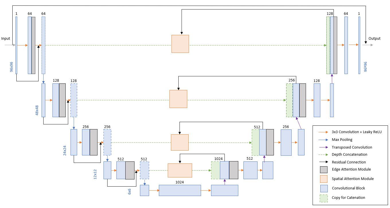
Figure 1: Network architecture of the proposed CEAN model with EAM and SAM. The network structure is based on Residual U-Net. EAM recovers the lost high spatial frequency information, particularly the areas corresponding to edge features. SAM allows the informative activations to be preserved through propagation, and the unwanted features to be progressively suppressed during training, in order to increase network robustness.
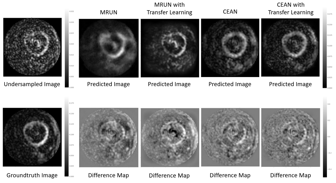
Figure 2: An example of the undersampled image, ground truth image, with corresponding predictions and their difference when compared to the ground truth acquired at b=600s/mm2 in one of the diffusion encoding directions in one of the healthy volunteers.
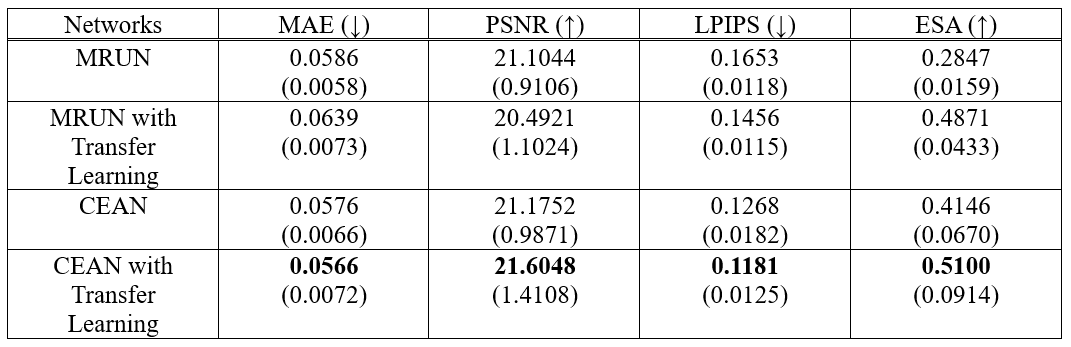
Table 1: Performance metrics with standard deviation, including Mean Absolute Error (MAE) relative to the ground truth, Peak Signal to Noise Ratio (PSNR), and Learned Perceptual Image Patch Similarity9 (LPIPS), a learned measure designed to quantify how perceptually similar the images are visually, and Edge Sharpness Assessment10 (ESA) measured on the free wall epicardium, for MRUN and our proposed network CEAN trained with acquired spiral data only, and MRUN and CEAN with transfer learning.
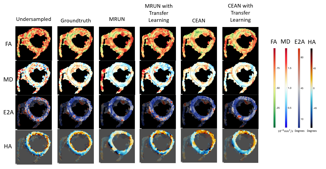
Figure 3: Example DT-CMR maps for Fractional Anisotropy (FA), Mean Diffusivity (MD), Sheetlet Angle (E2A) and Helix Angle (HA) from undersampled images, ground truth and network predictions. Results from CEAN with transfer learning most closely match the ground truth.
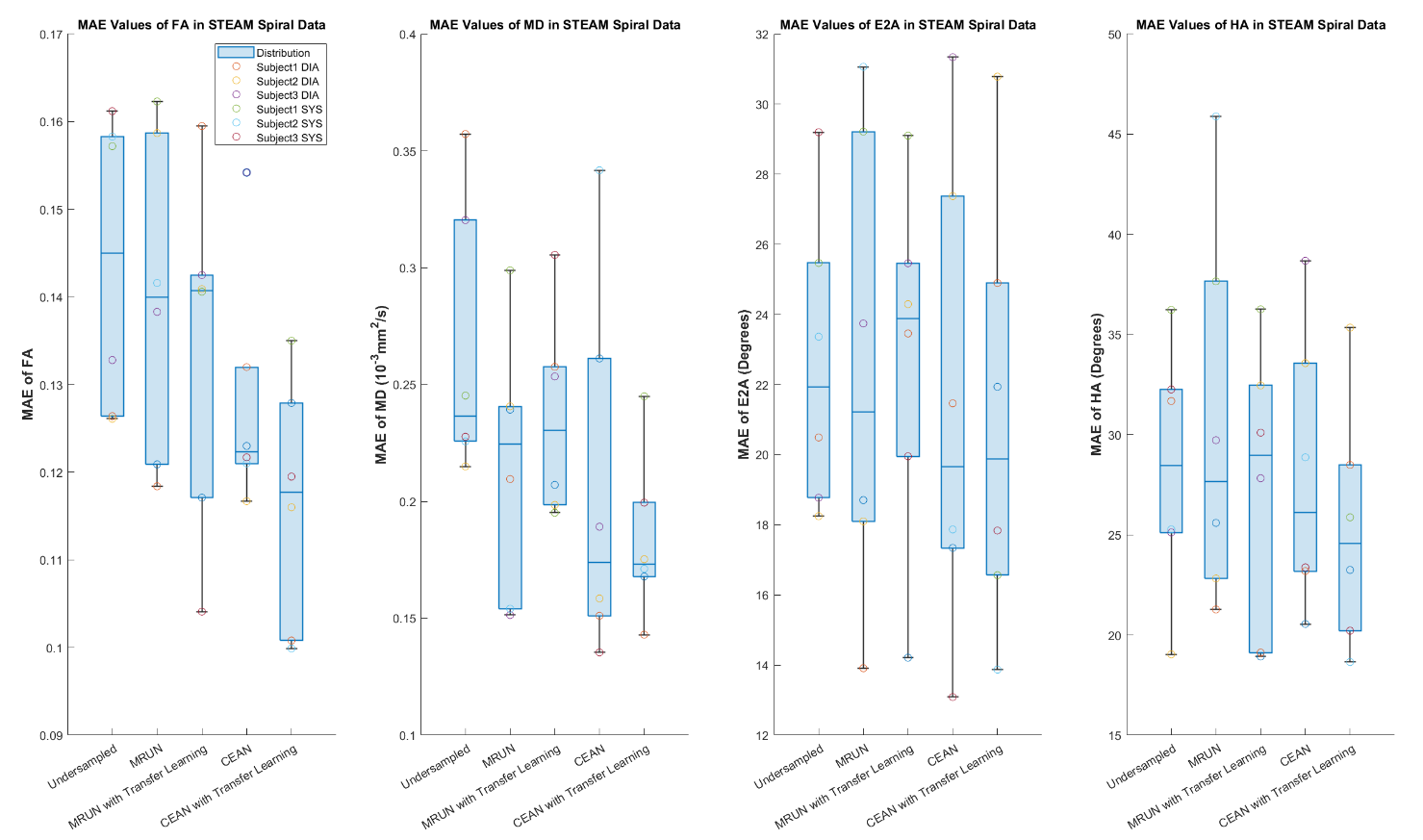
Figure 4: Comparison of pixelwise MAE in the test set for FA, MD, E2A and HA, with reference to the ground truth. Systolic and diastolic data are plotted together with boxplots summarising the parameters and cardiac phases.