0337
Accelerated Acquisition and Cross-Platform Reconstruction of Diffusion Tensor-Derived Indices Using Convolutional Neural Networks1Healthy Aging Research Center, Chang Gung University, Taoyuan, Taiwan, 2Department of Diagnostic Radiology, Chang Gung Memorial Hospital at Keelung, Keelung, Taiwan, 3Department of Medical Imaging and Radiological Sciences, Chang Gung University, Taoyuan, Taiwan
Synopsis
Keywords: AI/ML Image Reconstruction, Machine Learning/Artificial Intelligence, Diffusion tensor imaging, convolutional neural network, curve fitting, mean diffusivity, fractional anisotropy
Motivation: Diffusion-MRI faced limitations due to extended scan times and scanner/protocol variations.
Goal(s): This study aims to assess its ability to accelerate imaging procedures and unify data from diverse sources.
Approach: A convolutional neural network was employed to reconstruct diffusion-weighted images into diffusion tensor images. The effectiveness of reconstructed model was evaluated by normalized mean-square error (NMSE) and structural similarity index (SSIM).
Results: The CNN showed significantly better SSIM and lower NMSE in FA and MD (p < 0.001) compared to conventional methods. Moreover, the CNN model maintained strong performance when applied to other Scanners for FA and MD.
Impact: Through convolutional neural networks, images might be acquired fast and easily be harmonized across platforms . Subsequent research will further utilize deep/machine learning tools to investigate the impact of reconstructed image-segmented brain regions on the performance of classification models.
Introduction
Diffusion-MRI is a potential imaging biomarker for Neurodegenerative Disease [1]. However, it was often limited by prolonged acquisition time and variations between scanners and imaging protocols. Deep learning can be used to facilitate acquisition. In this investigation, we employ a Convolutional Neural Network to reconstruct Diffusion Tensor-derived indices as proposed by Ying et al [2]. This study seeks to explore its efficacy to accelerate the imaging processes and their capacity to harmonize data from different sources.Methods
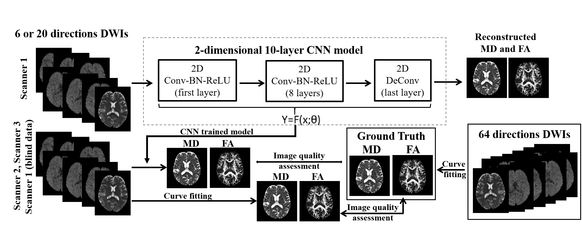
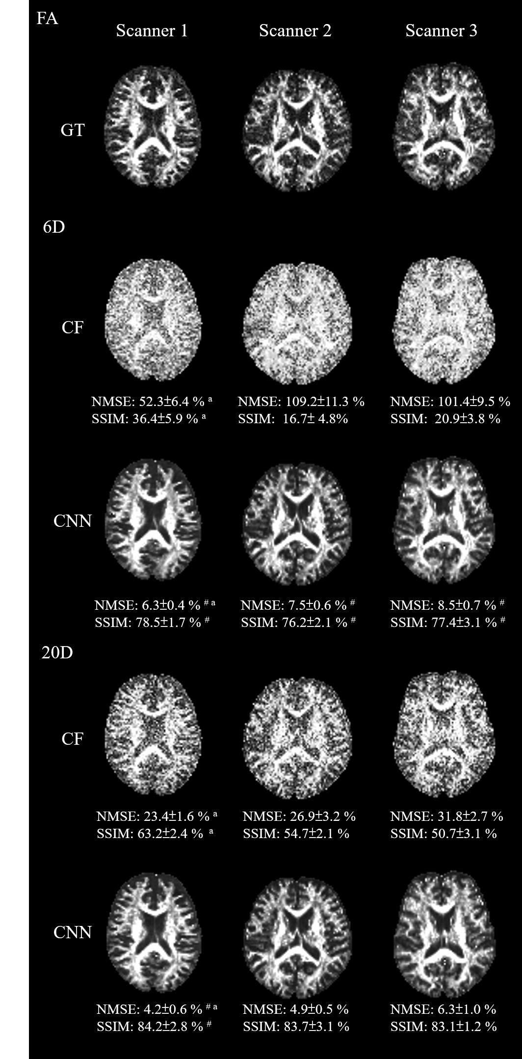
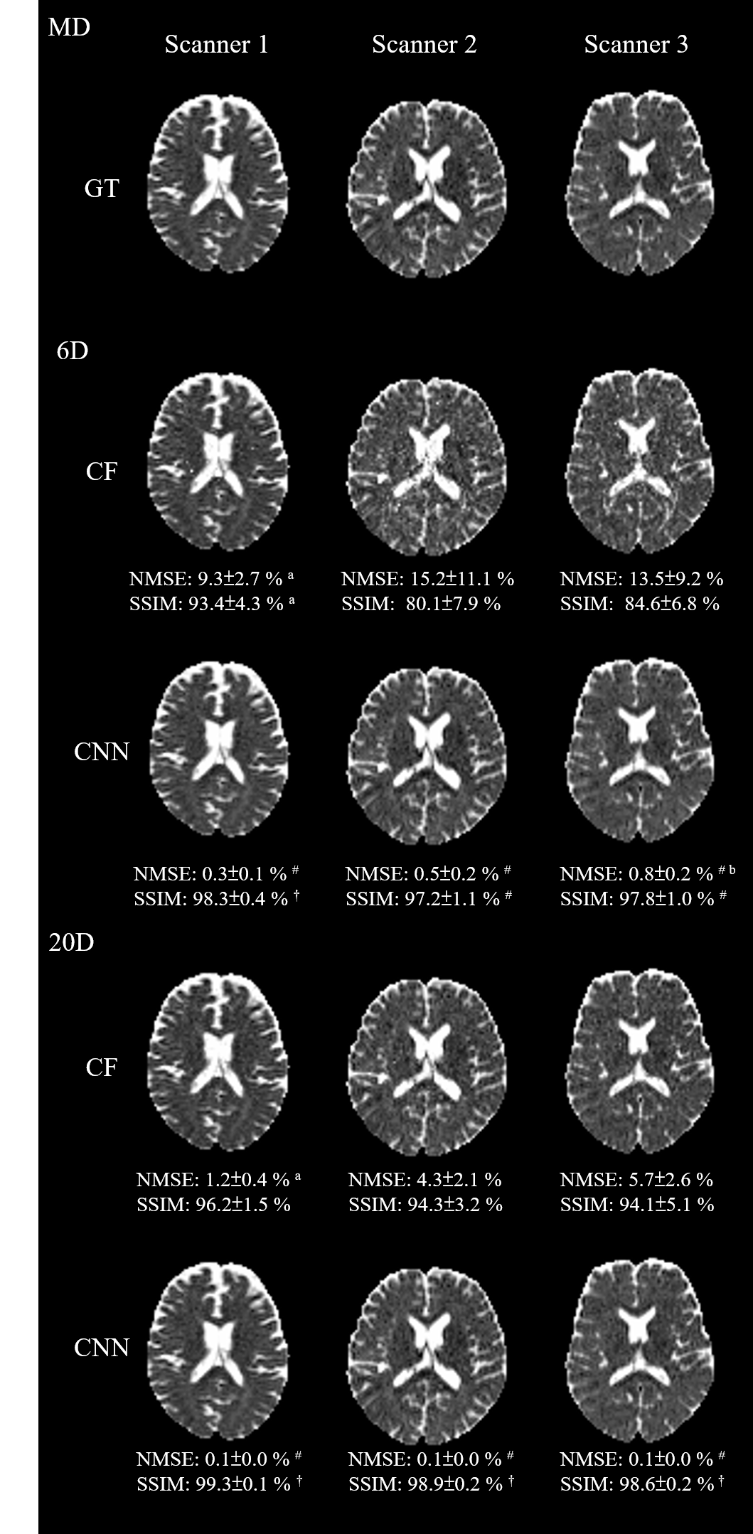
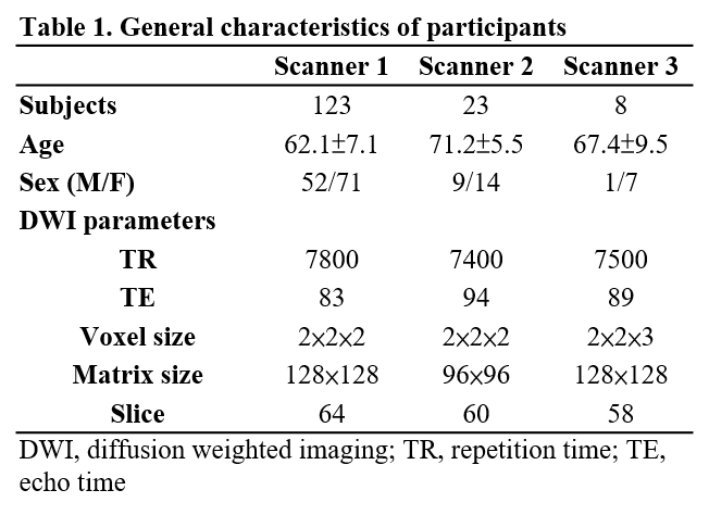
This research received approval from the Institutional Review Board of the Chang Gung Medical Foundation (Approval ID: 202000081B0). Images of normal subjects were acquired using 3T MR scanners: Trio and Skyra by Siemens, Germany (Scanner 1 and 2, respectively), and M750 by GE (Scanner 3). Diffusion-weighted images were acquired using a spin-echo echo-planar-imaging sequence, with a b-value of 1000 s/mm² and diffusion-weighting gradients along 64 non-collinear directions. The number of the participants and the imaging protocol of each scanner was in Table 1. Figure 1 illustrates the algorithm and Convolutional Neural Network structure. Subjects obtained from Scanner 1 were randomly divided into a training dataset (n = 60) and an independent blind dataset (n = 63). The images within the training dataset were used to train the Convolutional Neural Network. The diffusion tensor-derived indices were calculated, including mean diffusivity (MD) and fractional anisotropy (FA). To compare the reconstruction between scanners, diffusion-weighted images acquired from Scanner 2, Scanner 3, and the blind dataset of Scanner 1 were input into the trained model to compute diffusion tensor-derived indices. Index reconstructed by using conventional curve fit according to the diffusion encoding was calculated for comparison. To accelerate the acquisition, two sets of images were extracted from the acquired 64 diffusion directions: 6 or 20 diffusion encoding directions. This will speed up the acquisition by 10.66 times (from 64 to 6 directions) and 3.2 times (from 64 to 20 times). Index reconstructed by using curve fitting with 64 diffusion gradient directions was used a ground truth. To assess the effectiveness of the reconstruction, the following image quality assessments were calculated for each region: (1) Normalized Mean Square Error (NMSE). (2) Structural Similarity Index measure (SSIM) Analysis. The difference between the methods of curve fitting and convolutional neural network was examined by the Student’s T-Test. The difference among the scanners was evaluated by ANOVA. Post-hoc testing was carried out using Fisher's Least Significant Difference method, with p<0.05 considered statistically significant.Results
The maps of FA and MD were shown in Figure 2 and 3, respectively. These metrics were obtained either through curve fitting (second and fourth row) or using a convolutional neural network (third and fifth row). In both 6 and 20 diffusion encoding directions, those reconstructed by convolutional neural network tended to have a significantly higher SSIM and lower NMSE in both FA (Figure 2, p < 0.001) and MD (Figure 3, p <0.001), when compared with conventional curve fitting method. We also found that the convolutional neural network model trained on images collected by Scanner 1 still performed well when used to reconstruct from data collected by Scanner 2 (middle column of Figure 2 and 3) and Scanner 3 (right column of Figure 2 and 3). The SSIM and NMSE values showed similar results across the three scanners and exhibited significant improvements over the conventional curve fit method in FA and MD.Discussion
This study demonstrated that the convolutional neural network can be used to calculate the diffusion tensor derived index from different scanners and with different imaging protocols. When using a reduced number of diffusion encoding directions, the conventional curve fitting method tends to be affected with increased error and reduced similarity when compared to the ground truth. In contrast, the convolutional neural network -based approach showed enhanced image quality in both MD and FA. Both NMSE and SSIM confirm the superiority of the convolutional neural network approach. The acquisition time was reduced (from 64 to 6 or 20, respectively). Notably, the CNN model trained on data from Scanner 1 demonstrates robust performance when applied to data collected by Scanners 2 and 3. This cross-scanner applicability and under different imaging protocols underscores the effectiveness of the convolutional neural network approach. This presents significant potential for image harmonization. Our findings could potentially improve diffusion MRI's limitations to studies/scanner specific and expedite the data acquisition process. Future research will optimize deep learning structure to investigate the diagnostic performance of reconstructed images.Acknowledgements
The presents work was supported by the Imaging Core Laboratory of the Institute for Radiological Research and the Center for Advanced Molecular Imaging and Translation. The authors thank the Neuroscience Research Center (Chang Gung Memorial Hospital) and the Healthy Aging Research Center (Chang Gung University) for their invaluable support.References
1. Lu CS, et al. Alterations of diffusion tensor MRI parameters in the brains of patients with Parkinson's disease compared with normal brains: possible diagnostic use. Eur Radiol 2016;26(11):3978.
2. Li H, et al. SuperDTI: Ultrafast DTI and fiber tractography with deep learning. Magnetic resonance in medicine, 2021;86(6):3334-3347.
Figures