0336
Interleaved TMS-fMRI Explains Variability in TMS Response1Medical University of Vienna, Vienna, Austria, 2Max Planck Institute for Human Cognitive and Brain Sciences, Leipzig, Germany, 3Stanford University, Palo Alto, CA, United States
Synopsis
Keywords: Functional Connectivity, Brain Connectivity, brain stimulation
Motivation: TMS has become an invaluable asset in both research and clinical environments. However, variability in individual responses to TMS is a persistent issue, which limits its broader adoption.
Goal(s): The integration of TMS with fMRI through interleaved paradigms is a promising strategy for gaining insights into the factors that underlie this response variability.
Approach: Adopting an interleaved TMS-fMRI approach we explored the different factors of stimulation dose, sex differences, and cognitive state.
Results: Interleaved TMS-fMRI revealed individual dose-response patterns. Inherent sex differences were found between men and women. Precise timing of TMS relative to cognitive state demonstrated differential effects on relevant brain regions.
Impact: The findings represent a critical step toward addressing the challenge of response variability. TMS-fMRI promises to be a valuable tool for not only understanding factors that influence TMS response but also for potentially enhancing response rates in TMS applications.
Introduction
Transcranial Magnetic Stimulation (TMS) has become a highly effective treatment option for mental disorders1,2. However, the heterogeneity in response to TMS among individuals limits generalizability and effectiveness2. State-of-the-science hardware innovations and precise timing of magnetic stimulation pulses and accelerated image acquisition and stimulus presentation translate into a platform for gaining insights into individual factors that underlie the variability in neural response and clinical outcome. Leveraging the high spatial resolution of fMRI and the high temporal resolution of fMRI, we explored the contributions of stimulation dose, inherent sex differences, and cognitive state to the efficacy of TMS.Methods
All imaging was performed on a 3T Prisma scanner (Siemens). TMS-fMRI setup included a MagProX100 stimulator (MagVenture, Farum, Denmark), MRi-B91 MR-compatible TMS coil mounted on dedicated RF-coil3, MR-compatible optical neuronavigation to allow for coil- and subject-tracking during imaging. All functional images were acquired with 7-channel RF surface coils using EPI (TR/TE = 1000/38ms, flip angle = 60, voxel size = 3mm3 isotropic, 40 slices, Multiband Factor = 4)4.Dose-dependency of DLPFC TMS (Study 1). To examine the contribution of stimulation dose, 15 participants underwent TMS over the left DLPFC at 80%, 90%, 100%, and 110% of their motor threshold (MT) during concurrent fMRI. Two sessions were conducted: the first for anatomical data acquisition for neuronavigation, and the second for dose-response mapping. General Linear Models (GLMs) were computed for individual and group-level responses across each stimulation intensity.
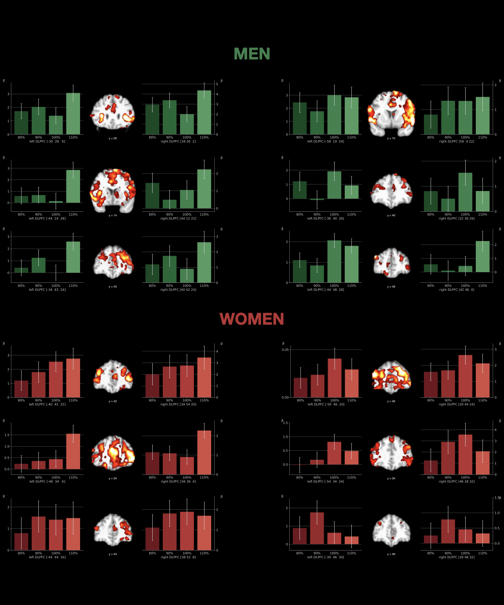
Individual Dose-Response & Sex Differences (Study 2). Following the procedure described in Study 1, we explored the inherent sex differences. The individual dose-response profiles of stimulation over the left DLPFC were assessed for 10 men and 10 women separately.
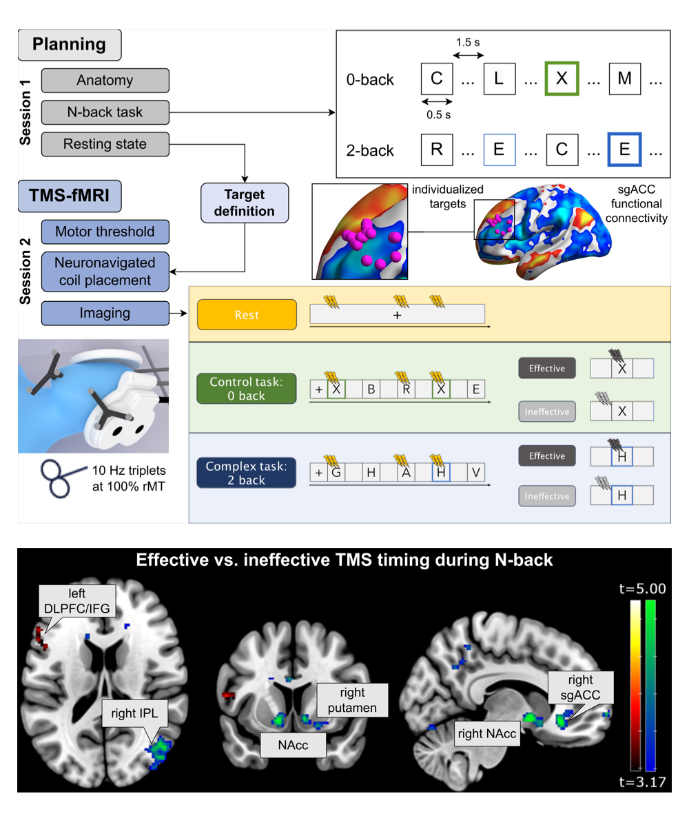
DLPFC-sgACC State-Dependency (Study 3). To investigate the influence of current cognitive state on target engagement, we adopted a chronometric interleaved TMS-fMRI approach during a working memory task. TMS was delivered to the left DLPFC based on individual highest anti-correlation with the sgACC5-7. During continuous imaging, 10 Hz triplets of TMS were precisely timed during an N-back task to interfere with different cognitive processes in a chronometric fashion. Stimulation was delivered either slightly before or after letter onset. This study included 15 participants (9 female, 6 male).
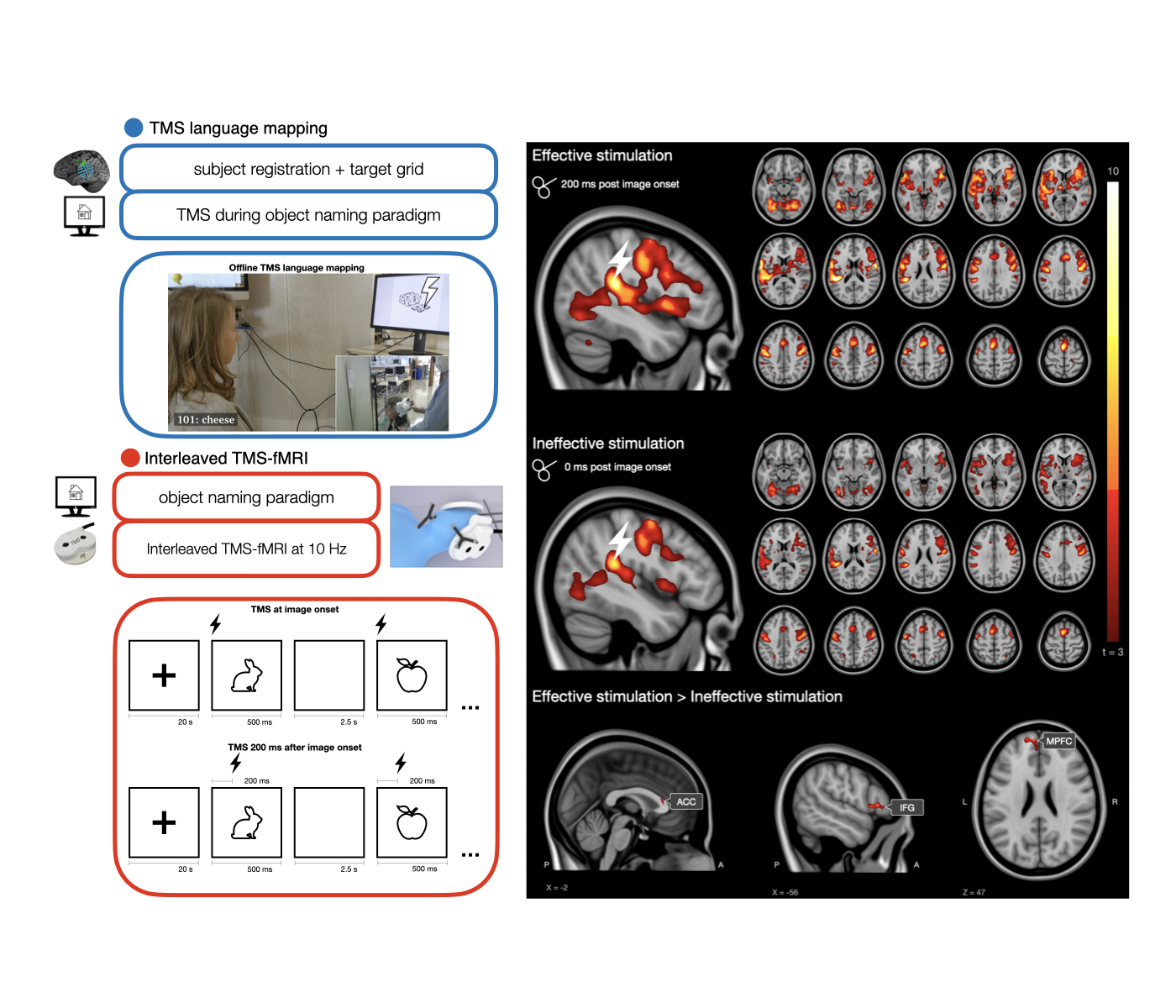
STG-IFG State-Dependency (Study 4). To assess TMS effects across the language network, we used a chronometric interleaved TMS-fMRI approach. Stimulation (bursts of 5 pulses at 10 Hz) was precisely timed to happen between acquired slices during an object naming task . Images were presented in blocks of 20 and the inter-image interval was set to 2.5 seconds. Stimulation occurred either directly at image onset (during conceptual preparation) or 200 ms after image onset (during linguistic processing)8.
Results
Regarding the dose-dependency of DLPFC TMS (Study 1), we show whole-brain network modulations with the strongest BOLD-response at the group-level for stimulation with 100% motor threshold. However, investigating individual dose response-relationships (Study 2) showed differences in response patterns: subjects that responded to subthreshold stimulation, subjects that required above threshold stimulation in order to show a significant BOLD-response and atypical response-curves. When investigating these results further for inherent sex differences, we find that women show more homogenous dose-response profiles compared to men. For chronometric TMS of the DLPFC (Study 3) we found statistically significant differences in TMS response with different timings. Areas showing these state-dependent effects included clinically relevant targets for depression treatment, e.g. the sgACC (peak: MNI 10, 30, -10; t=5.19, p=.000 ) and the NAcc (peak: MNI 10, 4, -6; t=6.46, p=.000). In the object naming task (Study 4), precise timing of TMS relative to the cognitive task phase demonstrated differential effects on the relevant brain regions. Specifically, stimulation during linguistic processing resulted in widespread activation in the language network, including at the stimulation target location (peak: MNI -56, -26, 16, t=6.26, p=.000) as well as in the inferior frontal gyrus (peak: MNI -54, 32, 4, t=4.37, p=.000).Conclusion
The integration of fMRI with TMS leverages the high spatial resolution of fMRI alongside the precise temporal resolution of TMS, presenting a significant methodological benefit in examining the variability of TMS responses. This combination has allowed us to uncover the intricate interplay between individual differences, cognitive states, and stimulation parameters. Our findings represent a critical step toward addressing the challenge of TMS response variability. As we move forward, TMS-fMRI promises to be a valuable tool for not only understanding the factors that influence TMS response but also for potentially enhancing response rates in TMS applications.Acknowledgements
No acknowledgement found.References
[1] Cole EJ et al. Stanford Neuromodulation Therapy (SNT): A Double-Blind Randomized Controlled Trial. AJP. 2022;179:132–141.[2] Chen L et al. Accelerated Repetitive Transcranial Magnetic Stimulation to Treat Major Depression: The Past, Present, and Future. Harvard Review of Psychiatry 2023;31(3):142-161.
[3] Navarro de Lara L et al. A novel coil array for combined TMS/fMRI experiments at 3 T. Magnetic Resonance in Medicine 2015;74(5):1492–1501.
[4] Tik M et al. Acute TMS/fMRI response explains offline TMS network effects - An interleaved TMS-fMRI study. Neuroimage, 2023. 267: p. 119833.
[5] Fox MD et al. Efficacy of transcranial magnetic stimulation targets for depression is related to intrinsic functional connectivity with the subgenual cingulate. Biol Psychiatry. 2012;72:595–603.
[6] Cash RFH et al. Using Brain Imaging to Improve Spatial Targeting of Transcranial Magnetic Stimulation for Depression. Biol Psychiatry. 2021;90:689–700.
[7] Siddiqi SH et al. Identification of Personalized Transcranial Magnetic Stimulation Targets Based on Subgenual Cingulate Connectivity: An Independent Replication. Biol Psychiatry. 2021;90:e55-e56
[8] Indefrey, P. The spatial and temporal signatures of word production components: a critical update. Frontiers in psychology, 2011; 2:255.
Figures
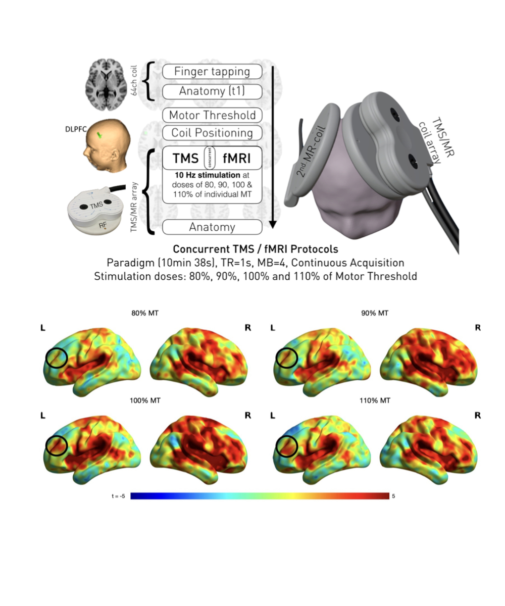
Top. For interleaved TMS/fMRI, tailor-made coil setup and an optimized multiband accelerated EPI sequence for high-resolution, artefact-free imaging of acute TMS effects. We used neuronavigation for defining the motor thresholds and DLPFC targets. We used two receive coil arrays, one mounted on the TMS coil and one on the contralateral side.
Bottom. Comparison of TMS induced network activation for real and sham. Unthresholded maps on the cortical surface depict stronger bilateral BOLD increase due to real stimulation as compared to sham stimulation.