0333
Aggregation of Connectivity Gradient in Hippocampus Induced by Long-Term Cognitive Training with Development1School of Physics, Zhejiang University, Hangzhou, China
Synopsis
Keywords: fMRI Analysis, fMRI (resting state), Connectivity gradient, Hippocampus, Development, Cognitive training
Motivation: The hippocampus-cortical connections have shown rapid developmental-changed nature during childhood and learning-adapted plasticity with skill acquirement.
Goal(s): However, little is known about the effect of development interacting with cognitive training on the hippocampal connectivity gradient during puberty.
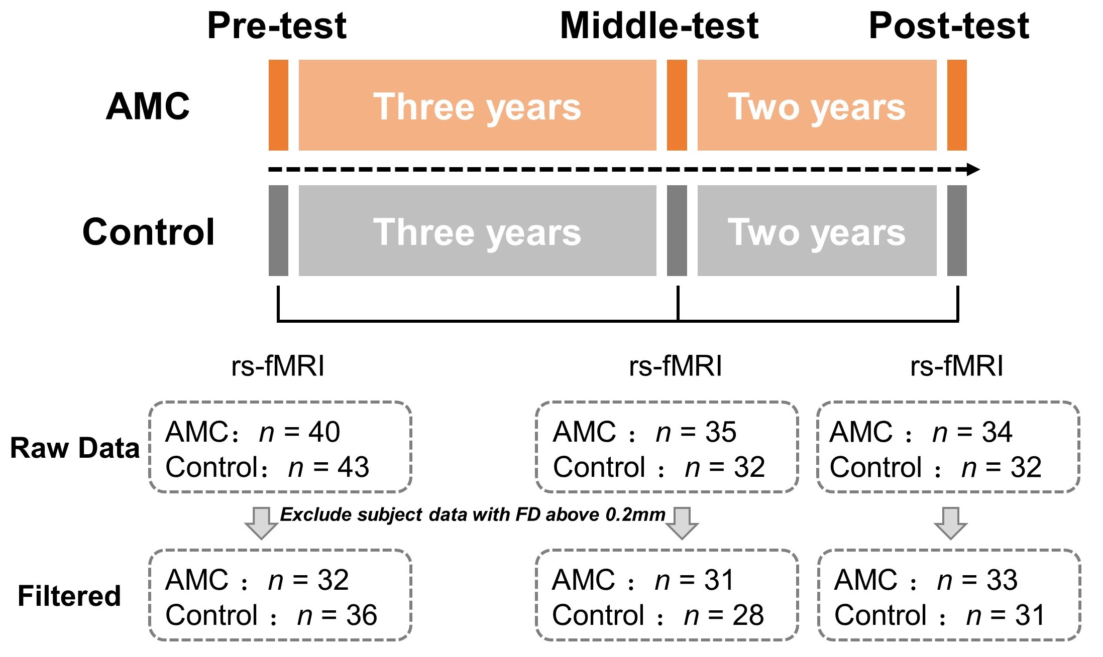
Approach: Here we employed longitudinal dataset (191 scans from training/control groups: n = 43/45) which collected neuroimaging data of school-age children across 0/3/5-year abacus mental calculation (AMC) training stages to explore this question.
Results: By calculating connectivity gradient of hippocampus, we observed significantly development-induced gradient aggregation of hippocampus, and training promoted that effect, which were resulted from changes in functional connectivity between hippocampus with different cortices.
Impact: These findings provide novel insights into development and training effects on function specialization of hippocampus during puberty from a largescale perspective of connectivity gradient, which may be helpful for better understanding of functionally atypical trait of hippocampal disorder for clinicians.
Introduction
The hippocampus is a brain region with complex structure located in the medial temporal lobe, which plays a key role in memory, navigation and learning 1–7. There exist rich functional connectivity (FC) between the hippocampus and cortex that change across development 8–10, which is critical for skill acquisition in line with problem-solving strategy shifting from procedural strategy to memory retrieval during childhood 11. Rapid structural changes of the hippocampal region mainly occur in infancy and early childhood, and the maturation of FCs between hippocampus and prefrontal cortex supports the improvement in episodic memory in late childhood 12. Previous studies have revealed the gradient nature of the hippocampus along the anterior-to-posterior long axis 13,14. However, little studies have explored the effects of development and cognitive training on the hippocampal connectivity gradients. In current study, we aim to investigate above questions by analyzing the neuroimages of across three abacus mental arithmetic (AMC) training stages collected from early to late childhood (7–12 years old).Methods
ParticipantsWe collected longitudinal resting-state fMRI data from three stages: pre-, middle-, and post-test, which requires trainees to train 2 hours every week while controls remain daily school life (Figure 1). We excluded neuroimaging data with mean framewise displacement above 0.2 mm. It yields 43 participants in the training group (23 females, mean ± SD = 6.83 ± 0.50) with 96 scans, as well as 45 participants in the control group (21 females, mean ± SD = 6.98 ± 0.49) with 95 scans. There were no significant group differences in age (t[86] = -1.414, p = 0.162) and gender (χ2 = 0.409, p = 0.522).
Hippocampal gradient calculation
We extracted time series from a total of 16584 voxels of 214 brain regions, including 210 cortical areas and 4 hippocampal subregions 15, and then calculated FC matrix. The connectivity gradient was calculated from obtained FC matrix as previous study 16. After calculating the individual gradient components, we aligned all individual gradients to that average gradient template generated from pre-test data for further analysis.
Multiple indicators to measure hippocampal gradient
We tried to use multiple indicators to delineate gradient characteristics of each hippocampal subregion affected by development and training. Indicators of hippocampal subregions in voxel-wise gradient are depicted by gradient range, median, standard deviation (SD), as well as two-dimensional (2D) SD, 2D distance.
Linear mixed model
Linear mixed model (LMM) is used to explore the imapct of development and training on hippocampal gradients or FC between hippocampus and cortex. The formula of the LMM is as follows: gradient indicator/value/FC ~ training time + group + sex + head motion + training time*group + (group|subID), while sex and head motion are controlled as covariates.
Results
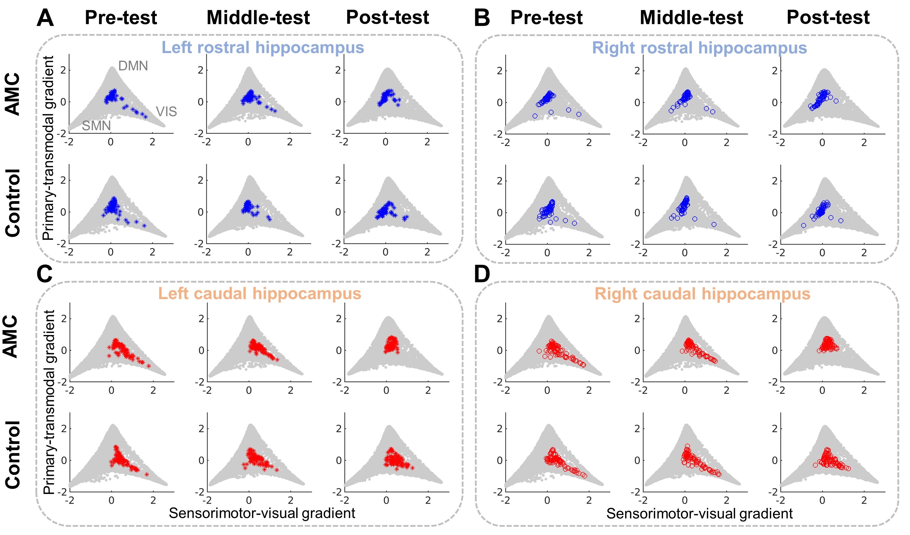
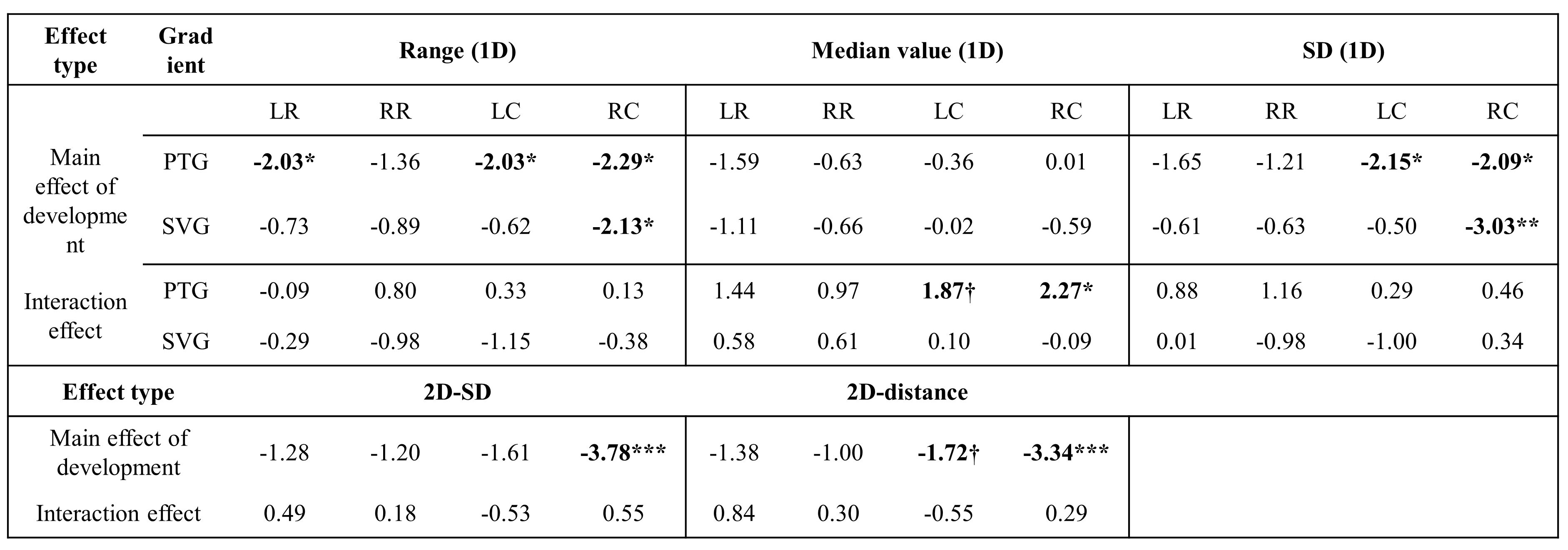
Aggregation of hippocampal gradient induced by development and trainingWe found that the hippocampal gradient presented a hierarchical organization along anterior-to-posterior long axis. With development, these voxel-wise gradient points that originally extend toward the visual area gradually shrink away from this tip, which appeared more aggregation in the training group (Figure 2). And these observations were confirmed by multiple gradient indicators using LMM, including significant main effect of development in gradient range, SD, 2D SD and 2D distance, as well as the significant interaction effect in gradient median value (Table 1).
Changes in FCs underlies the gradient aggregation across development
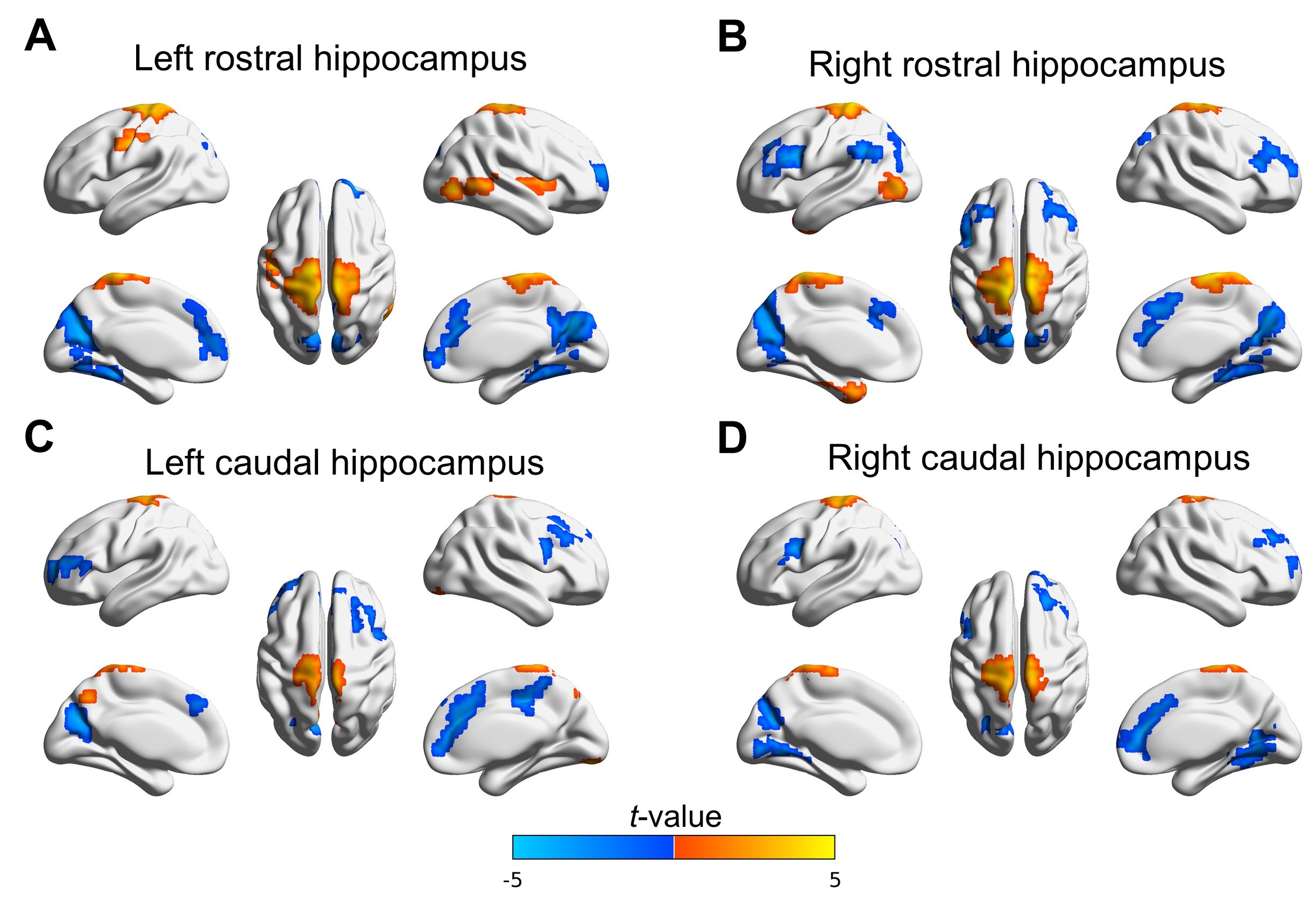
We applied LMM to investigate developmental changes of FCs between hippocampus and cortex. The significant main effect of development in gradients were resulted from strengthened FCs between hippocampus with sensorimotor areas, as well as weakened FCs with anterior cingulate cortex, medial parieto-occipital sulcus (p < 0.05, cluster size > 50 voxels, Figure 3).
Training effect on voxel-wise hippocampus gradient
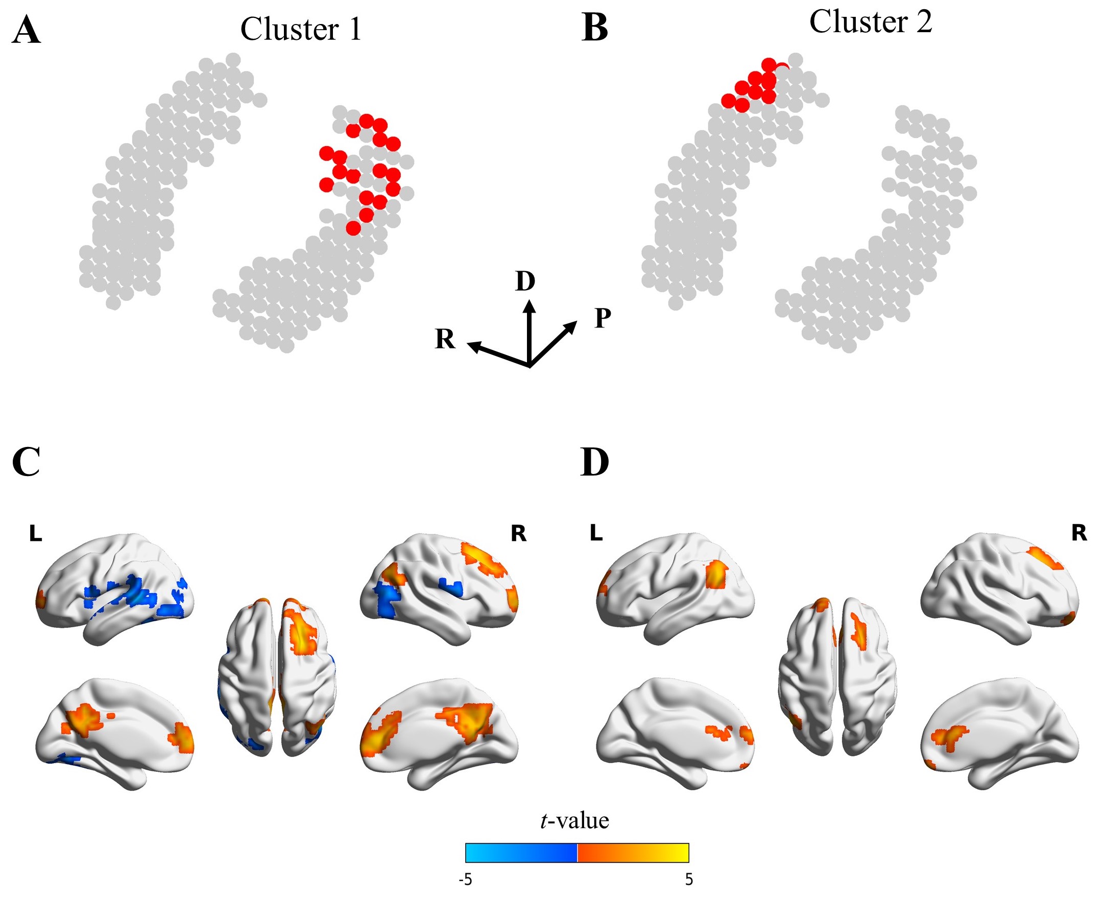
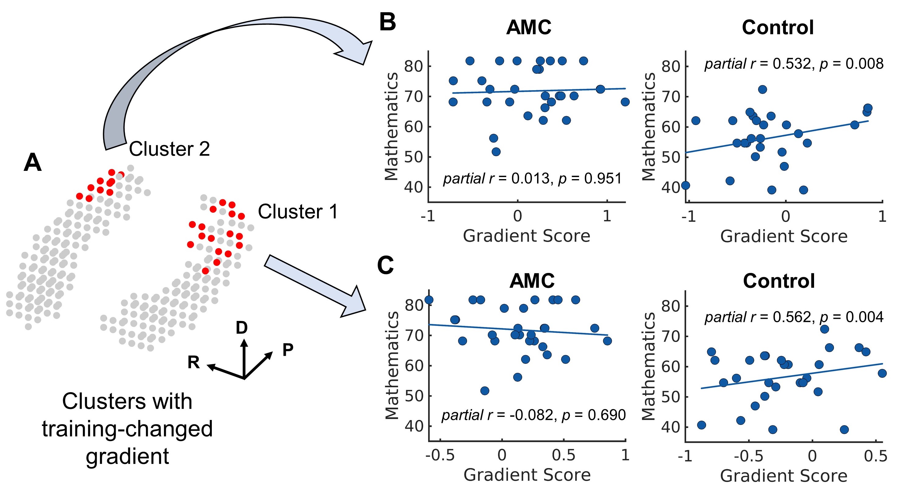
We observed a significant interaction effect of development and training of primary-transmodal gradient in the caudal hippocampus (p < 0.05, cluster size > 10 voxels, Figure 4). Using seed-based FC analysis, we found that group difference in caudal hippocampus gradient mainly originated from the enhanced FCs with anterior cingulate cortex, posterior cingulate cortex, and right dorsolateral prefrontal cortex (p < 0.05, cluster size > 50 voxels, Figure 4).
Relation to behavior
The results of partial correlation controlling for sex and head motion showed that the average gradient value of cluster in right caudal hippocampus was positively correlated with the mathematical scores in the control group (partial r = 0.532, p < 0.01, Figure 5), while no significant correlation in the training group (partial r = 0.013, p = 0.951). And we observed similar results in left caudal hippocampus.
Acknowledgements
We are grateful to the Chinese Abacus and Mental Arithmetic Association for their kind support, as well as to the children, parents, and teachers for their participation in this study. This work was supported by the National Natural Science Foundation of China (32071096).References
1. Eichenbaum H, Yonelinas AP, Ranganath C. The medial temporal lobe and recognition memory. Annu Rev Neurosci. 2007;30:123-152.
2. Eichenbaum H. The role of the hippocampus in navigation is memory. J Neurophysiol. 2017;117(4):1785-1796.
3. Poppenk J, Moscovitch M. A hippocampal marker of recollection memory ability among healthy young adults: contributions of posterior and anterior segments. Neuron. 2011;72(6):931-937.
4. Strange BA, Witter MP, Lein ES, Moser EI. Functional organization of the hippocampal longitudinal axis. Nat Rev Neurosci. 2014;15(10):655-669.
5. Lisman J, Buzsáki G, Eichenbaum H, Nadel L, Ranganath C, Redish AD. Viewpoints: how the hippocampus contributes to memory, navigation and cognition. Nat Neurosci. 2017;20(11):1434-1447.
6. Voss JL, Bridge DJ, Cohen NJ, Walker JA. A closer look at the hippocampus and memory. Trends Cogn Sci. 2017;21(8):577-588.
7. Xue G. The neural representations underlying human episodic memory. Trends Cogn Sci. 2018;22(6):544-561.
8. Damoiseaux JS, Viviano RP, Yuan P, Raz N. Differential effect of age on posterior and anterior hippocampal functional connectivity. NeuroImage. 2016;133:468-476.
9. Blankenship SL, Redcay E, Dougherty LR, Riggins T. Development of hippocampal functional connectivity during childhood. Hum Brain Mapp. 2017;38(1):182-201.
10. Calabro FJ, Murty VP, Jalbrzikowski M, Tervo-Clemmens B, Luna B. Development of hippocampal–prefrontal cortex interactions through adolescence. Cereb Cortex. 2020;30(3):1548-1558.
11. Qin S, Cho S, Chen T, Rosenberg-Lee M, Geary DC, Menon V. Hippocampal-neocortical functional reorganization underlies children’s cognitive development. Nat Neurosci. 2014;17(9):1263-1269.
12. Ghetti S, Bunge SA. Neural changes underlying the development of episodic memory during middle childhood. Dev Cogn Neurosci. 2012;2(4):381-395.
13. Vos de Wael R, Larivière S, Caldairou B, et al. Anatomical and microstructural determinants of hippocampal subfield functional connectome embedding. Proc Natl Acad Sci U S A. 2018;115(40):10154-10159.
14. Masouleh SK, Plachti A, Hoffstaedter F, Eickhoff S, Genon S. Characterizing the gradients of structural covariance in the human hippocampus. NeuroImage. 2020;218:116972.
15. Fan L, Li H, Zhuo J, et al. The human brainnetome atlas: a new brain atlas based on connectional architecture. Cereb Cortex. 2016;26(8):3508-3526.
16. Margulies DS, Ghosh SS, Goulas A, et al. Situating the default-mode network along a principal gradient of macroscale cortical organization. Proc Natl Acad Sci U S A. 2016;113(44):12574-12579.
Figures