0318
SimulScan and Partial Least Squares: Linking speech and swallowing dynamics to brain function1Carle Illinois College of Medicine, University of Illinois Urbana Champaign, Urbana, IL, United States, 2Purdue University, West Lafayette, IN, United States, 3Bioengineering, University of Illinois Urbana Champaign, Urbana, IL, United States, 4Beckman Institute for Advanced Science and Technology, University of Illinois Urbana Champaign, Urbana, IL, United States
Synopsis
Keywords: fMRI Analysis, fMRI (task based), speech, swallowing, fMRI analysis, dynamic imaging
Motivation: SimulScan enables the joint imaging of dynamic oropharyngeal movements during speech and swallowing along with their central control from functional MRI.
Goal(s): The large, complex dataset requires memory-efficient analysis to find the uncover the underlying relationships between the dynamic and functional imaging data.
Approach: Here we develop a memory-efficient implementation of partial least square (PLS) and apply it to a blocked tongue tapping task.
Results: The PLS method separates out correlated motions and brain function for different components of the task.
Impact: SimulScan with PLS analysis can enable the visualization of central control of complex processes such as speech and swallowing. This approach will enable the in-depth study of healthy and disordered speech and swallowing mechanisms in age and disease.
INTRODUCTION
SimulScan is a method that acquires both dynamic MRI of the oropharyngeal region along with functional MRI signals of the brain, simultaneously1,2. With this scan, we can examine the fast dynamic movements of the tongue, lips, velum, larynx, and pharynx while also getting functional MRI of the whole brain with a reasonable TR. The acquisition enables the precise study of central control of oropharyngeal structure movements in speech and swallowing tasks. However, the dimensionality of the data makes analysis challenging. In an approximately 10-minute scan, over 10,000 dynamic images are generated from the midsagittal plane and nearly 300 functional MRI 3D volumes are also acquired. In this work, we leverage the partial least squares (PLS) framework3,4 to find underlying latent variables linking the brain function to the dynamic imaging.METHODS
The PLS method allows us to combine the dynamic and functional MRI data to understand underlying driving latent variables shared between the oropharyngeal motions and brain functional signals. A core component of PLS is performing singular value decomposition (SVD) on the combined dynamic and functional MRI data. However, the default MATLAB implementation of SVD was empirically determined to require more than 10x the RAM of the size of the input matrix, which is prohibitively expensive with inputs that are typically larger than 15GB. To circumvent this RAM requirement, we developed a Python program to efficiently perform SVD on MATLAB matrices with less than 2x RAM consumption5 and return the result to the main MATLAB process for further processing.A healthy participant underwent a SimulScan acquisition with dynamic imaging of a 30-cm FOV, matrix size 225, 2D mid-sagittal scan acquired at 50.3 ms per frame, with 10,640 dynamic images acquired in a 9-minute scan. Simultaneous with this a functional MRI acquisition with 2.5x2.5x3 mm resolution and 38 slices acquired axially were obtained with a TR of 1.9 sec, TE of 25 ms, with 280 fMRI volumes acquired in the time series. To demonstrate that functional signal is obtained with this acquisition, the subject performed tongue tapping with 20 s off and 20 s on blocks repeated for the 9 minutes, cued by a flashing checkerboard.
RESULTS
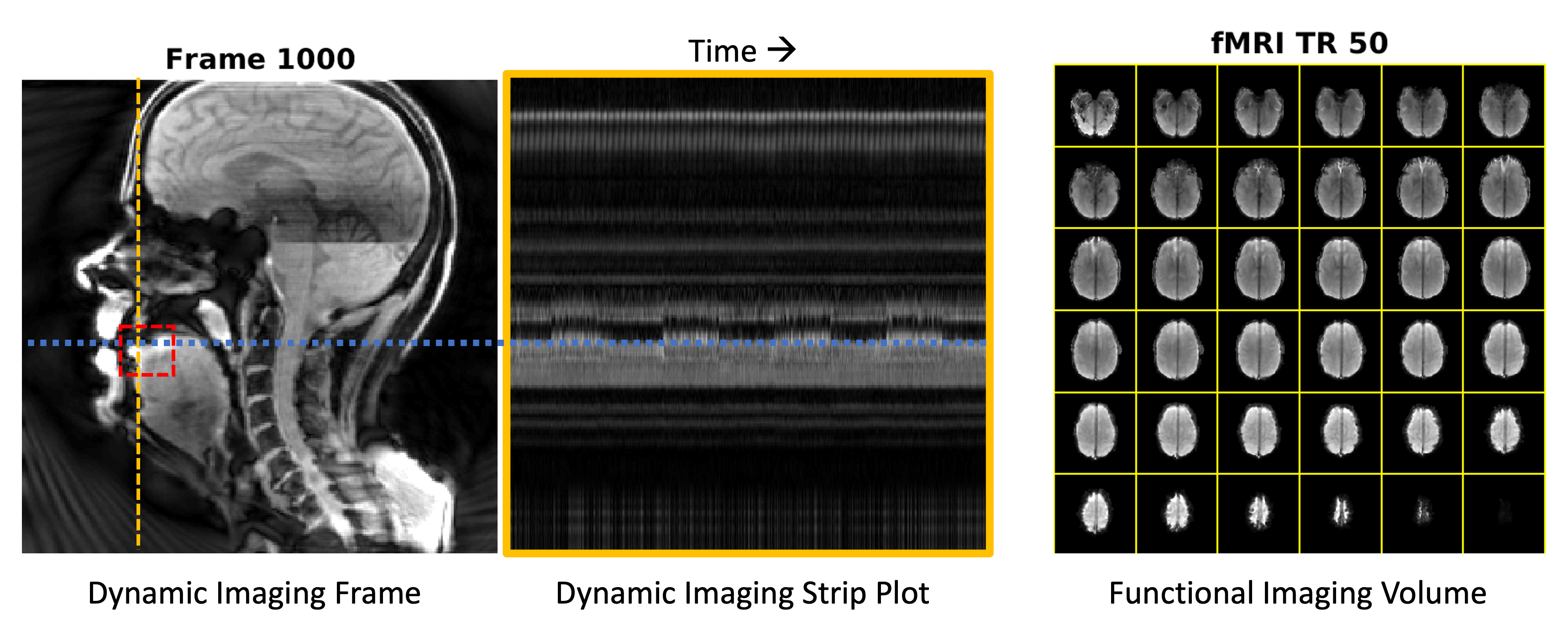
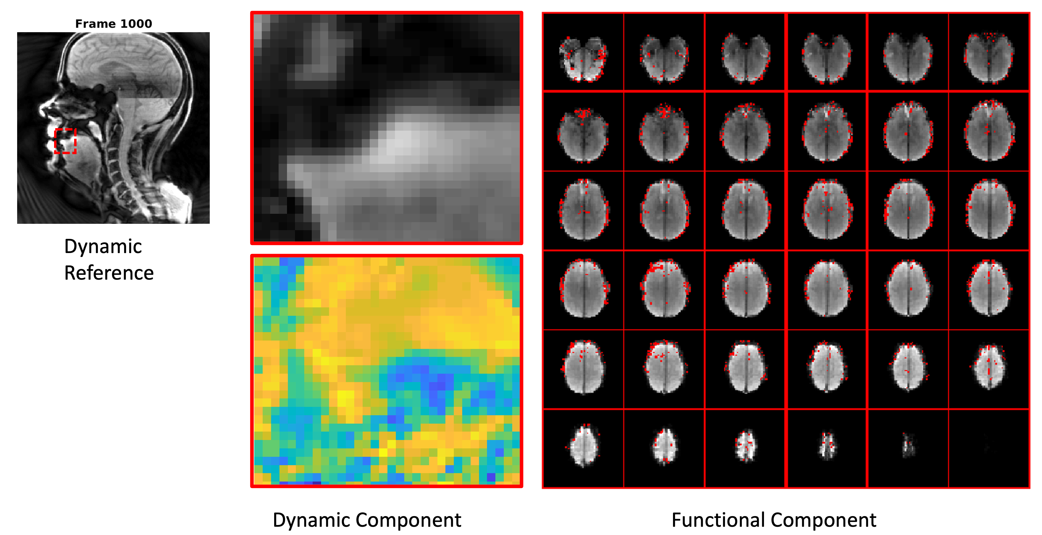
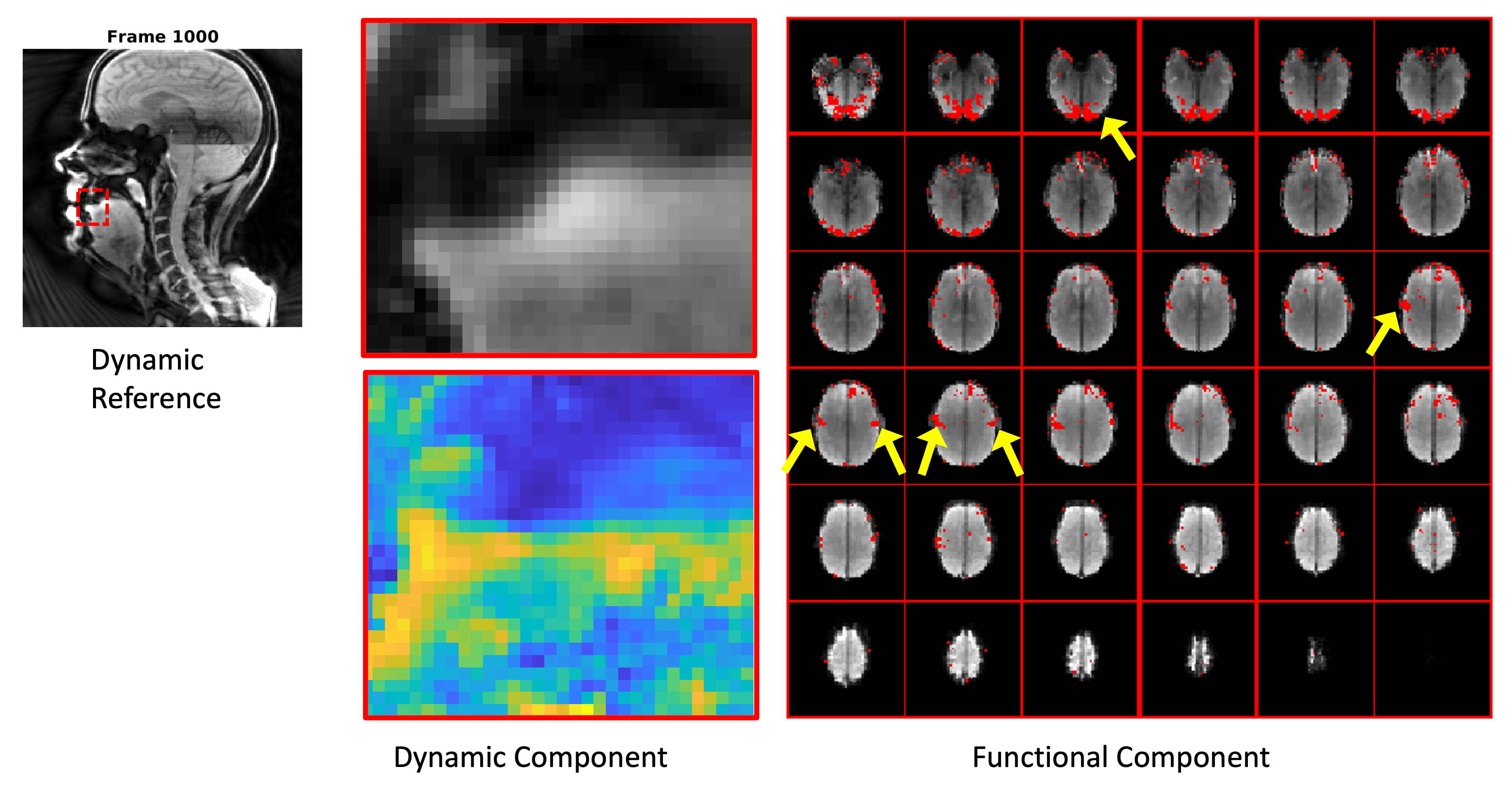
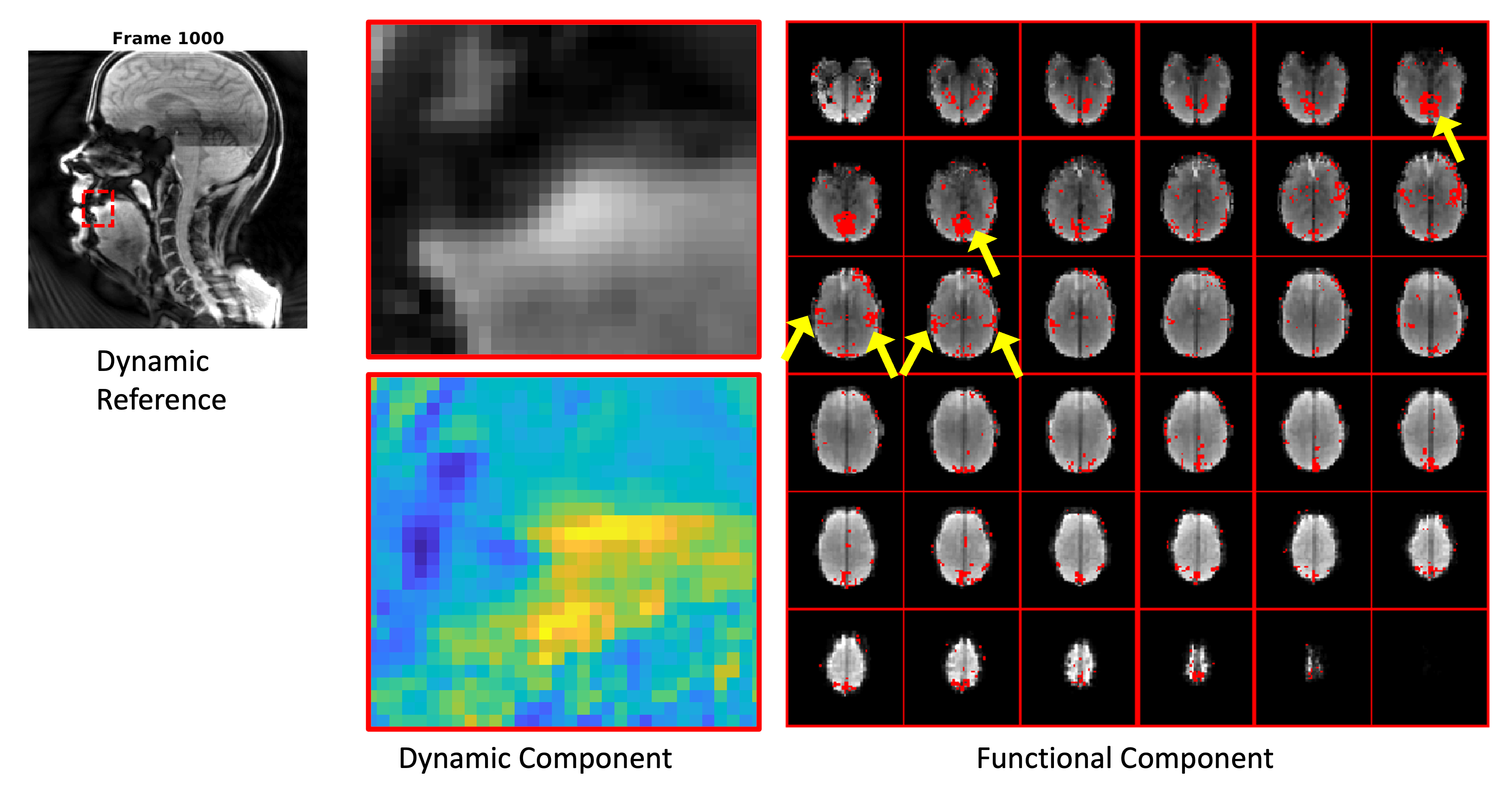
Dynamic data was visualized, see Figure 1, and a region of interest was formed around the tongue tip. The dynamic data was transformed into an estimate of motion by grouping dynamic images that were acquired within a specific functional imaging TR and taking the standard deviation over time within each voxel. This standard deviation signal, indicating motion during the task, was convolved with a hemodynamic response function and formed the dynamic input to our PLS method. The functional MRI volumes were also used as input to the PLS method. The resulting PLS analysis yielded several components: the first component is shown in Figure 2. The second component, reflecting the dynamics of the tongue tip, is shown in Figure 3. You can also see in Figure 3 that there is neural activation in the tongue region of the motor cortex and also visual activation showing in this component. Figure 4 displays the third component, which has additional primary somatosensory activations and different visual processing areas. Additionally in this component, the dynamic motions are more posterior, away from the tongue tip.DISCUSSION
The PLS method identifies underlying latent variables that are driving dynamic changes in both the functional MRI signal and the dynamic images, which in this case were the variance of the signal intensity over time convolved with the hemodynamic response function. This enables a linking between the dynamic information contained in both data to enable visualization of the brain function driving the dynamic motions.CONCLUSION
Although the task used in the current work was simple tongue tapping, the results serve to validate the data analysis approach and demonstrate that the PLS method can associate brain function with specific dynamic information in the SimulScan sequence. Future applications will include more complex speech and swallowing tasks which are important to further explore for the diagnosis and treatment of speech and swallowing disorders.Acknowledgements
Research reported in this publication was supported by the National Institute On Aging of the National Institutes of Health under Award Number R01AG078513. The content is solely the responsibility of the authors and does not necessarily represent the official views of the National Institutes of Health.References
1. Paine TL, Conway CA, Malandraki GA, Sutton BP. Simultaneous dynamic and functional MRI scanning (SimulScan) of natural swallows. Magn Reson Med 2011;65(5):1247-1252.2.
2. Sutton BP, Malandraki GA, Jin R, Marchini C, Anderson A, Arnold P, Liang ZP. Accelerating dynamic imaging in SimulScan: Simultaneous functional MRI and dynamic imaging of tongue motion. Intl Soc. Magn Reson Med, 2023. P. 116.3.
3. McIntosh AR, Lobaugh NJ. Partial least squares analysis of neuroimaging data: applications and advances. NeuroImage 2004;23 Suppl 1:S250-263.4.
4. Krishnan A, Williams LJ, McIntosh AR, Abdi H. Partial Least Squares (PLS) methods for neuroimaging: a tutorial and review. NeuroImage 2011;56(2):455-475.
5. dask package accessed at: https://docs.dask.org/en/latest/generated/dask.array.linalg.svd_compressed.html
Figures