0316
Effect of iron content on GSH levels in the basal ganglia and midbrain in young healthy controls1Department of Radiology, Ruijin Hospital, Shanghai Jiao Tong University School of Medicine, Shanghai, China, Shanghai, China, 2Faculty of Medical Imaging Technology, College of Health Science and Technology, Shanghai Jiao Tong University School of Medicine, Shanghai, China, 3Philips Healthcare, Shanghai, China, Shanghai, China, 4Shanghai Key Laboratory of Magnetic Resonance, College of Physics and Electronic Science, East China Normal University, Shanghai, China, 5Department of Radiology, Wayne State University, Detroit, MI, USA, Detroit, MI, United States
Synopsis
Keywords: Parkinson's Disease, Parkinson's Disease, Glutathione, MEGA-PRESS, Deep gray matter
Motivation: It is important to characterize the relationship between oxidative stress and iron deposition in neurodegenerative diseases.
Goal(s): To investigate if there is a correlation between the glutathione (GSH) levels and brain iron in young healthy controls.
Approach: Taking advantage of proton magnetic resonance spectroscopy (1H-MRS) with MEscher-Garwood Point RESolved Spectroscopy (MEGA-PRESS) and quantitative susceptibility mapping to detect GSH and iron levels.
Results: An intriguing phenomenon was found that as the iron content increased in the putamen, substantia nigra, and red nucleus, the GSH level showed an increasing trend in the basal ganglia and midbrain region respectively.
Impact: The relationship between oxidative
stress and excessive iron deposition is complicated. This preliminary study
offers new insight to investigate the time sequence in iron homeostasis and
oxidative stress.
Introduction
Glutathione (GSH) is an antioxidant that plays a critical role in physiological functions. Altered GSH levels are associated with neurodegenerative diseases such as Parkinson's disease (PD)1. Increases in iron on the other hand is the hallmark of PD patients. There is increasing evidence that the interaction between oxidative stress, mitochondrial dysfunction and abnormal cellular iron homeostasis may play a role in the pathogenesis of PD2. Therefore, it is imperative to characterize the correlation between GSH levels and iron content in healthy individuals. In this preliminary study, we evaluated GSH and iron levels in the basal ganglia and midbrain regions in young individuals using 1H-MRS with spectral editing using MEscher-Garwood Point RESolved Spectroscopy (MEGA-PRESS)3 and quantitative susceptibility mapping (QSM).Methods
This study was approved by the local ethics committee (approval number: KY2022-266) and written informed consent was obtained from all participants. Twenty-two young healthy individuals were enrolled and scanned on a 3T Philips MRI scanner (Elision X, Philips Healthcare) equipped with a 32-channel head coil. QSM data were acquired using a 3D multi-echo strategically acquired gradient echo (STAGE) imaging protocol4, the imaging parameters were: an in-plane resolution = 0.67 × 1 × 1.34 mm3, (interpolated to 0.67 × 0.67 × 1.34 mm3), 5 echoes, TR/TE/△TE = 27/2.7/4.7 ms, flip angles (FA) = 6o and 30o, pixel bandwidth = 362 Hz/pixel. GSH data were acquired using a MEGA-PRESS sequence with the following parameters: Volume of interest (VOI) = 30 × 30 × 30 mm3 (basal ganglia) or 25 × 35 × 30 mm3 (midbrain). The location and the corresponding GSH peak of VOIs are showed in Figure 1. spectral bandwidth = 2000 Hz, readout duration: 512 ms, TR/TE = 2000/130 ms, edit on/off frequencies = 4.56/1.44 ppm, nine dynamics with 16 NSA for each dynamic.The deep gray matter (putamen, substantia nigra, and red nucleus) was auto-segmented from the QSM data using SPIN software (SpinTech MRI, Inc., Bingham Farms, Ml, USA). Spectral-edited data were processed using GANNET 3.1 to get the GSH and creatine (Cr) concentrations5. Spectral data with fitting errors greater than 15% for basal ganglia or 20% for midbrain were considered unreliable and excluded. Visual inspection of the remaining edited spectrum was done to confirm the correctness of the result. Finally, the relative quantification results were expressed by the ratio of GSH/Cr.
The correlation between GSH/Cr level and iron content was evaluated with Pearson correlation analysis (level of significance p < 0.05). This statistical analysis was performed using IBM SPSS Statistics (version 24; IBM Corp., Armonk, NY).
Results
The demographic and imaging features of the participants are showed in Table 1. The correlations of the level of GSH and the iron content are showed in Figure 2. Specifically, the level of GSH in basal ganglia increased with the higher the iron content of the putamen (r = 0.5543, p = 0.0138); the GSH levels in the midbrain showed an increasing trend with the increase iron content of the substantia nigra and red nucleus, but it not reach statistical significance.Discussion and Conclusion
In this study, an intriguing phenomenon was found that with the increase of iron content in the deep gray matter of basal ganglia (putamen) and midbrain (substantia nigra and red nucleus), there was a corresponding upward trend in the level of GSH. Based on these preliminary results, we speculated that this increase in GSH levels may serve as a compensatory response to counteract the potential oxidative effects of elevated iron in the brains of healthy participants aged between 21 and 34 years. This aligns with Fitzmaurice et al.'s perspective, which suggests a compensatory elevation in the antioxidant GSH due to higher levels of endogenous oxidative stress in this brain area6.It’s worth noting that GSH depletion could occur if brain iron content exceeds a certain threshold, possibly resembling the conditions observed in PD. This hypothesis needs to be further tested in future research. Although the etiology of PD remains unclear, biochemical abnormalities, including oxidative stress and excessive iron, have been identified in the PD brain, which potentially playing an important role in the pathogenesis of PD. Our preliminary study offers new insight into investigating the time sequence in iron homeostasis and oxidative stress in neurodegenerative diseases.
Acknowledgements
This work was supported, in part, by the National Natural Science Foundation of China (grant number: 82271954, 81971576); Chinese National Science & Technology Pillar Program (grant number: 2022YFC2009900/2022YFC2009905) and the Innovative Research Team of High-level Local Universities in Shanghai.
References
1. Sofic E, Lange KW, Jellinger K, Riederer P. Reduced and oxidized glutathione in the substantia nigra of patients with Parkinson's disease. Neurosci Lett 1992;142(2):128-130.
2. Picca A, Calvani R, Coelho-Junior HJ, Landi F, Bernabei R, Marzetti E. Mitochondrial Dysfunction, Oxidative Stress, and Neuroinflammation: Intertwined Roads to Neurodegeneration. Antioxidants 2020;9(8):647.
3. Mescher M, Merkle H, Kirsch J, Garwood M, Gruetter R. Simultaneous in vivo spectral editing and water suppression. NMR Biomed 1998;11(6):266-272.
4. Chen Y, Liu S, Wang Y, Kang Y, Haacke EM. STrategically Acquired Gradient Echo (STAGE) imaging, part I: Creating enhanced T1 contrast and standardized susceptibility weighted imaging and quantitative susceptibility mapping. Magn Reson Imaging 2018;46:130-139.
5. Edden RA, Puts NA, Harris AD, Barker PB, Evans CJ. Gannet: A batch-processing tool for the quantitative analysis of gamma-aminobutyric acid–edited MR spectroscopy spectra. J Magn Reson Imaging 2014;40(6):1445-1452.
6. Maschke M, Weber J, Dimitrova A, et al. Age-related changes of the dentate nuclei in normal adults as revealed by 3D fast low angle shot (FLASH) echo sequence magnetic resonance imaging. J Neurol 2004;251(6):740-746.
7. Fitzmaurice PS, Ang L, Guttman M, Rajput AH, Furukawa Y, Kish SJ. Nigral glutathione deficiency is not specific for idiopathic Parkinson's disease. Movement Disorders 2003;18(9):969-976.
Figures
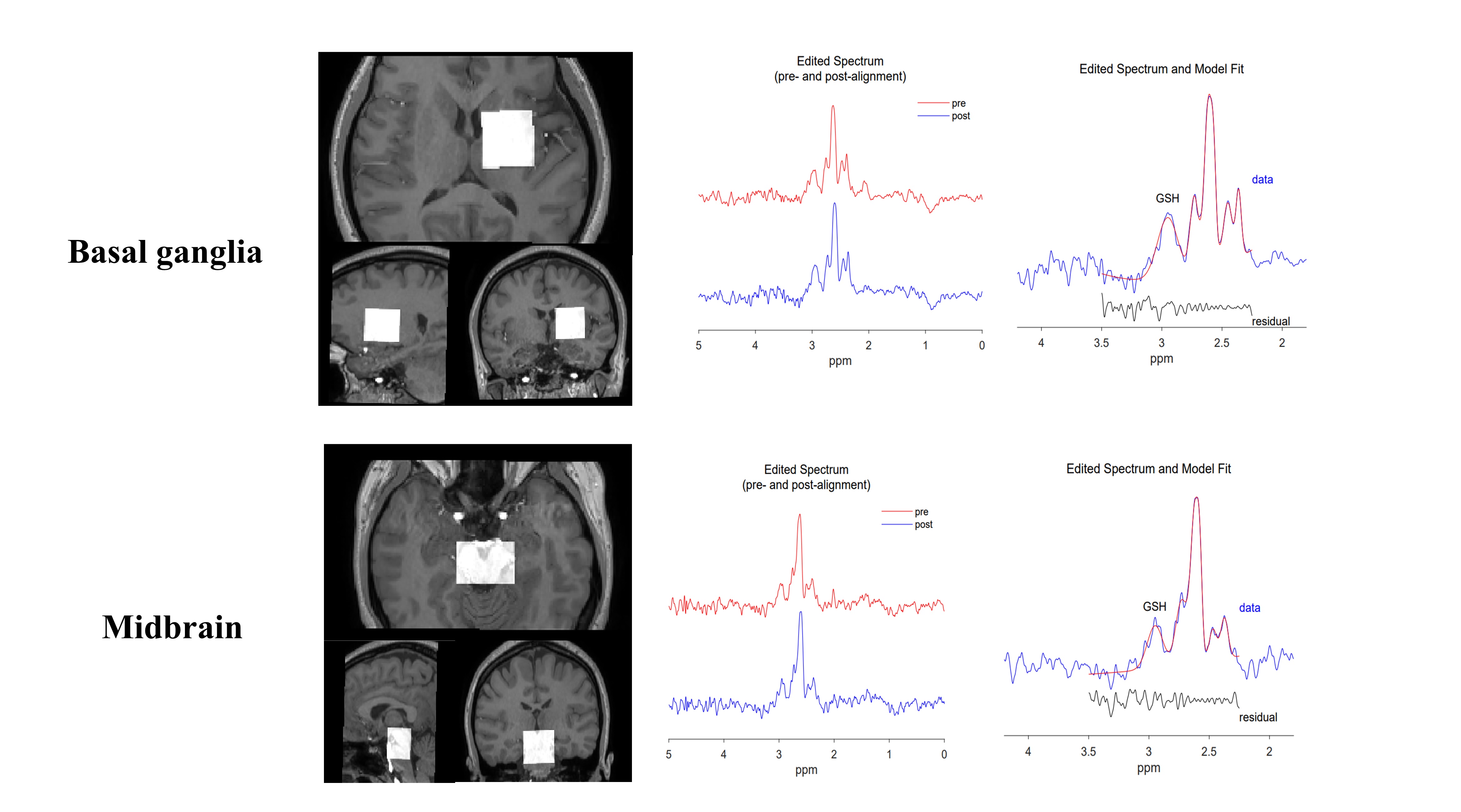
Figure 1. The location the proton magnetic resonance spectroscopy (1H-MRS) volume of interest in the basal ganglia and midbrain regions and the corresponding GSH peak from a representative subject. The edited spectrum is depicted in blue, the estimated metabolite model in red, and the residual in black.
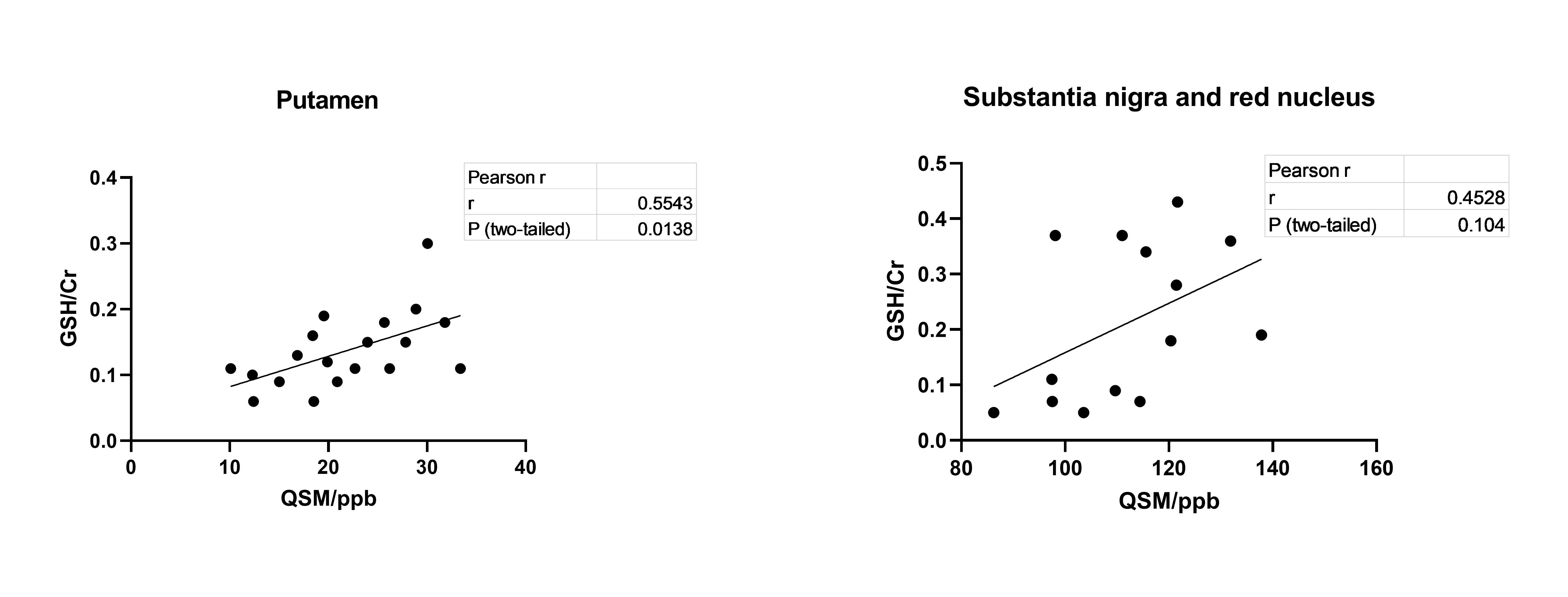
Figure 2. The relationship between the glutathione/creatine ratio (GSH/Cr) in the volume of interest (basal ganglia and midbrain regions) and the iron content in the putamen (basal ganglia region) and substantia nigra and red nucleus (midbrain region).
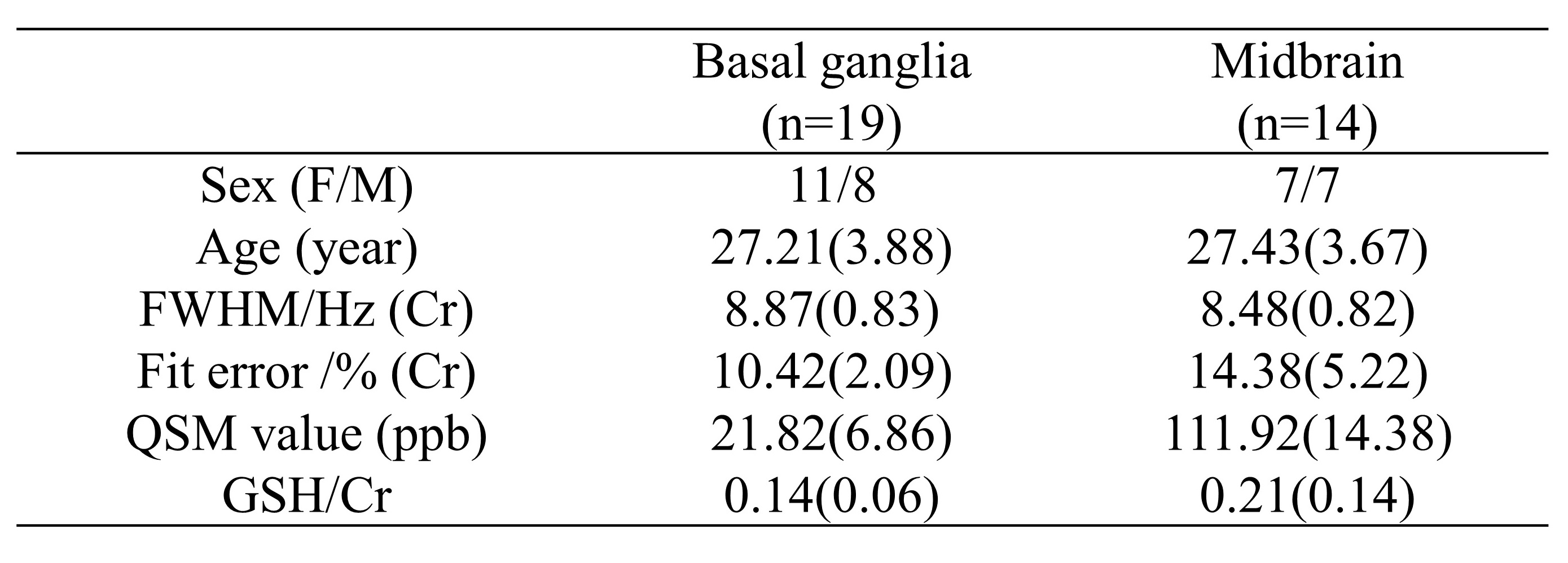
Table 1. The demographic and imaging features of the participants.
Note: FWHM, Full Width Half Maximum; QSM: Quantitative Susceptibility Mapping; ppb: partial per billion; GSH: glutathione; Cr: Creatine.