0315
Subcortical structures alterations in Parkinson's disease patients with depression by Vertex-based morphological analysis1The First Affiliated Hospital of Dalian Medical University, Dalian, China
Synopsis
Keywords: Parkinson's Disease, Neurodegeneration
Motivation: Subcortical structure is critical to the pathogenesis of Parkinson's disease (PD) and depression.
Goal(s): shape analysis can precisely localise regional shape deformations in the subcortical gray matter and detect changes that are not found in VBM and volumetric analyses.
Approach: This study explored the shape change of subcortical gray matter nuclei in Parkinson's disease patients with depression (PDD).
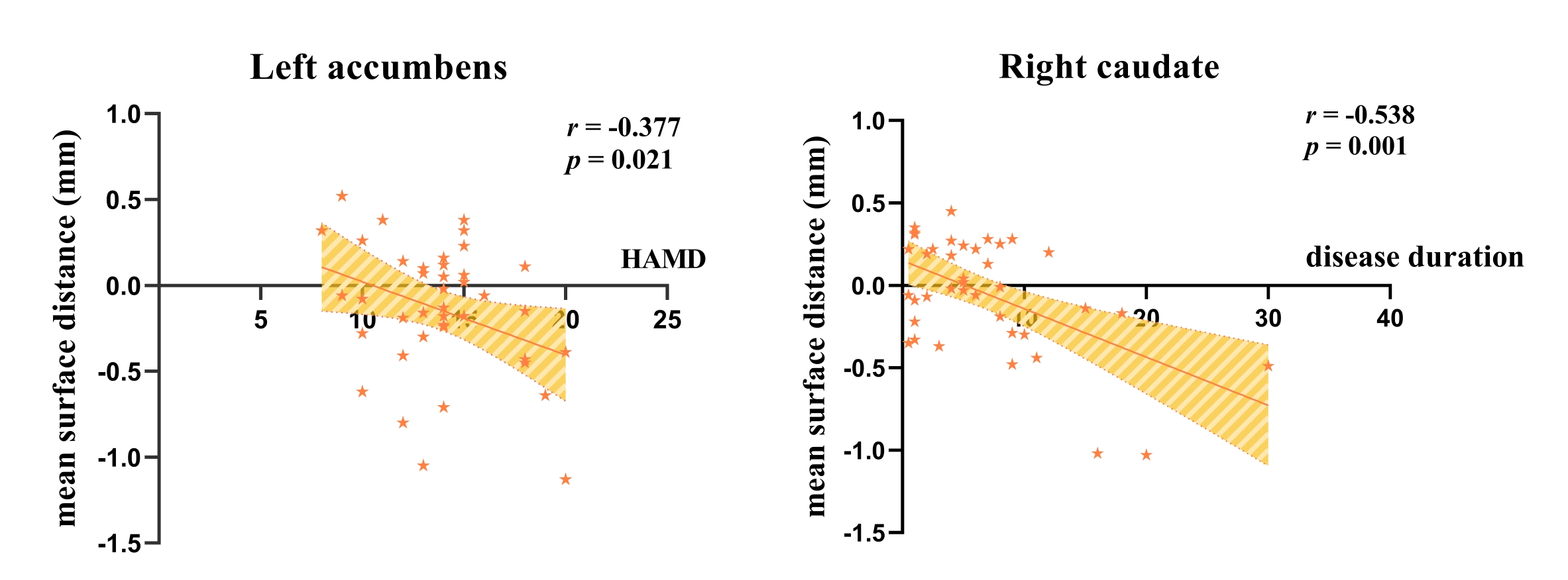
Results: PDD patients have multiple subcortical morphological atrophy, and local expansion areas were also found in several nuclei. The shape change of left nucleus accumbens was negatively correlated with Hamilton depression scale (HAMD) scores, and the shape change of right caudate nucleus was negatively correlated with disease duration.
Impact: This study suggested that PDD patients have multiple subcortical morphological atrophy, and local expansion areas were also found in several nuclei. Partial correlation analysis showed that the shape change of left nucleus accumbens was negatively correlated with HAMD scores.
Introduction
Depression is the most common psychiatric disease in Parkinson's disease (PD), occurring in approximately half of the patients. Subcortical structure is critical to the pathogenesis of Parkinson's disease (PD) and depression. Although studies exploring cerebral modification related to PD often focus on the striatum[1], it is common knowledge that pathophysiological modifications go far beyond putamen and caudate nucleus, reaching many other subcortical nuclei and cortical areas. However, the effect of subcortical structural abnormalities on PD with depression (PDD) remains unknown. The structure volume is a global measurement and thus it cannot quantify localized morphometrics. Using shape analysis to investigate PD can accurately locate the regional atrophy or expansion of subcortical structures, and provide a sensitive, hopefully correlating with disease duration and with the severity of depressive symptoms.Materials and Methods
This study included 40 PDD patients, 26 PD patients without depression (PDND) patients and 33 healthy controls (HC) matched by sex, age and education. We used the Hamilton Depression Scale (HAMD), with a HAMD score of > 7 in the PDD group and a HAMD score of ≤7 in the PDND group. All participants underwent neuropsychological examination and 3T MRI to acquire 3D-T1 sequence. Segmentation was performed using FIRST. The following subcortical nuclei were automatically segmented: left and right nucleus accumbens, amygdala, caudate nucleus, hippocampus, globus pallidus, putamen, and thalamus. A permutation-based non-parametric analysis of subcortical shape data and general linear models comprise age, gender and education as covariates. Results were corrected for multiple comparisons using threshold-free cluster enhancement methods (P<0.05). The mean surface distances values of local areas of nuclei with significant differences are extracted for subsequent analysis to evaluate the correlation between subcortical nucleus volume and HAMD scores and clinical data.Result
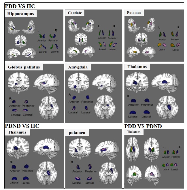
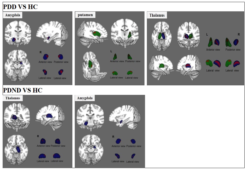
Compared with HC group, there were inhomogeneous atrophy of local areas on the surface of multiple nuclei in PDD group, including bilateral hippocampal (left P=0.016, right P=0.043), bilateral putamen (left P<0.001, right P=0.001), bilateral caudate (left P=0.019, right P=0.002), left nucleus accumbens (P=0.013), right globus pallidus (P=0.002), right thalamus (P=0.028) and right amygdala (P=0.017). The local expansion area was also found in the bilateral thalamus (left P=0.024, right P=0.001), the left putamen (P=0.01) and the right amygdala (P=0.007). In PDND group, compared with HC, there were local atrophy areas in the right putamen (P=0.015) and the right thalamus (P=0.016), while there were small expansion areas in the right thalamus (P=0.039) and the right amygdala (P=0.038). Compared with PDND group, there are extensive atrophy areas in bilateral thalamus (left P=0.023, right P=0.04) in PDD group (Figure 1-2). In partial correlation analysis, the regional shape deformation of the left nucleus accumbens was negatively correlated with the HAMD score (r=-0.377, P=0.021), and the regional shape deformation of the right caudate nucleus was negatively correlated with disease duration (r=-0.538, P=0.001) (Figure 3).Discussion
In this study, we investigated the relationship between changes in subcortical structural volume and depression symptoms in PDD patients using vertexwise shape analyses. Although there are surface atrophy of multiple subcortical nuclei in PDD patients, we found both atrophy and expansion effects in several nuclei (thalamus, amygdala, and putamen). In the literature, it has been repeatedly reported that the greatest dopamine depletion in PD is found in the head of the caudate nucleus and the bilateral caudal putamen of PD patients[2]. The clusters of local atrophy we found in the striatum were compatible with these areas of maximum dopamine depletion. We found that there were significant amygdala and hippocampus atrophy in PDD, and there was a small expansion area in the right amygdala, which was consistent with previous findings[3]. it is suggested the functional hyperactivity in the right amygdala may be a potential compensation mechanism for its volume decrease. These findings confirmed the existence of morphological abnormalities of amygdala and hippocampus in the early stage of PDD pathology to some extent. A study found that decreased activation in the left mediodorsal thalamus and in medial prefrontal cortex in PD patients with depression compared to those without depression. In addition, VBM analysis showed increased volume of MD thalamic nuclei bilaterally in the PD depressed group[4].Conclusion
In conclusion, we confirmed that there were multiple subcortical nuclei surface atrophy in patients with PDD, and some nuclei were also found to be locally expanded. These findings contribute to a deeper understanding of the mechanisms underlying the neurological symptoms of patients with PDD. Our results also show that shape analysis can reveal the surface deformation of subcortical structures that can not be detected by traditional volume analysis.Acknowledgements
We would like to thank all participants and staff involved in this research.References
[1] Pitcher TL, Melzer TR, Macaskill MR, Graham CF, Livingston L, Keenan RJ, Watts R, Dalrymple-Alford JC, Anderson TJ. Reduced striatal volumes in Parkinson's disease: a magnetic resonance imaging study. Transl Neurodegener. 2012 Aug 21;1(1):17. doi: 10.1186/2047-9158-1-17.
[2] Nemmi F, Sabatini U, Rascol O, Péran P. Parkinson's disease and local atrophy in subcortical nuclei: insight from shape analysis. Neurobiol Aging. 2015 Jan;36(1):424-33. doi: 10.1016/j.neurobiolaging.2014.07.010. Epub 2014 Jul 23.
[3] Yao Z, Fu Y, Wu J, Zhang W, Yu Y, Zhang Z, Wu X, Wang Y, Hu B. Morphological changes in subregions of hippocampus and amygdala in major depressive disorder patients. Brain Imaging Behav. 2020 Jun;14(3):653-667. doi: 10.1007/s11682-018-0003-1.
[4] Cardoso EF, Maia FM, Fregni F, Myczkowski ML, Melo LM, Sato JR, Marcolin MA, Rigonatti SP, Cruz AC Jr, Barbosa ER, Amaro E Jr. Depression in Parkinson's disease: convergence from voxel-based morphometry and functional magnetic resonance imaging in the limbic thalamus. Neuroimage. 2009 Aug 15;47(2):467-72. doi: 10.1016/j.neuroimage.2009.04.059. Epub 2009 May 3.
Figures